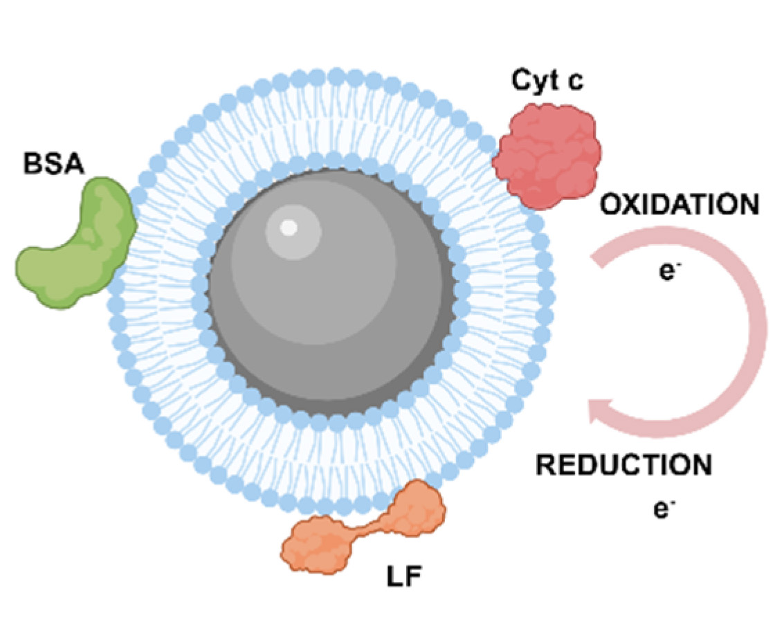
The dynamic behavior of lipids at membrane interfaces is essential for maintaining the structural stability and functional specificity of biomembranes, with its regulation closely linked to protein interactions and the redox microenvironment. Understanding the interaction between proteins and phospholipid membranes, as well as the impact of redox processes on membrane structures, is therefore crucial for clarifying membrane functions and mechanisms. This study developed a phospholipid bilayer-on-plasmonic nanoparticle (BOM) configuration using an extruder. Based on this configuration, we conducted a systematic investigation of the dynamic interactions and concentration-dependent effects of bovine serum albumin (BSA), cytochrome c (Cyt c), and lactoferrin (LF) on zwitterionic dioleoylphosphatidylcholine (DOPC) bilayers coated silver nanoparticles (DOPC@Ag) via surface-enhanced Raman spectroscopy (SERS). Furthermore, by leveraging a non-physiological artificial electron-transport chain model, we elucidated the redox-dependent regulatory mechanisms of Cyt c on both synthetic phospholipid membranes and natural cellular membranes. Our observations provide insight into the lipid-protein-redox interaction network, offering suggestions for membrane biosensor design, targeted drug delivery systems, and the study of membrane-related physiological and pathological processes.
- Open Access
- Article
SERS Studies on the Dynamic Response of Membrane Interface Lipids to Proteins: Concentration Effect and Redox State Conversion
- Ji Sha 1,2,
- Xin Wang 1,
- Yan Zhou 1,
- Yifan Chen 1,
- Junyi Zhao 1,
- Lili Cong 1,3,
- Jingjing Chang 2,*,
- Shuping Xu 1,4,*
Author Information
Received: 31 Mar 2026 | Revised: 25 May 2026 | Accepted: 31 May 2026 | Published: 16 Jun 2026
Abstract
Graphical Abstract
Keywords
surface-enhanced Raman spectroscopy | phospholipid membrane | lipid-protein interaction | redox | cytochrome c
References
- 1.
Jelokhani-Niaraki, M. Membrane proteins: Structure, function and motion. Int. J. Mol. Sci. 2022, 24, 468.
- 2.
Vance, J.A.; Devaraj, N.K. Membrane mimetic chemistry in artificial cells. J. Am. Chem. Soc. 2021, 143, 8223–8231.
- 3.
Almén, M.S.; Nordström, K.J.; Fredriksson, R.; Schiöth, H.B. Mapping the human membrane proteome: A majority of the human membrane proteins can be classified according to function and evolutionary origin. BMC Biol. 2009, 7, 50.
- 4.
Kusumi, A.; Nakada, C.; Ritchie, K.; Murase, K.; Suzuki, K.; Murakoshi, H.; Kasai, R.S.; Kondo, J.; Fujiwara, T. Paradigm shift of the plasma membrane concept from the two-dimensional continuum fluid to the partitioned fluid: High-speed single-molecule tracking of membrane molecules. Annu. Rev. Biophys. Biomol. Struct. 2005, 34, 351–378.
- 5.
Simons, K.; Gerl, M.J. Revitalizing membrane rafts: New tools and insights. Nat. Rev. Mol. Cell Biol. 2010, 11, 688–699.
- 6.
Fajardo, V.A.; McMeekin, L.; LeBlanc, P.J. Influence of phospholipid species on membrane fluidity: A meta-analysis for a novel phospholipid fluidity index. J. Membr. Biol. 2011, 244, 97–103. https://doi.org/10.1007/s00232-011-9401-7.
- 7.
Crosio, M.A.; Wilke, N. The neutral Red probe location in large unilamellar vesicles: Influence of membrane composition and phase states. J. Photochem. Photobiol. A Chem. 2024, 452, 163–171. https://doi.org/10.1016/j.jphotochem.2024.115615.
- 8.
Bahja, J.; Dymond, M.K. Does membrane curvature elastic energy play a role in mediating oxidative stress in lipid membranes? Free Radic. Biol. Med. 2021, 171, 191–202. https://doi.org/10.1016/j.freeradbiomed.2021.05.021.
- 9.
Anitha, S.; Jayasree, R.; Kulanthaivel, L.; Subbaraj, G.K.; Muthuvel, R. Advanced techniques for analyzing the protein–lipid interactions. In Biochemical Techniques for Analyzing Protein-Lipid Interactions; Patel, A., Ed.; Springer Nature Singapore: Singapore, 2024; pp. 157–175.
- 10.
Arrondo, J.L.R.; Goñi, F.M. Chapter 13 Infrared Spectroscopic Studies of Lipid-Protein Interactions in Membranes. In New Comprehensive Biochemistry; Watts, A., Ed.; Elsevier: Amsterdam, The Netherlands, 1993; Volume 25, pp. 321–349.
- 11.
Tatulian, S.A. Analysis of protein–protein and protein–membrane interactions by isotope-edited infrared spectroscopy. Phys. Chem. Chem. Phys. 2024, 26, 21930–21953. https://doi.org/10.1039/D4CP01136H.
- 12.
Heider, S.; Reimhult, E.; Metzner, C. Real-time analysis of protein and protein mixture interaction with lipid bilayers. Biochim. Et Biophys. Acta Biomembr. 2018, 1860, 319–328. https://doi.org/10.1016/j.bbamem.2017.10.024.
- 13.
Ouyang, H.; Moore, D.J.; Sills, R.H.; Mendelsohn, R. FT-IR studies of sickle hemoglobin interaction with phosphatidylserine. Spectrosc. Int. J. 2004, 18, 407–413. https://doi.org/10.1155/2004/805318.
- 14.
Huster, D. Solid-state NMR spectroscopy to study protein lipid interactions. Biochim. Et Biophys. Acta-Mol. Cell Biol. Lipids 2014, 1841, 1146–1160. https://doi.org/10.1016/j.bbalip.2013.12.002.
- 15.
Drücker, P.; Gerke, V.; Galla, H.J. Importance of phospholipid bilayer integrity in the analysis of protein-lipid interactions. Biochem. Biophys. Res. Commun. 2014, 453, 143–147. https://doi.org/10.1016/j.bbrc.2014.09.079.
- 16.
Bhowmik, D.; Mote, K.R.; MacLaughlin, C.M.; Biswas, N.; Chandra, B.; Basu, J.K.; Walker, G.C.; Madhu, P.K.; Maiti, S. Cell-membrane-mimicking lipid-coated nanoparticles confer Raman enhancement to membrane proteins and reveal membrane-attached amyloid-β conformation. ACS Nano 2015, 9, 9070–9077. https://doi.org/10.1021/acsnano.5b03175.
- 17.
Tu, A.T. Raman Spectroscopy in Biology: Principles and Applications; John Wiley & Sons Hoboken: Hoboken, NJ, USA, 1982.
- 18.
Ma, H.; Li, L.; Bi, X.; Li, P.; Bao, Y.; Wang, X.; Guo, H.; Chen, Z.; Zhang, W.; Wu, Z.; et al. Surfaced-enhanced Raman spectroscopy (SERS) 50 Years: Theories, applications and perspectives. J. Light Scatt. 2025, 37, 357–514. https://doi.org/10.13883/j.issn1004-5929.202503001. (In Chinese)
- 19.
Zhu, H.; Zhang, J.; Dai, X.; Mesias, V.S.D.; Chi, H.; Wang, C.; Yeung, C.S.; Chen, Q.; Liu, W.; Huang, J. Tunable lipid-coated nanoporous silver sheet for characterization of protein-membrane interactions by surface-enhanced Raman scattering (SERS). Anal. Bioanal. Chem. 2023, 415, 3243–3253.
- 20.
Xu, G.; Li, W.; Xie, H.; Zhu, J.; Song, L.; Tang, J.; Miao, Y.; Han, X.X. In situ monitoring of membrane protein electron transfer via surface-enhanced resonance Raman spectroscopy. Anal. Chem. 2024, 96, 6–11. https://doi.org/10.1021/acs.analchem.3c04700.
- 21.
Yin, H.Y.; Vergeade, A.; Shi, Q.; Zackert, W.E.; Gruenberg, K.C.; Bokiej, M.; Amin, T.; Ying, W.Z.; Masterson, T.S.; Zinkel, S.S.; et al. Acetaminophen inhibits cytochrome c redox cycling induced lipid peroxidation. Biochem. Biophys. Res. Commun. 2012, 423, 224–228. https://doi.org/10.1016/j.bbrc.2012.05.058.
- 22.
Vladimirov, G.K.; Nesterova, A.M.; Levkina, A.A.; Osipov, A.N.; Teselkin, Y.O.; Kovalchuk, M.V.; Vladimirov, Y.A. The dynamics of the formation of cytochrome c complexes with anionic lipids and the mechanism of the production of lipid radicals catalyzed by these complexes. Biol. Membr. 2020, 37, 287–298. https://doi.org/10.31857/S0233475520040088.
- 23.
Gorbenko, G.P.; Trusova, V.M.; Molotkovsky, J.G.; Kinnunen, P.K.J. Cytochrome c induces lipid demixing in weakly charged phosphatidylcholine/phosphatidylglycerol model membranes as evidenced by resonance energy transfer. Biochim. Et Biophys. Acta-Biomembr. 2009, 1788, 1358–1365. https://doi.org/10.1016/j.bbamem.2009.03.007.
- 24.
Zhao, J.; Zhao, L.; Xu, W.; Lu, Z.; Xu, S. Fabrication of high-negatively charged bicelle-mediated supported lipid bilayer. Langmuir 2024, 40, 8083–8093.
- 25.
An, H.H.; Han, W.B.; Kim, Y.; Kim, H.S.; Oh, Y.; Yoon, C.S. Preparation of SERS active Ag nanoparticles encapsulated by phospholipids. J. Raman Spectrosc. 2014, 45, 292–298. https://doi.org/10.1002/jrs.4461.
- 26.
Giancaspro, J.; Scollan, P.; Rosario, J.; Miller, E.; Braziel, S.; Lee, S. Structural determination of model phospholipid membranes by Raman spectroscopy: Laboratory experiment. Biochem. Mol. Biol. Educ. 2022, 50, 181–192.
- 27.
Bangham, A.D. Diffusion of univalent ions across the lamellae of swollen phospholipids. Journal of Molecular Biology 1965, 13, 238–252.
- 28.
Gaber, B.P.; Peticolas, W.L. Quantitative interpretation of biomembrane structure by Raman-spectroscopy. Biochim. Et Biophys. Acta-Biomembr. 1977, 465, 260–274. https://doi.org/10.1016/0005-2736(77)90078-5.
- 29.
Chen, X.; Al-Mualem, Z.A.; Baiz, C.R. Lipid Landscapes: Vibrational spectroscopy for decoding membrane complexity. Annu. Rev. Phys. Chem. 2024, 75, 283–305.
- 30.
Jin, S.; Lednev, I.K.; Jung, Y.M. Recent trends in surface-enhanced Raman spectroscopy-based biosensors: Label-free early disease diagnosis. J. Phys. Chem. C 2024, 128, 8861–8873. https://doi.org/10.1021/acs.jpcc.4c0201
- 31.
Greig, J.C.; Tipping, W.J.; Graham, D.; Faulds, K.; Gould, G.W. New insights into lipid and fatty acid metabolism from Raman spectroscopy. Analyst 2024, 149, 4789–4810. https://doi.org/10.1039/D4AN00846D.
- 32.
Li, J.B.; Cheng, W.N.; Wang, X.L.; Zhang, H.J.; Jin, J.; Ji, W.; Han, X.X.; Zhao, B. Electron transfer of Cytochrome c on surface-enhanced Raman scattering-active substrates: material dependence and biocompatibility. Chem. A Eur. J. 2017, 23, 9034–9038. https://doi.org/10.1002/chem.201702307.
- 33.
Zhu, J.Y.; Jiang, M.W.; Ma, H.; Zhang, H.J.; Cheng, W.N.; Li, J.B.; Cai, L.J.; Han, X.X.; Zhao, B. Redox-State-Mediated Regulation of Cytochrome c Release in Apoptosis revealed by surface-enhanced Raman scattering on Nickel substrates. Angew. Chem. Int. Ed. 2019, 58, 16499–16503. https://doi.org/10.1002/anie.201909638.
- 34.
Zivanovic, V.; Kochovski, Z.; Arenz, C.; Lu, Y.; Kneipp, J. SERS and cryo-EM directly reveal different liposome structures during interaction with gold nanoparticles. J. Phys. Chem. Lett. 2018, 9, 6767–6772. https://doi.org/10.1021/acs.jpclett.8b03191.
- 35.
Zivanovic, V.; Seifert, S.; Drescher, D.; Schrade, P.; Werner, S.; Guttmann, P.; Szekeres, G.P.; Bachmann, S.; Schneider, G.; Arenz, C.; et al. Optical nanosensing of lipid accumulation due to enzyme inhibition in live cells. ACS Nano 2019, 13, 9363–9375. https://doi.org/10.1021/acsnano.9b04001.
- 36.
Zivanovic, V.; Milewska, A.; Leosson, K.; Kneipp, J. Molecular structure and interactions of lipids in the outer membrane of living cells based on surface-enhanced Raman scattering and liposome models. Anal. Chem. 2021, 93, 10106–10113. https://doi.org/10.1021/acs.analchem.1c00964.
- 37.
Lee, P.C.; Meisel, D. Adsorption and surface-enhanced Raman of dyes on silver and gold sols. J. Phys. Chem. 1982, 86, 3391–3395.

This work is licensed under a Creative Commons Attribution 4.0 International License.
 Suite 4002 Level 4, 447 Collins Street, Melbourne, Victoria 3000, Australia
 General Inquiries: info@sciltp.com