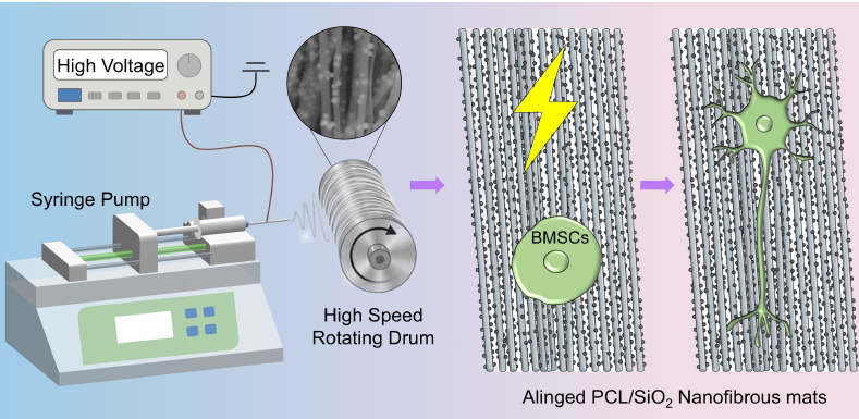
Peripheral nerve injury (PNI) often leads to disability and chronic pain, with limited options available to promote regeneration and functional recovery. Bone marrow mesenchymal stem cells (BMSCs) are considered promising candidates for cell therapy in PNI repair. However, the effective induction of BMSCs towards neurogenesis and the directed migration of cells remain challenging. Here, we constructed a biomimetic environment by combining uniaxially aligned polycaprolactone (PCL) nanofibers containing silica nanoparticles (SiO2 NPs) with electrical stimulation (ES). A proper portion of SiO2 NPs was uniformly integrated into uniaxially aligned PCL nanofibers to create nanoscale protrusions on fiber surfaces. Such fibrous mats showed uniaxially aligned morphology, uniform fiber diameter, and improved wettability. BMSCs were then cultured on both the nanofibers with smooth surfaces (PCL) or those decorated with nanoscale protrusions (PCL/SiO2), followed by treatment with or without ES, with glass slides used as a control. All the fibrous mats showed good cell viability, and the uniaxially aligned fibers induced better cell extension and alignment in comparison to the control group. Both the contact guidance provided by the nanoscale protrusions on fiber surfaces and the biochemical signal from ES contributed to BMSC differentiation, with a combination of both promoting the greatest differentiation into neural-like cells. Immunofluorescence micrographs demonstrated significantly increased expression of NF200 and S100β, along with a higher proportion of NF200‑positive cells compared with S100β, indicating the preferred differentiation of BMSCs to neuron-like cells under such conditions. Additionally, in migration assays the greatest number and longest migration distance of BMSCs were exhibited on the PCL/SiO2 nanofibers with ES. These studies offer valuable strategies for the manipulation of stem cell behavior in PNI repair.
- Open Access
- Article
Synergistic Effects of Topographical Guidance and Electrical Stimulation on Modulation of BMSC Behaviors Using Electrospun Nanofibers Decorated with Nanoscale Protrusions
- Yuying Yan 1,
- Binbin Sun 2,
- Melissa LD Rayner 3,
- Yuanfei Wang 1,4,
- Tong Wu 1,4,*
Author Information
Received: 14 Apr 2026 | Revised: 31 May 2026 | Accepted: 05 Jun 2026 | Published: 08 Jun 2026
Abstract
Graphical Abstract
Keywords
bone marrow mesenchymal stem cells | nanofibers | nanoscale protrusions | electrical stimulation | neural differentiation
References
- 1.
Ahmad, P.; Estrin, N.; Farshidfar, N.; Zhang, Y.; Miron, R. Isolation methods of exosomes derived from dental stem cells. Int. J. Oral Sci. 2025, 17, 846–855.
- 2.
Chen, L.; He, D.; Zhang, Y. Differentiation of mesenchymal stem cells from rat bone marrow into dopaminergic neuron-like cells in vitro. Blood 2007, 110, 4067.
- 3.
Wei, Z.; Fan, B.; Liu, Y.; Ding, H.; Tang, H.; Pan, D.; Shi, J.; Zheng, P.; Shi, H.; Wu, H.; et al. MicroRNA changes of bone marrow-derived mesenchymal stem cells differentiated into neuronal-like cells by schwann cell-conditioned medium. Neural Regen. Res. 2019, 14, 1462–1469.
- 4.
Simsar, E.; Cheng, P.; Dogruel, T.; Donta, M.; Jung, J.; Asante, N.; Sakaguchi, D.; Mallapragada, S.; Kidambi, P.; Metin, U. Few-layered conductive graphene foams for electrical transdifferentiation of mesenchymal stem cells into schwann cell-like phenotypes. Adv. Healthc. Mater. 2026, 15, e02204.
- 5.
Li, J.; Zhang, D.; Guo, S.; Zhao, C.; Wang, L.; Ma, S.; Guan, F.; Yao, M. Dual-enzymatically cross-linked gelatin hydrogel promotes neural differentiation and neurotrophin secretion of bone marrow-derived mesenchymal stem cells for treatment of moderate traumatic brain injury. Int. J. Biol. Macromol. 2021, 187, 200–213.
- 6.
Wan, X.; Liu, Z.; Li, L. Manipulation of stem cells fates: The master and multifaceted roles of biophysical cues of biomaterials. Adv. Funct. Mater. 2021, 31, 2010626.
- 7.
Liu, N.; Ning, X.; Zhang, X.; Zhou, Z.; Fu, M.; Wang, Y.; Wu, T. Gradient galectin-1 coating technology: Bionic multichannel nerve guidance conduits promote neural cell migration. Adv. Technol. Neurosci. 2024, 1, 276–289.
- 8.
Sousa, M.; Arab-Tehrany, E.; Cleymand, F.; Mano, J. Surface micro- and nanoengineering: Applications of layer-by-layer technology as a versatile tool to control cellular behavior. Small 2019, 15, e1901228.
- 9.
Zhang, X.; Guo, M.; Guo, Q.; Liu, N.; Wang, Y.; Wu, T. Modulating axonal growth and neural stem cell migration with the use of uniaxially aligned nanofiber yarns welded with NGF-loaded microparticles. Mater. Today Adv. 2023, 17, 100343.
- 10.
Chen, R.; Wang, Y.; Yu, C.; Zhang, X.; Wang, Y.; Yu, T.; Wu, T. Bioactive glass-reinforced hybrid microfibrous spheres promote bone defect repair via stem cell delivery. Adv. Funct. Mater. 2025, 7, 240–253.
- 11.
Xue, J.; Wu, T.; Li, J.; Zhu, C.; Xia, Y. Promoting the outgrowth of neurites on electrospun microfibers by functionalization with electrosprayed microparticles of fatty acids. Angew. Chem. Int. Ed. Engl. 2019, 58, 3948–3951.
- 12.
Li, Z.; Zhou, Y.; Li, T.; Zhang, J.; Tian, H. Stimuli-responsive hydrogels: Fabrication and biomedical applications. Biomater. Sci. 2022, 3, 20200112.
- 13.
Yao, L.; Yan, Y.; Zhang, H.; Zhang, X.; Lan, J.; Chen, Y.; Rayner, M.; Wang, Y.; Wu, T. Engineered nanofiber-based nerve guidance conduit facilitates the restoration of peripheral nerve injury through enhanced vascularization. Small 2026, 22, e14312.
- 14.
Wei, S.; Xiong, F.; Gu, H.; Zhang, Z.; Xuan, H.; Jin, Y.; Xue, Y.; Li, B.; Feng, W.; Yuan, H. Highly aligned electroactive ultrafine fibers promote the differentiation of mesenchymal stem cells into schwann-like cells for nerve regeneration. Int. J. Biol. Macromol. 2024, 279, 135388.
- 15.
Wu, J.; Hong, Y. Enhancing cell infiltration of electrospun fibrous scaffolds in tissue regeneration. Bioact. Mater. 2016, 1, 56–64.
- 16.
Xue, J.; Xie, J.; Liu, W.; Xia, Y. Electrospun nanofibers: New concepts, materials, and applications. Acc. Chem. Res. 2017, 50, 1976–1987.
- 17.
Fang, J.; Nan, L.; Song, K.; Weng, Z.; Shan, J.; Shahin, V.; Liu, J.; Qian, Y. Application and progress of bionic scaffolds in nerve repair: A narrative review. Adv. Technol. Neurosci. 2024, 1, 43–50.
- 18.
Wang, J.; Zhou, Z.; Zhang, X.; Fu, M.; Fang, K.; Wang, Y.; Wu, T. One-Step manufacture and crosslinking of gelatin/polygonum sibiricum polysaccharide bioactive nanofibrous sponges for rapid hemostasis and infected wound healing. Adv. Fiber Mater. 2025, 7, 1148–1164.
- 19.
Chen, S.; Zhang, X.; Guo, Q.; Yan, Y.; Fu, M.; Wang, Y.; Wu, T. Complex bioactive nanofibrous dura mater repairs traumatic brain injury. Neural Regen. Res. 2026, 21, 4275–4289.
- 20.
Chaudry, A.; Wu, J.; Wang, H.; Mo, X.; Bhutto, M.; Sun, B. Research and application of conductive nanofiber nerve guidance conduits for peripheral nerve regeneration: A narrative review. Adv. Technol. Neurosci. 2025, 2, 47–57.
- 21.
Dethe, M.; Prabakaran, A.; Ahmed, H.; Agrawal, M.; Roy, U.; Alexander, A. PCL-PEG copolymer based injectable thermosensitive hydrogels. J. Control. Release 2022, 343, 217–236.
- 22.
Wang, X.; Chen, S.; Chen, X.; Wu, J.; Huang, Z.; Wang, J.; Chen, F.; Liu, C. Biomimetic multi-channel nerve conduits with micro/nanostructures for rapid nerve repair. Bioact. Mater. 2024, 41, 577–596.
- 23.
Wang, H.; Zhang, P.; Lu, P.; Cai, X.; Wang, G.; Xu, X.; Liu, Y.; Huang, T.; Li, M.; Qian, T.; et al. Neural tissue-engineered prevascularization in vivo enhances peripheral neuroregeneration via rapid vascular inosculation. Mater. Today Bio. 2023, 21, 100718.
- 24.
Wang, Y.; Zhang, X.; Yao, L.; Yan, Y.; Wang, Y.; Wu, T. “Cell climbing stones”-varying the surfaces of electrospun nanofibers with protrusions as secondary structures to manipulate neural cell behaviors. Nanoscale Horiz. 2025, 10, 2411–2421.
- 25.
Liu, Y.; Zhang, X.; Wang, Y.; Guo, M.; Sheng, J.; Wang, Y.; Wu, T. Promoting neurite outgrowth and neural stem cell migration using aligned nanofibers decorated with protrusions and galectin-1 coating. Chem. Commun. 2023, 59, 10753–10756.
- 26.
Wang, X.; Xu, Q.; Xu, Y.; Yuan, L.; Guan, X.; Ma, S.; Zhang, D.; Liu, X.; Li, J.; Zhang, T.; et al. Portable absorbable electrical stimulation system for enhanced peripheral nerve repair. Adv. Funct. Mater. 2025, 35, 2417839.
- 27.
Yi, Z.; Lin, Y.; Jing, R.; Feng, X.; Lu, X.; Tian, D.; Lin, H.; Zhao, L. Dual biomimetic nanofiber conduits enable synergistic NGF delivery and endogenous piezoelectric stimulation for peripheral nerve regeneration. Adv. Fiber Mater. 2026, 8, 338–358.
- 28.
Guan, W.; Liu, Y.; Jia, M.; Wang, L.; Gao, H.; Sun, S.; Shang, Y.; Shen, H.; Yang, J.; Jin, N.; et al. Magnetic-topological multistage synergy: Anisotropic ovalbumin scaffolds loaded with magnetically-responsive neural cells for long-distance peripheral nerve regeneration. Bioact. Mater. 2026, 57, 36–53.
- 29.
Zhang, J.; Li, F.; Gao, X.; Qiu, W.; Xia, B.; He, S.; Zhang, Y.; Huang, X.; Liu, B.; Huang, J.; et al. Bamboo-inspired composite conduit accelerates peripheral nerve regeneration through synergistic oriented structure and piezoelectricity. Adv. Mater. 2026, 38, e09425.
- 30.
Zhang, N.; Yao, X.; Zhang, Q.; Zhang, C.; Zheng, Q.; Wang, Y.; Shan, F. Electrical stimulation promotes peripheral nerve regeneration by upregulating glycolysis and oxidative phosphorylation. Biochim. Biophys. Acta Mol. Basis Dis. 2025, 1871, 167804.
- 31.
Yan, X.; Liu, J.; Huang, J.; Huang, M.; He, F.; Ye, Z.; Xiao, W.; Hu, X.; Luo, Z. Electrical stimulation induces calcium-dependent neurite outgrowth and immediate early genes expressions of dorsal root ganglion neurons. Neurochem. Res. 2014, 39, 129–141.
- 32.
Yuan, L.; Zhu, Y.; Li, J.; Jiang, C.; Xu, Q.; Wang, X.; Liu, X.; Wang, X.; Zhang, A.; Zhang, T.; et al. Mesenchymal stem cell-driven neurotrophic bioelectronic platform (MSC-NBP) potentiated peripheral nerve regeneration. Adv. Funct. Mater. 2025, 36, e23049.
- 33.
Yang, Y.; Yin, X.; Wang, H.; Qiu, W.; Li, L.; Li, F.; Shan, Y.; Zhao, Z.; Li, Z.; Guo, J.; et al. Engineering a wirelessly self-powered and electroconductive scaffold to promote peripheral nerve regeneration. Nano Energ. 2023, 107, 108145.
- 34.
Qin, C.; Yue, Z.; Forster, R.; Chen, J.; Wallace, G. On demand, wireless electrochemical release of brain derived neurotrophic factor. Electrochem. Commun. 2023, 157, 107626.
- 35.
Huang, Y.; Jing, W.; Li, Y.; Cai, Q.; Yang, X. Composites made of polyorganophosphazene and carbon nanotube up-regulating osteogenic activity of BMSCs under electrical stimulation. Colloids Surf. B Biointerf. 2021, 204, 111785.
- 36.
Cheng, H.; Huang, Y.; Chen, W.; Che, J.; Liu, T.; Na, J.; Wang, R.; Fan, Y. Cyclic strain and electrical co-stimulation improve neural differentiation of marrow-derived mesenchymal stem cells. Front. Cell Dev. Biol. 2021, 9, 624755.
- 37.
Min, G.; Peng, Y.; Wang, W.; Wang, T.; Zhang, Y.; Yin, Z.; Lv, F.; Dong, X.; Xu, S.; Xu, K. Biodegradable dual-stimuli hydrogel scaffoldoid synergizing piezoionic and lithium-ion release for critical-sized bone defect regeneration. Adv. Funct. Mater. 2025, 36, 2515477.
- 38.
Han, Z.; Wang, F.; Xiong, W.; Meng, C.; Yao, Y.; Cui, W.; Zhang, M. Precise cell type electrical stimulation therapy via force-electric hydrogel microspheres for cartilage healing. Adv. Mater. 2025, 37, e2414555.
- 39.
Soltani, A.; Azimzadeh, A.; Behboodi, S.; Mamdoohi, M.; Kajbafzadeh, A.-M.; Slavin, K.; Rahimi-Movaghar, V.; Hassannejad, Z. Electrical stimulation enhances sciatic nerve regeneration using a silk-based conductive scaffold beyond traditional nerve guide conduits. Sci. Rep. 2024, 14, 15196.
- 40.
Greasley, S.; Page, S.; Sirovica, S.; Chen, S.; Martin, R.; Riveiro, A.; Hanna, J.; Porter, A.; Jones, J. Controlling particle size in the stöber process and incorporation of calcium. J. Colloid Interf. Sci. 2016, 469, 213–223.
- 41.
Stöber, W.; Fink, A.; Bohn, E. Controlled growth of monodisperse silica spheres in the micron size range. J. Colloid Interf. Sci. 1968, 26, 62–69.
- 42.
Wu, T.; Xue, J.; Xia, Y. Engraving the surface of electrospun microfibers with nanoscale grooves promotes the outgrowth of neurites and the migration of schwann cells. Angew. Chem. Int. Ed. Engl. 2020, 59, 15626–15632.
- 43.
Parisi, G.; Szewczyk, P.; Narayan, S.; Ura, D.; Knapczyk-Korczak, J.; Stachewicz, U. Multifunctional piezoelectric yarns and meshes for efficient fog water collection, energy harvesting and sensing. Small Sci. 2024, 4, 00021.
- 44.
Parisi, G.; Szewczyk, P.; Narayan, S. Photoresponsive electrospun fiber meshes with switchable wettability for effective fog water harvesting in variable humidity conditions. ACS Appl. Mater. Interfaces 2023, 33, 15.
- 45.
Huang, J.; Li, J.; Li, S.; Yang, X.; Huo, N.; Chen, Q.; Wang, W.; Yang, N.; Wang, Y.; Zhou, N. Netrin-1-engineered endothelial cell exosomes induce the formation of pre-regenerative niche to accelerate peripheral nerve repair. Sci. Adv. 2024, 10, eadm8454.
- 46.
Dutta, K.; Saikia, A.; Singh, A. Transforming lignin into polymer film with improved physiochemical properties by modification with itaconic acid and grafting with polycaprolactone. Int. J. Biol. Macromol. 2025, 305, 141226.
- 47.
Hyde, E.; Moreno-Atanasio, R.; Neville, F. Fabrication of magnetic core PEI-silica shell particles. Mater. Res. Bull. 2017, 96, 222–232.
- 48.
Sundaram, V.; Schütza, V.; Schröter, N.; Backhaus, A.; Bilsing, A.; Joneck, L.; Seelbach, A.; Mutschler, C.; Gomez-Sanchez, J.; Schäffner, E.; et al. Adipo-glial signaling mediates metabolic adaptation in peripheral nerve regeneration. Cell Metab. 2023, 35, 2136–2152.
- 49.
Guo, J.; Guo, Z.; Huang, Y.; Ma, S.; Yan, B.; Pan, C.; Jiang, Z.; Wang, F.; Zhang, Z.; Da, Y.; et al. Blockage of MLKL prevents myelin damage in experimental diabetic neuropathy. Proc. Natl. Acad. Sci. USA 2022, 119, e2121552119.
- 50.
Cohen, C.; Popovic, M.; Klooster, J.; Weil, M.; Möbius, W.; Nave, K.; Kole, M. Saltatory conduction along myelinated axons involves a periaxonal nanocircuit. Cell 2020, 180, 311–322.
- 51.
Joe, H.; Seo, H.; Dolkas, J.; Jawala, M.; Hullugundi, S.; Chung, Y.; Patel, H.; Chernov, A.; Shubayev, V. TIMP-1 associates with myelin membrane and preserves myelin in injured peripheral nerve. Neurobiol. Dis. 2025, 209, 106892.
- 52.
Egger, R.; Tupikov, Y.; Elmaleh, M.; Katlowitz, K.; Benezra, S.; Picardo, M.; Moll, F.; Kornfeld, J.; Jin, D.; Long, M. Local axonal conduction shapes the spatiotemporal properties of neural sequences. Cell 2020, 183, 537–548.
- 53.
Sun, Y.; Zhang, H.; Zhang, Y.; Liu, Z.; He, D.; Xu, W.; Li, S.; Zhang, C.; Zhang, Z. Li-Mg-Si bioceramics provide a dynamic immuno-modulatory and repair-supportive microenvironment for peripheral nerve regeneration. Bioact. Mater. 2023, 28, 227–242.
- 54.
Xiao, L.; Sun, Y.; Liao, L.; Su, X. Response of mesenchymal stem cells to surface topography of scaffolds and the underlying mechanisms. J. Mater. Chem. B 2023, 11, 2550–2567.
- 55.
Yu, L.; Wu, Q.; Jin, F.; Zhang, Y.; Li, M.; Javanmardi, N.; Zhu, H.; Wang, Y.; Yu, X.; Zhou, G.; et al. Self-sustained biomimetic bioelectronic accelerated metabolic reprogramming of bone regeneration. Biomaterials 2026, 325, 123572.

This work is licensed under a Creative Commons Attribution 4.0 International License.
 Suite 4002 Level 4, 447 Collins Street, Melbourne, Victoria 3000, Australia
 General Inquiries: info@sciltp.com