
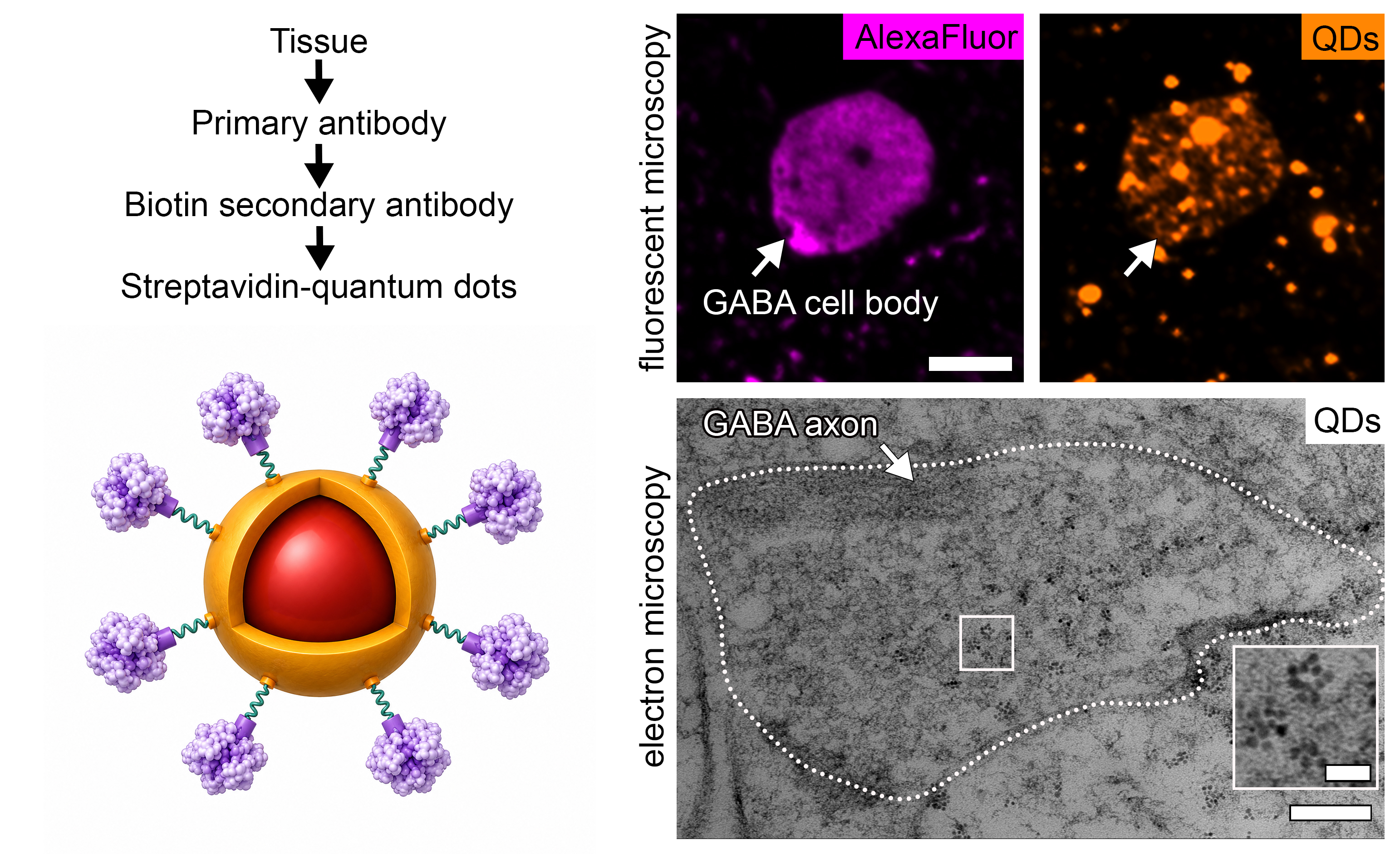
Detecting molecules across different spatial scales remains a major challenge in biological samples, particularly in complex tissues such as brain, where linking molecular identification and nanoscale structural detail is essential for understanding function. Fluorescence microscopy provides selective labeling of specific molecules but cannot fully resolve many cellular and subcellular features, whereas electron microscopy reveals ultrastructure without molecular specificity. Bridging these modalities is difficult because commonly used fluorescent probes such as dyes and proteins cannot provide electron contrast. Quantum dots are particularly useful as they retain fluorescence while providing sufficient electron density for ultrastructural visualization. Here, we show that streptavidin-functionalized CdSe/CdS core/shell quantum dots can function as dual-modal probes for labeling γ-aminobutyric acid (GABA) neurons in brain tissue. The quantum dots produce bright and stable fluorescence signals for optical detection while their inorganic composition generates strong electron contrast in ultrathin sections examined by transmission electron microscopy. This work demonstrates that functionalized CdSe/CdS quantum dots can serve as stable dual-modal probes for fluorescence and electron microscopy in complex tissues, highlighting the potential of semiconductor nanocrystals for multifunctional imaging applications.
- Open Access
- Communication
Imaging of Brain Tissue Using CdSe/CdS Core/Shell Quantum Dots for Correlative Fluorescence and Electron Microscopy
Author Information
Received: 15 May 2026 | Revised: 05 Jul 2026 | Accepted: 25 Jul 2026 | Published: 14 Aug 2026
Abstract
Graphical Abstract
References
- 1.
Rah, J.C.; Choi, J.H. Finding Needles in a Haystack with Light: Resolving the Microcircuitry of the Brain with Fluorescence Microscopy. Mol. Cells 2022, 45, 84–92.
- 2.
Yin, C.; Li, J.; Meng, K.; Zhang, J.; Chen, M.; Chen, R.; Hu, Y.; Wang, S.; Xie, S. Fluorescent Labeling Methods for Brain Structure Research. Molecules 2026, 31, 817.
- 3.
Luján, R.; Rubio, M.E. Editorial: Immunoelectron Microscopy: Placing Molecular Functions within a Neuronal Context. Front. Neuroanat. 2022, 16, 1043371.
- 4.
Jones, J.C.R. Pre- and Post-Embedding Immunogold Labeling of Tissue Sections. Methods Mol. Bio. 2016, 1474, 291–307.
- 5.
Lucocq, J. Particulate Markers for Immunoelectron Microscopy. In Fine Structure Immunocytochemistry; Griffiths, G., Ed.; Springer: Berlin/Heidelberg, Germany, 1993; pp. 279–306.
- 6.
Humbel, B.M.; Sibon, O.C.; Stierhof, Y.D.; Schwarz, H. Ultra-Small Gold Particles and Silver Enhancement as a Detection System in Immunolabeling and in Situ Hybridization Experiments. J. Histochem. Cytochem. 1995, 43, 735–737.
- 7.
Stierhof, Y.-D.; Humbel, B.M.; Hermann, R.; Otten, M.T.; Schwarz, H. Direct Visualization and Silver Enhancement of Ultra-Small Antibody-Bound Gold Particles on Immunolabeled Ultrathin Resin Sections. Scanning Microsc. 1992, 6, 12.
- 8.
Petralia, R.S.; Wang, Y.X. Review of Post-Embedding Immunogold Methods for the Study of Neuronal Structures. Front. Neuroanat. 2021, 15, 763427.
- 9.
Mayer, G.; Leone, R.D.; Hainfeld, J.F.; Bendayan, M. Introduction of a Novel HRP Substrate-Nanogold Probe for Signal Amplification in Immunocytochemistry. J. Histochem. Cytochem. 2000, 48, 461–469.
- 10.
Greytak, A.B.; Allen, P.M.; Liu, W.; Zhao, J.; Young, E.R.; Popović, Z.; Walker, B.J.; Nocera, D.G.; Bawendi, M.G. Alternating Layer Addition Approach to CdSe/CdS Core/Shell Quantum Dots with near-Unity Quantum Yield and High on-Time Fractions. Chem. Sci. 2012, 3, 2028–2034.
- 11.
Chen, O.; Zhao, J.; Chauhan, V.P.; Cui, J.; Wong, C.; Harris, D.K.; Wei, H.; Han, H.S.; Fukumura, D.; Jain, R.K.; et al. Compact High-Quality CdSe-CdS Core-Shell Nanocrystals with Narrow Emission Linewidths and Suppressed Blinking. Nat. Mater. 2013, 12, 445–451.
- 12.
Murray, C.B.; Norris, D.J.; Bawendi, M.G. Synthesis and Characterization of Nearly Monodisperse CdE (E = S, Se, Te) Semiconductor Nanocrystallites. J. Am. Chem. Soc. 1993, 115, 8706–8715.
- 13.
Nisman, R.; Dellaire, G.; Ren, Y.; Li, R.; Bazett-Jones, D.P. Application of Quantum Dots as Probes for Correlative Fluorescence, Conventional, and Energy-Filtered Transmission Electron Microscopy. J. Histochem. Cytochem. 2004, 52, 13–18.
- 14.
Smith, A.M.; Nie, S. Chemical Analysis and Cellular Imaging with Quantum Dots. Analyst 2004, 129, 672–677.
- 15.
Cotta, M.A. Quantum Dots and Their Applications: What Lies Ahead? Appl. Nano Mater. 2020, 3, 4920–4924.
- 16.
Susumu, K.; Uyeda, H.T.; Medintz, I.L.; Pons, T.; Delehanty, J.B.; Mattoussi, H. Enhancing the Stability and Biological Functionalities of Quantum Dots via Compact Multifunctional Ligands. J. Am. Chem. Soc. 2007, 129, 13987–13996.
- 17.
Walther, T. Electron Microscopy of Quantum Dots. J. Microsc. 2015, 257, 171–178.
- 18.
Ogunkoya, Y.; Nickel, B.M.; Gay, V.L.; Murray, S.A. Using Quantum Dots to Visualize Clathrin Associations. Biotech. Histochem. 2009, 84, 109–115.
- 19.
Bera, D.; Qian, L.; Tseng, T.K.; Holloway, P.H. Quantum Dots and Their Multimodal Applications: A Review. Materials 2010, 3, 2260–2345.
- 20.
Killingsworth, M.C.; Bobryshev, Y.V. Correlative Light- and Electron Microscopy Using Quantum Dot Nanoparticles. J. Vis. Exp. 2016, 114, e54307.
- 21.
Uematsu, M.; Mikami, K.; Nakamura, A.; Takahashi, R.; Yokota, T.; Hirokawa, K.; Uchihara, T. Parallel Gold Enhancement of Quantum Dots 565/655 for Double-Labelling Correlative Light and Electron Microscopy on Human Autopsied Samples. Sci. Rep. 2022, 12, 6113.
- 22.
Deerinck, T.J. The Application of Fluorescent Quantum Dots to Confocal, Multiphoton, and Electron Microscopic Imaging. Toxicol. Pathol. 2008, 36, 112–116.
- 23.
Giepmans, B.N.G.; Adams, S.R.; Ellisman, M.H.; Tsien, R.Y. The Fluorescent Toolbox for Assessing Protein Location and Function. Science 2006, 312, 217–224.
- 24.
Pérez-Garza, J.; Orea, J.; Deane, Z.; Raimondi, G.; Tripp, R.; Charles, I.; Ostroff, L. Ultraplex Microscopy: Versatile Highly-Multiplexed Molecular Labeling and Imaging across Scale and Resolution. bioRxiv 2024. https://doi.org/10.1101/2024.08.17.605585.
- 25.
Wang, X.; Chen, S.; Thota, S.; Wang, Y.; Tan, H.; Tang, M.; Quan, Z.; Zhao, J. Anisotropic Arm Growth in Unconventional Semiconductor CdSe/CdS Nanotetrapod Synthesis Using Core/Shell CdSe/CdS as Seeds. J. Phys. Chem. C 2019, 123, 19238–19245.
- 26.
Bayer, E.A.; Wilchek, M. Application of Avidin—Biotin Technology to Affinity-Based Separations. J. Chromatogr. A 1990, 510, 3–11.
- 27.
Michael Green, N. Avidin and Streptavidin. Methods Enzymol. 1990, 184, 51–67.
- 28.
Chen, Y.; Ren, H.L.; Liu, N.; Sai, N.; Liu, X.; Liu, Z.; Gao, Z.; Ning, A.N. A Fluoroimmunoassay Based on Quantum Dot−Streptavidin Conjugate for the Detection of Chlorpyrifos. J. Agric. Food Chem. 2010, 58, 8895–8903.
- 29.
Cardoso Dos Santos, M.; Algar, W.R.; Medintz, I.L.; Hildebrandt, N. Quantum Dots for Förster Resonance Energy Transfer (FRET). Trends Anal. Chem 2020, 125, 115819.
- 30.
Clegg, R.M. Fluorescence Resonance Energy Transfer. Curr. Opin. Biotechnol. 1995, 6, 103–110.
- 31.
Medintz, I.L.; Clapp, A.R.; Mattoussi, H.; Goldman, E.R.; Fisher, B.; Mauro, J.M. Self-Assembled Nanoscale Biosensors Based on Quantum Dot FRET Donors. Nat. Mater. 2003, 2, 630–638.
- 32.
Lu, X.; Hou, X.; Tang, H.; Yi, X.; Wang, J. A High-Quality CdSe/CdS/ZnS Quantum-Dot-Based FRET Aptasensor for the Simultaneous Detection of Two Different Alzheimer’s Disease Core Biomarkers. Nanomaterials 2022, 12, 4031.
- 33.
Medintz, I.L.; Mattoussi, H. Quantum Dot-Based Resonance Energy Transfer and Its Growing Application in Biology. Phys. Chem. Chem. Phys. 2008, 11, 17–45.
- 34.
Clapp, A.R.; Medintz, I.L.; Mauro, J.M.; Fisher, B.R.; Bawendi, M.G.; Mattoussi, H. Fluorescence Resonance Energy Transfer Between Quantum Dot Donors and Dye-Labeled Protein Acceptors. J. Am. Chem. Soc. 2003, 126, 301–310.
- 35.
McDonald, A.J. Functional Neuroanatomy of the Basolateral Amygdala: Neurons, Neurotransmitters, and Circuits. In Handbook of Behavioral Neuroscience; Elsevier: Amsterdam, The Netherlands, 2020; Volume 26, pp. 1–38.
- 36.
McDonald, A.J. Immunohistochemical Identification of γ-Aminobutyric Acid-Containing Neurons in the Rat Basolateral Amygdala. Neurosci. Lett. 1985, 53, 203–207.
- 37.
Jiang, C.; Chen, Y.; Sun, T. From the Gut to the Brain, Mechanisms and Clinical Applications of γ-Aminobutyric Acid (GABA) on the Treatment of Anxiety and Insomnia. Front. Neurosci. 2025, 19, 1570173.
- 38.
Shamirian, A.; Samareh Afsari, H.; Hassan, A.; Miller, L.W.; Snee, P.T. In Vitro Detection of Hypoxia Using a Ratiometric Quantum Dot-Based Oxygen Sensor. ACS Sens. 2016, 1, 1244–1250.
- 39.
Tosat-Bitrián, C.; Avis-Bodas, A.; Porras, G.; Borrego-Hernández, D.; García-Redondo, A.; Martín-Requero, A.; Palomo, V. CdSe Quantum Dots in Human Models Derived from ALS Patients: Characterization, Nuclear Penetration Studies and Multiplexing. Nanomaterials 2021, 11, 671.
- 40.
Le, P.; Lim, S.J.; Baculis, B.C.; Chung, H.J.; Kilian, K.A.; Smith, A.M. Counting Growth Factors in Single Cells with Infrared Quantum Dots to Measure Discrete Stimulation Distributions. Nat. Commun. 2019, 10, 909.
- 41.
Zrazhevskiy, P.; Gao, X. Quantum Dot Imaging Platform for Single-Cell Molecular Profiling. Nat. Commun.2013, 4, 1619.
- 42.
Wu, J.K.; Tian, Z.Q.; Zhang, Z.L.; Liu, A.A.; Tang, B.; Zhang, L.J.; Chen, Z.L.; Pang, D.W. Purification of Quantum Dot-Based Bioprobes via High-Performance Size Exclusion Chromatography. Talanta 2016, 159, 64–73.

This work is licensed under a Creative Commons Attribution 4.0 International License.

 Suite 4002 Level 4, 447 Collins Street, Melbourne, Victoria 3000, Australia
 General Inquiries: info@sciltp.com