
Autophagy is integral in the protection and survival of cardiomyocytes in heart disease. If impaired, it can lead to apoptosis of healthy cardiac tissue. This review discusses the role of autophagy in heart disease. Primary mechanisms and their benefits for cellular homeostasis in cardiomyocytes will be explored as well as the effects of dysregulated autophagy on aggravating conditions and the potential therapeutic approaches to modulate autophagy to improve outcomes in heart disease. Autophagy degrades damaged cellular components during myocardial infarction, increasing energy levels to reduce Ischemic/Reperfusion injury. However, over-activation of autophagy can contribute to this injury by forming excessive autophagosomes, which destabilize mitochondria, consequently accelerating cell apoptosis and cardiac injury. In atherosclerosis, disrupted autophagy promotes cell apoptosis, exacerbating inflammation and worsening atherogenesis. In contrast, tightly regulated autophagy promotes cell survival in vascular smooth muscle cells. Autophagy helps adaptively remodel the heart and compensate for cardiac overload. Abnormal levels of autophagy can increase the progression of cardiac hypertrophy to heart failure by degrading crucial cellular components. Autophagy plays various roles in many heart diseases, making it a promising target in developing new therapies to recover and minimize heart disease damage. When exploring pharmacological treatments for diabetic cardiomyopathy, a large percentage are autophagy modulators, which have the potential to be utilized to promote cardiac repair in cardiovascular disease.
- Open Access
- Review
The Role of Autophagy and Potential Drug Targets in Cardiovascular Disease
- Vaseega Sathianandan 1,
- Tayyiba Azam 2,*
Author Information
Received: 19 Dec 2025 | Revised: 09 Feb 2026 | Accepted: 09 Feb 2026 | Published: 07 Aug 2026
Abstract
Keywords
autophagy | cardiovascular disease | pharmacological modulation | myocardial infarction | diabetic cardiomyopathy
1. Introduction
Heart disease is a significant worldwide health problem, with cardiovascular disease (CVD) being the most common cause of death globally [1]. From the year 1993 to 2019, the worldwide prevalence of CVD has almost doubled, acting as a tremendous economic burden on healthcare systems such as the National Health Service [1,2]. This highlights the importance of understanding the mechanisms regulating cardiac health in heart diseases and the need to develop new therapeutic approaches to combat these diseases. One promising area of research is autophagy. This cytoprotective mechanism in cardiomyocytes, helps minimize cardiac damage. Autophagy is a tightly regulated, adaptive process; it contributes to cellular homeostasis by facilitating cell survival through the removal of damaged organelles and proteins, and by inducing apoptosis to reduce tissue damage. Although autophagy initially plays a protective role, its dysregulation can turn it maladaptive. This paradoxical role makes autophagy a significant mechanism in preventing and potentially exacerbating heart diseases such as cardiac hypertrophy, heart failure, myocardial infarction and atherosclerosis. This review aims to explore the mechanisms of autophagy, key heart diseases and the emerging therapies targeting autophagy in cardiac repair.
2. The Mechanisms of Autophagy
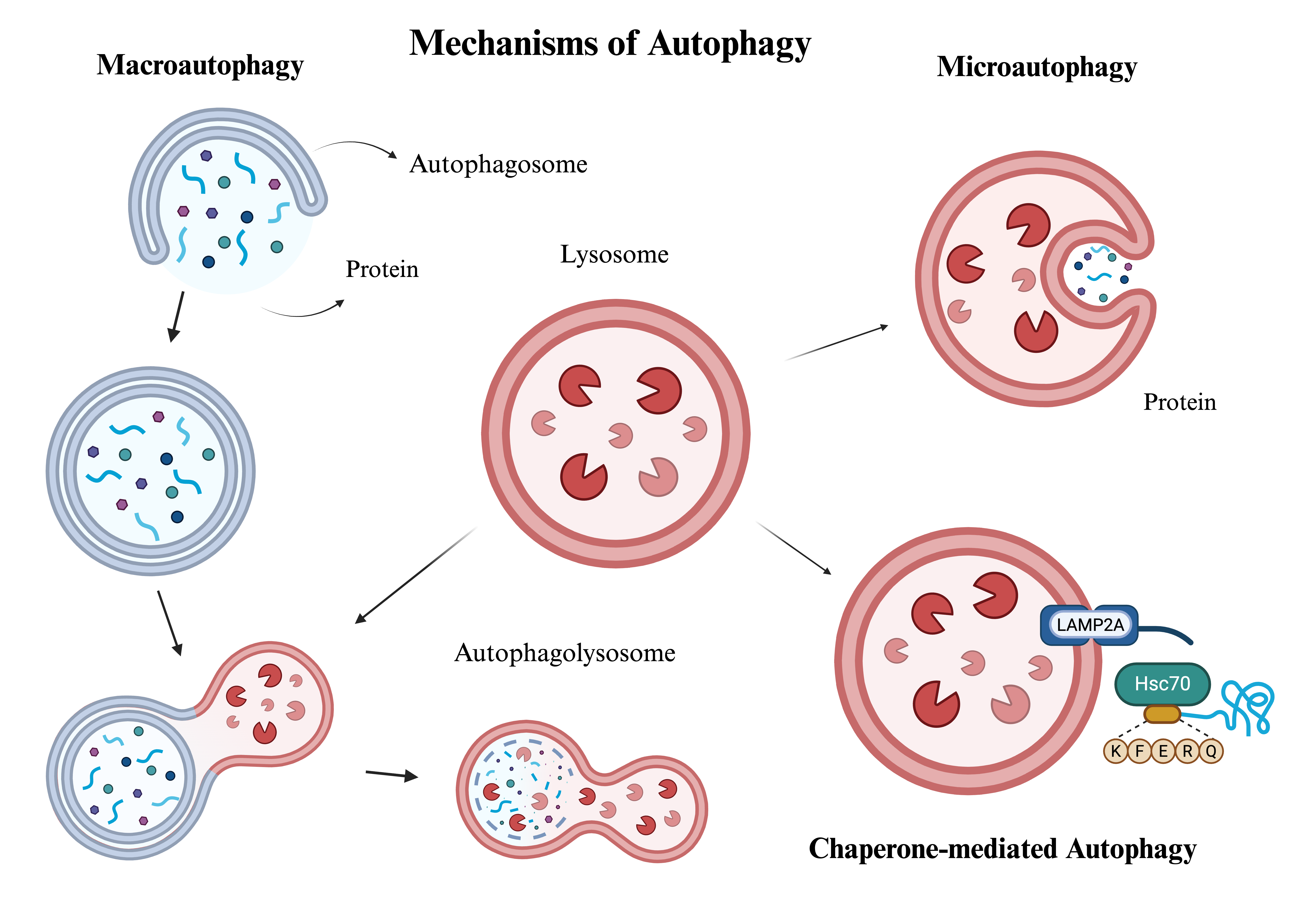
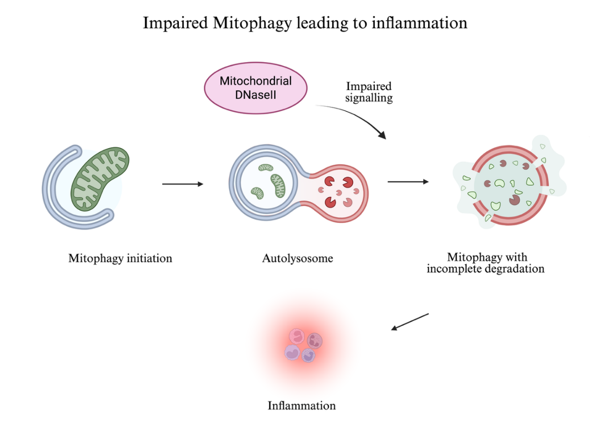
Autophagy is a complex and adaptive mechanism that preserves cardiac function by tightly regulating the degradation of cytoplasmic components. Autophagy plays many intricate roles in cardiac homeostasis, from maintaining ATP levels in stressed cardiomyocytes to regulating the quality control of cellular components [3]. These functions are mediated by three main types of autophagy, each with a distinct mechanism of action (Figure 1). Macroautophagy plays a crucial role in the turnover and autophagy of damaged organelles and in adaptation to cellular stress, thereby sustaining cellular homeostasis and metabolism [3,4]. This process entails the formation of autophagosomes, a double-membraned cytosolic vesicle that fuses with lysosomes for degradation, forming autolysosomes [3,4,5]. Lysosomes also play an integral role in microautophagy, where they form membrane invaginations to directly engulf and facilitate the cellular components [3,4]. Similarly, chaperone-mediated autophagy also involves the direct transport of cytoplasmic material to the lysosome [3,4,6,7]. This is mediated by the KFERQ sequence, which allows proteins to bind to the chaperone Hsc70, which delivers them to lysosomal-associated membrane protein (LAMP) type 2A for degradation in the lumen [3,4,6,7]. The intricate process of autophagy is established by the biosynthesis and maturation of the autophagosome [6]. This involves many steps in its linear progression, regulated by the ATG proteins; these include initiation, nucleation, expansion, and closure of the vesicle [3,6]. The ULK1 complex—which consists of ULK1, ATG101, FIP200, and ATG13- promotes the initiation of the phagophore, an essential precursor for autophagosome formation [3,6,8]. The molecular interactions between the unc-51-like autophagy-activating kinase (ULK1) and the mechanistic target of rapamycin complex one (mTORC1) are pivotal for maintaining autophagy and regulating cardiomyocyte function [6]. mTORC1 phosphorylates ULK1 and ATG13, hence suppressing autophagy in the presence of abundant amino acids and growth factor signals. When these nutrient supplies deplete, mTORC1 levels fall, allowing ULK1 to activate autophagy [3,5,8]. Contrarily, adenosine monophosphate-activated protein kinase (AMPK) elevates autophagy by phosphorylating ULK1, causing its dissociation from mTORC1 [3]. AMPK is sensitive to intracellular AMP/ATP levels [9]. In response to depleted energy stores, AMPK triggers the Tumour Suppressor Complex TSC1/2, which acts as an indirect inhibitor of MTORC1 [8]. During heart disease, cardiomyocytes are under stress, cells become nutrient-deprived, and mTORC1 activity diminishes [3,8]. Macroautophagy is initiated as mTORC1 is released from ULK1 and ATG13, leaving them moderately dephosphorylated [9]. Dysregulation in the induction of autophagy and imbalance in these mechanisms can critically exacerbate heart disease. To mediate the nucleation and expansion of the autophagosome membrane, ULK1 phosphorylates certain constituents of the PI3K complex (Figure 2), which consists of Beclin1, ATG14, VPS34, VPS15, and AMBRA1 [3,8]. The elongation of autophagosomes is facilitated by microtubule-associated protein 1 light chain 3 (LC3) and ATG12-ATG5-ATG16, key components of the Atg conjugation system, which also plays an integral role in the closure of the autophagosome [3,10]. By facilitating the fission and degradation of autophagosomal membranes, this system promotes the fusion of the lysosome and the autophagosome, forming an autolysosome [3,9]. Autophagic flux is defined as the entirety of the autophagy process from initiation to the lysosomal degradation of cellular matter. This intricately regulated process is an essential cardioprotective mechanism that is activated by homeostatic imbalance [6]. An increase in autophagosomes alone does not accurately represent effective breakdown whereas an increase in autophagic flux indicates successful completion of autophagy [6]. Whilst this process is essential for the disintegration of cytoplasmic components, excessive initiation can lead to detrimental consequences, such as promoting cardiac hypertrophy [11]. Autophagy can be nonselective, in which autophagosomes, when faced with cardiac distress, take up random cellular components for breakdown [6]. Selective autophagy regulates the degradation of distinct organelles; based on the composition of the targeted substrate, it can be divided into distinct types [6,8]. Mitophagy is a key type of selective autophagy that directs the catabolic recycling of damaged mitochondria and plays a pivotal role in maintaining cardiac stability [11]. Whilst mitophagy reduces cell death by eliminating mitochondrial damage and recycling their cellular components, impaired mitophagy can trigger inflammatory responses in cardiac tissue [3,4]. This aggravates the progression of heart disease. Lipophagy is initiated when lipid droplets accumulate, serving as a primary mechanism for the selective degradation of lipid deposits in cardiac tissue [3].
3. The Role of Autophagy in Cardiac Hypertrophy and Heart Failure
Cardiac hypertrophy is the adaptive remodeling of the heart in response to stressors such as hypertension, myocardial infarction, and heart valve disorders, contributing to the maladaptive thickening or enlargement of the cardiac muscle [6,13]. Despite starting as an adaptive process compensating for elevated cardiac overload, cardiac hypertrophy can become detrimental, as it can be a major contributor to heart failure and arrhythmia [13]. It is mainly distinguished by excess biosynthesis, growth in cardiomyocyte size and the thickening of ventricular walls [13]. Autophagy plays an integral role in the breakdown of damaged cellular components and misfolded and aged proteins in hypertrophic cells, as their toxic accumulation can damage cardiomyocytes [11]. In damaged heart tissue, mitophagy mitigates the detrimental effects of reactive oxygen species (ROS) on cardiomyocytes by degrading dysfunctional mitochondria [11]. Specifically, a study on PINK1-mediated mitophagy suggests that it can play a protective role in cardiac hypertrophy by inhibiting the mtDNA-cGAS-STING pathway [14]. In this study, PINK1/STING (stimulator of interferon genes) double-knockout mice had a substantially reduced PINK1 expression in TAC-induced hypertrophy [14]. When mitochondrial DNA was released into the cytosol in mice, triggering cGAS-STING signalling, cardiac inflammation was activated, exacerbating cardiac hypertrophy [14]. However, when cardiac-specific PINK1 is overexpressed, it inhibits the cGAS-STING signalling [14]. This prevents the detrimental effects of cGAS-STING signalling, reinforcing the idea that PINK1-mediated mitophagy can be a protective mechanism in cardiac hypertrophy [14]. This role of autophagy in heart disease is further demonstrated in a study examining ultrastructural findings in cardiomyocytes from patients with dilated cardiomyopathy (DCM). This study found an abundance of autophagic vacuoles in their heart tissue using transmission electron microscopy (TEM), demonstrating that DCM patients undergo the initiation of the autophagy pathway during cardiac recovery. In this study, patients with positive left ventricular reverse remodelling (LVRR) exhibited a substantially elevated autolysosome-to-autophagosome ratio, as well as increased numbers of autophagic vacuoles and lysosomes, compared with the LVRR-negative group [15]. These findings of a favorable clinical outcome reinforce the idea that effective debris clearance through autophagy is a protective mechanism of the heart. The initial role of adaptive autophagy in cardiac hypertrophy is thought to compensate for increased stress on the ventricular wall due to elevated oxygen demand [4]. Gradually, however, this protective role can turn maladaptive if autophagy becomes dysregulated in heart tissue. When the contractile function of the heart declines significantly due to prolonged deterioration of its ability to deliver blood, and it cannot meet the current demand for oxygen, heart failure may occur [16]. Heart failure is a critical condition in which the heart receives an insufficient supply of oxygen, often due to a loss of the ability to pump blood efficiently [16]. There are many factors in the progression of this illness, some being chronic cardiac stress, pressure-induced injury, or volume overload conditions like hypertension [16]. Autophagy can regulate inflammation and maintain ATP levels in cardiac tissue; by improving myocyte contractility, it can play a key role in preventing heart failure [8,10]. Autophagy is often described as a double-edged sword. If autophagy is unregulated, it becomes maladaptive. Autophagy in cardiomyocytes is a highly regulated and complex process, and a disruption in the coordination between autophagy and cellular components is often the cause of its dysfunction. In heart disease, where oxidative stress damages cardiac tissue, mitophagy plays a pivotal role in the breakdown and turnover of mitochondrial components; this is facilitated by mitochondrial DNase II (Figure 2). Impaired signalling between mitochondrial DNase II and mitophagy can lead to the incomplete degradation of mitochondrial DNA, leading to sterile inflammation, a key factor in the development of heart failure [3]. In addition to dysregulated mitophagy under oxidative stress, the various complex mechanisms of general autophagy also contribute to cardiac pathology. In cases of pressure overload, autophagy can mediate the progression of cardiac hypertrophy to heart failure by promoting adverse cardiac remodelling [4]. In an experiment on transgenic mice, the overexpression of the autophagy regulator Beclin1 in cardiomyocytes exacerbated cardiac remodeling and hypertrophy. Meanwhile, partial loss of Beclin1 through haploinsufficiency reduced autophagy and partially protected the heart [4]. Furthermore, studies suggest that, under pressure overload, whilst minimal autophagy helps alleviate cardiomyocyte injury, excessive autophagic repair is detrimental to the heart [10]. They exacerbate injury by promoting macrophage engulfment and degradation of dead cells, thereby activating inflammation [10]. This occurs because, while autophagy can degrade dysfunctional cellular components and contribute to cardiac injury, excessive autophagy can break down functioning organelles and proteins, ultimately impairing cardiomyocyte recovery [10]. These findings further support the paradoxical role of autophagy in cardiac tissue, ranging from the removal and recycling of damaged cardiomyocytes to promote repair, to its facilitation of progression to heart failure. This maladaptive development is further reinforced by a recent study exploring the mechanisms of microRNAs and their impact on autophagy and heart disease [17]. The study highlights increased expression of MicroRNA-34c-5p in the cardiac tissue of isoprenaline (ISO)-infused mice [17]. At a molecular level, miR-34c-5p downregulated autophagy in the cardiomyocytes, leading to progression of hypertrophy [17]. This was mediated by miR-34c-5p, which reduced the expression of autophagy-related gene 4B (ATG4B), suppressed the autophagic mechanism, and thereby induced cardiac hypertrophy [17]. This downregulation of autophagic activity has the potential to serve as a novel therapeutic target for cardiac hypertrophy in the future [17].
4. The Role of Autophagy in Atherosclerosis
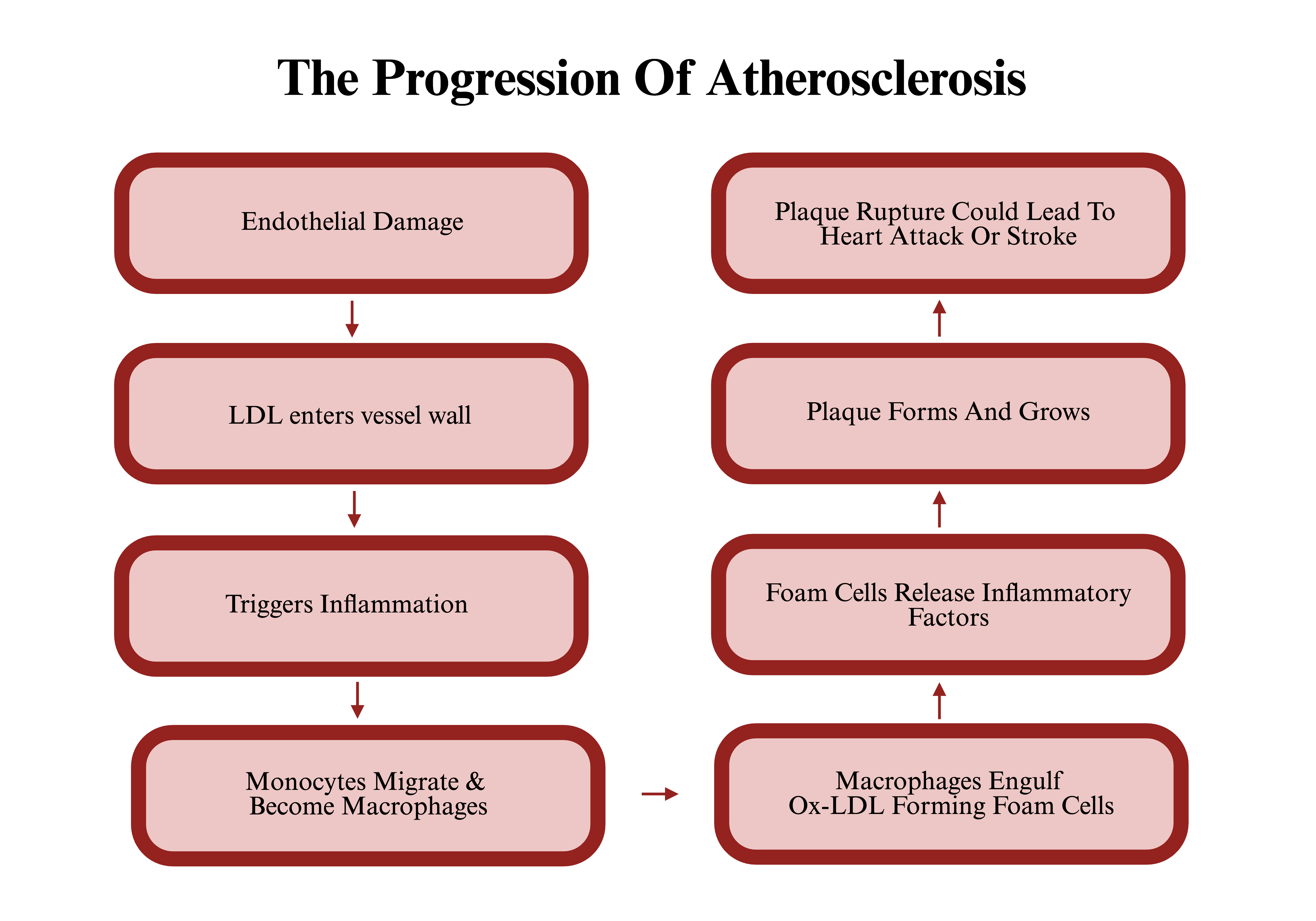
Atherosclerosis is a multifactorial cardiovascular disease and the leading cause of death worldwide [18]. This disease is characterized by the retention and accumulation of lipoproteins in the subendothelial vascular wall, triggering chronic inflammation and encouraging plaque development [18,19]. Autophagy contributes towards cellular protection, repair and survival within cardiovascular walls by degrading dysfunctional protein aggregates and necrotic organelles [4]. In cardiac vasculature, damage to the endothelial lining activates an inflammatory response, as infiltrating low-density lipoproteins (LDL) are oxidized (ox-LDL), releasing a cascade of pro-inflammatory factors (Figure 3) [8,20]. OxLDL signals for monocyte migration; here, they differentiate into macrophages that engulf accumulated lipoproteins and dysfunctional proteins, transitioning into foam cells [19]. Foam cells are lipid-rich macrophages that promote inflammation by releasing pro-inflammatory factors and enhancing the formation of lipid core within plaque [19]. This process is driven by the accumulation of foam cells, inflammatory mediators, and damaged cells that release their cytoplasmic contents, thereby accelerating the progression of atherosclerosis [19]. Studies suggest that autophagy is triggered by endoplasmic reticulum stress, oxidized low-density lipoprotein, and reactive oxygen species (ROS) [20]. As mentioned previously, recurrent endothelial cell damage is a major driver of atherosclerotic disease, contributing to vascular intimal thickening due to plaque accumulation [21]. In response to endothelial stress, autophagy acts as a cytoprotective mechanism by breaking down damaged mitochondrial components, reducing oxidative stress, and promoting plaque stability, consequently preventing the exacerbation of atherosclerosis [20]. This idea is supported by in vitro studies using shear stress-exposed bovine aortic endothelial cells; this showed increased expression of key autophagic markers -including Atg3 and Atg5- and this consequently corresponded with an increase in mitochondrial turnover [19,22]. In the genetic suppression and inhibition of Atg3 or Atg5 using 3-methyladenine, there was a decrease in autophagy. This led to a maladaptive endothelial phenotype characterized by increased inflammatory signalling, reduced nitric oxide availability, and elevated ROS production, demonstrating Autophagy’s significance in maintaining endothelial lining stability. Furthermore, autophagy plays a beneficial role in resolving endoplasmic reticulum stress; when misfolded proteins accumulate, autophagy is triggered, protecting the lesion from necrosis [19]. This is supported by a study in apolipoprotein E–deficient (Apoe−/−) mice, in which disruption of macrophage autophagy triggered the activation of inflammasomes, thereby mediating the progression of atherosclerosis and resulting in larger atherosclerotic lesions [19]. Beyond its role in regulating stress and inflammation, autophagy also plays a crucial role in lipid metabolism in foam cells, specifically through an adaptive form known as lipophagy. Lipophagy selectively delivers lipid droplets to the lysosome for degradation through lipolysis [23]. By facilitating the efflux of cholesterol, lipophagy is imperative in reducing foam cell formation, as it reduces the accumulation of excess lipids within macrophages—a major driver of plaque development [24]. The concept that macroautophagy contributes to maintaining plaque stability is reinforced by a study in which mice deficient in Atg5 (an essential component of autophagosome formation) were analyzed [23]. Findings showed that the formation of atherosclerotic plaques was greatly increased in the absence of macrophage autophagy [23]. In addition to its role in foam cells, autophagy also regulates vascular smooth muscle cells (VSMC). Autophagic mechanisms mediate this by regulating phenotypic switching, limiting apoptosis, and protecting cells against phosphate-induced calcification. While autophagy can be protective against atherogenesis-promoting factors in VSMCs, extreme stress and elevated autophagy can be maladaptive and contribute to autophagic death in VSMCs [25]. This is further exacerbated as a reduction in VSMCs leads to reduced production of collagen—an important component of the fibrous caps in plaques- resulting in plaque instability [25]. This idea is supported by a study on ApoE−/− mice fed a 10-week high-fat diet, in which the important autophagy gene Atg7 was selectively deleted in VSMCs. This study demonstrated that defective autophagy in VSMCs can promote faster plaque development—exacerbating atherosclerosis [25]. Dysregulation of autophagy shifts its role from protective to harmful, especially in advanced vascular plaques [26]. By damaging cardiac tissues and accelerating their deterioration, it can aggravate heart disease. Studies exploring Autophagy’s maladaptive role in atherosclerotic progression suggest the cholesterol crystals in macrophages with impaired autophagy excessively activate the inflammasome, which secretes more IL-1β—a pro-inflammatory cytokine. In addition to this, macrophages deficient in ATG5 produce more IL-1β than general macrophages [27]. Both these mechanisms suggest that defective autophagy can directly and excessively trigger inflammation via increased IL-1β signalling and the overactivation of the inflammasome, thereby promoting plaque progression [27]. Apart from its inflammatory consequences, impaired autophagy also plays a maladaptive role within the vascular walls, leading to apoptotic cell death [28]. When cardiac tissue is exposed to extreme oxidative stress, autophagy can no longer efficiently remove damaged mitochondria [28]. The build-up of incomplete mitochondrial degradation -specifically cytochrome c- can activate the intrinsic apoptotic pathway in VSMCs [28]. Apoptotic cells can contribute to atherogenesis as they become necrotic and contribute towards inflammation and plaque instability [25]. Furthermore, autophagic mechanisms, when combined with oxidative stress, lead to ceroid formation -an insoluble protein complex composed of oxidised lipids—which is a prominent component of vascular lesions [28]. The lysosomal accumulation of ceroid, a mechanism intended to help in its breakdown, can impair the lysosomal hydrolase function, promoting apoptosis and further dysregulation of autophagy [28]. This dysregulation leads to increased accumulation of damaged mitochondrial components, exacerbating oxidative stress and increasing the formation of ceroid-containing lysosomes [28]. As a result, this cycle further contributes to the progression of unstable atherosclerotic lesions.
5. Autophagy in Myocardial Infarction (MI) and Ischemia/Reperfusion (I/R)
Myocardial infarction (MI) is the sudden death of cardiomyocytes due to prolonged lack of oxygen in cardiac tissue, typically as a consequence of the abrupt atherothrombotic occlusion of a coronary artery [8]. Cardiac tissue suffering from ischemia -insufficient oxygenated blood supply—must recover its oxygen levels rapidly in order to minimize the damage of cardiomyocytes, which can radiate from the subendocardium to the epicardium. Myocardial ischemia/reperfusion injury (MIRI) is the damage sustained by cardiac tissue after blood supply is restored following a period of ischemia [26]. During this, autophagy occurs in two distinct waves: one in ischemia and another in reperfusion, each with a varied effect on cardiomyocytes [29]. The heart is a structurally complex organ and consists of numerous cardiomyocytes. However, autophagy is crucial in maintaining cellular homeostasis as these cells have limited regenerative potential [30]. Autophagy controls protein and organelle regulation whether these compounds are in excess or becoming aged [30]. Despite initially having a beneficial role in MI and MIRI, it becomes maladaptive when key genes required for autophagy mechanisms are lost [30]. This change is important for understanding the adaptive role of Autophagy, initially supporting the maintenance of energy stores through ATP production during early ischemia and later contributing to dysfunctional cellular degradation during reperfusion [30]. This leads to the accumulation of dysfunctional cellular compounds, thereby exacerbating these conditions [30]. In MI, the lack of oxygenated blood flow depletes ATP in cells, disrupts ion pumps, causes calcium overload, and leads to membrane damage. As a result, these cardiomyocytes are necrosized [31]. Following necrosis in the aftermath of myocardial infarction, the myocardium initiates a regenerative response; this involves an overlap of the phases of inflammation, fibrosis, and angiogenesis within the damaged cardiac tissue [32]. The ischemic and hypoxic myocardium, over hours to days, initiates an inflammatory response, leading to the infiltration and activation of immune cells, which trigger a pro-inflammatory cascade [32]. Whilst this immune response is crucial for initiating myocardial repair, excessive inflammation can worsen cardiomyocyte injury. In response to inflammation, autophagy acts as a suppressor, driven to alleviate the progression of injury in the myocardium [31]. During the ischemic phase, low ATP is detected by AMPK, a key regulator of cellular energy, which is triggered by ATP depletion and initiates autophagy through ULK1-mediated signalling [30]. Furthermore, Beclin1 is another key factor in activating the initial stages of autophagy, which is essential in autophagosome formation and regulation of autophagy [13]. When initiated, autophagy mediates the exocytosis of fatty acids and amino acids, which are recycled to be utilized in the production of ATP in the tricarboxylic acid cycle (TCA cycle) [30]. This compensates for the critical condition of the myocardium due to low energy supply in I/R injury [30]. Disruption of the AMPK activation pathway impairs autophagy and can exacerbate myocardial tissue damage [3]. In addition to this, hypoxia-inducible factor 1 alpha (HIF-1α), triggered by low oxygen perfusion or excessive oxidative stress, induces mitophagy as a cytoprotective mechanism [30]. In ischemia, autophagy can be both selective and adaptive to meet cellular metabolic needs and remove damaged mitochondrial components, which, if not cleared, can induce apoptosis through the release of harmful ROS [4]. The idea that autophagy plays a cytoprotective role during MI and ischemia in myocardial ischemic injury is supported by numerous studies. One study found that inhibiting autophagy with the drug bafilomycin A1 can increase infarct size in animal models of MI [31]. Beyond its protective role, some researchers suggest autophagy also serves as an adaptive mechanism in MI by mediating the degradation of protein aggregates [31]. If these clumps accumulate, they can disrupt cell function and potentially lead to necrosis [31]. During Ischemic/Reperfusion (I/R) injury, there is increased stress on mitochondria, which abnormally increases mitochondrial fission [33]. This creates a variety of functional and dysfunctional mitochondria, leading them to lose their ability to refuse, and the damaged mitochondria are degraded via mitophagy [33]. When mitochondrial fission becomes impaired, for example, through the expression of the dominant-negative mutant gene Drp1K38A, mitophagy becomes disrupted, leading to more dysfunctional mitochondria accumulating, and this deteriorates cardiac health [33]. This highlights Autophagy’s dual role in heart disease; its protective role becomes maladaptive when the general mitochondrial degradation cascade is dysregulated [33]. Thereby, mitophagy demonstrates a key mechanism through which autophagy protects the heart in I/R by limiting the accumulation of damaged organelles and clearing oxidative stress [33]. Its maladaptive role is demonstrated during MIRI, where autophagy occurs in waves: the first occurs during ischemia, and the second, a heightened wave, during reperfusion, when the myocardium is suddenly reoxygenated [29]. During the second wave, due to increased autophagic activity, more autophagosomes are observed, which can be detrimental to the myocardium [29]. Consequently, this build-up of autophagosomes is maladaptive due to increased ROS levels and its facilitation in the disruption of mitochondrial membrane, a key trigger in the process of cell apoptosis [29]. Studies indicate that during I/R, autophagy activity is compromised [29]. This contributes to disruption in the formation of autophagosomes and the breakdown of lysosomes, partly driven by oxidative stress, which upregulates Beclin1 and downregulates LAMP-2, a key mediator in the fusion of lysosomes and autophagosomes. This combination leads to excessive accumulation of autophagosomes and becomes increasingly detrimental to cardiomyocyte function [29]. This highlights the harmful role autophagy can play in damaging the myocardium when dysfunctional. This is further supported by a study in mice with only one functional Beclin1 gene, which reduced autophagic flux and, consequently, decreased ischemic damage, suggesting that Beclin1-driven autophagy levels can contribute to myocardial damage [3]. Together, these findings highlight the complex nature of autophagy and the importance of its tight regulation in maintaining cardiomyocyte health. Acting as both a shield and a sword, autophagy determines the fate of cells during MI and I/R injury. Several current pre-clinical pharmacological research focuses on inducing autophagy in cardiovascular disease; however, existing evidence suggests that in some cardiovascular diseases, excessive activation of autophagy can be harmful. Therefore, modulation of the autophagy pathway may enhance cardiomyocyte survival by attenuating autophagy in specific phases of MI and I/R injury, maximizing its beneficial role in cardiac tissue recovery.
6. Stage-Specific Autophagy Targeting
Autophagy is an intricate mechanism, the net effect of it in cardiovascular diseases often depends on the stage of the disease. It is not a single pathway that can be increased or decreased to have a set linear effect on these diseases, as each stage in myocardial injury has distinct metabolic demands and stressors. This exemplifies the major complexity of autophagy, and the difficulties in developing a drug that precisely modulates and utilises its role therapeutically for cardiovascular diseases. A stage specific model can be proposed to understand how autophagy can shift from an adaptive process to a maladaptive one in cardiovascular diseases. During the early stages of cardiovascular disease, enhancing autophagic flux may be beneficial in maximising tissue recovery as it acts as an adaptive cardioprotective response to early stressors. In this stage autophagy acts to alleviate injury through the removal of damaged mitochondria and misfolded proteins, minimising toxic accumulation [11,13]. In the various cardiovascular diseases, these early compensatory mechanisms can limit injury, including stabilisation of atherosclerotic plaques and the recycling of metabolic substrates to replenish depleting ATP in I/R injury [16,25]. However, these illnesses can progress and worsen, creating prolonged stress on the heart. In this stage autophagy role can be varied, it may be insufficient, excessive or dysregulated, depending on the illness or cell type. This is reflected in cardiac hypertrophy where a decrease in autophagy regulator Beclin1 reduced autophagy and attenuated myocardial injury, suggesting that upregulating autophagy does not guarantee a better outcome [4]. In parallel, mitophagy can reduce cell death by removing toxic accumulation of damaged mitochondria and proteins; However in prolonged cardiac injury under oxidative stress, it can become impaired promoting inflammation and exacerbating cardiac tissue injury [4]. Autophagy remains operational and partially beneficial, but aspects of the disease can dysregulate autophagy creating an imbalance between excessive digestion of cellular material and protective clearance. Furthermore, Autophagy has the potential to turn detrimental in advanced cardiovascular disease stages, for example in prolonged I/R injury, increasingly unstable atherosclerotic plaques and decompensated heart failure. Although autophagy may appear upregulated in this stage of the disease, for example through elevated LC3, flux may be blocked. This leads to the accumulation of autophagosomes, due to final stages of autophagy being impaired, in this instance restoring flux or limiting excessive activation of autophagy may help recover cardiac tissue. This is suggested in I/R, where the disruption of regular autophagy, due to oxidative stress, can lead to increased initiation of autophagy but impaired fusion of lysosomes and autophagosomes, causing failure of successful autophagic flux [29]. This can damage myocardium further. This elevated cellular stress can contribute to autophagic dysregulation, potentially rendering autophagy harmful or ineffective. However, restoring autophagic flux in stages where autophagy is dysregulated could be beneficial, this is exemplified by CARD9 which binds to and sequesters rubicon, allowing autophagy to return to regular levels, thereby limiting myocardial damage [34]. The complexity of initiating, restoring or inhibiting autophagic flux dependent on stages of cardiovascular disease makes it difficult to develop effective pharmacological strategies. Stage specific autophagy targeting could be utilized by supporting autophagy when autophagic flux is completed effectively, restoring flux when autophagosomes accumulate and inhibiting excessive initiation in overactive autophagy which exacerbates cardiac injury.
7. Potential Therapies Targeting Autophagy for Cardiac Repair
Autophagy plays a critical role in the repair of myocardial damage by regulating endothelial cells, cardiomyocytes, and VSMCs [8]. As a tightly regulated intracellular pathway, autophagy is complex and dynamic, balancing cell survival and cell death [8]. It works to minimize myocardial damage, making it a suitable target for reducing the progression of heart disease [8]. However, currently there is no clinically approved autophagy targeting drugs for cardiovascular disease, only drugs that indirectly modulate autophagy. As shown in Table 1, several therapeutic approaches may modulate autophagy to improve cardiovascular outcomes. Whilst numerous studies in vivo animal models, ex vivo cardiac tissue and in vitro cellular systems show the effect of autophagy on cardiovascular diseases, applying this to human models is difficult. Human cardiovascular diseases develop over many years, with various comorbidities, these fundamental differences limit direct translation as the experimental models may not accurately represent the autophagy activation in human disease. In addition, the evidence of autophagy modulators reducing myocardial damage is predominantly observed in drugs that indirectly regulate autophagy. Working to directly regulate autophagic mechanisms in the heart can advance future therapeutic approaches to improve cardiac health. Despite autophagy being a promising therapeutic target, its role in supporting cardiomyocytes involves intricately coordinated mechanisms. Even a slight dysregulation of this process can contribute to myocardial injury, creating major hurdles when developing safe autophagy-mediated therapeutic approaches. For example, sometimes lysosome-autophagosome fusion fails, leading to a buildup of autophagic vacuoles and autophagy-associated proteins in cells, such as LC3 and p62—this represents the onset of autophagy-mediated damage in the myocardium [29]. Elevated LC3 reflects the successful initiation of autophagy; however, when autophagic vacuole degradation is impaired, this indicates incomplete autophagic flux. This exemplifies how autophagy may appear activated whilst being defective. This disruption of the autophagic mechanism triggers apoptosis, rendering autophagy maladaptive; this is a difficult problem, as in human patients, it is hard to determine whether autophagy is harmful or beneficial to cardiomyocytes [29]. In experimental and animal studies, monitoring autophagic flux can be achieved using tools such as autophagy flux assays, but in humans, there is no distinct clinical test that demonstrates autophagy as functional and active [29]. This makes it difficult to optimize therapies that target autophagy without potentially compromising cardiac health [29]. Scientists proposed a combined approach to counteract this limitation; they utilized the measurement of circulating LC3 levels in the blood, which is an autophagosome marker, and integrated this with in vivo imaging to observe the autophagy markers directly in the cardiac tissues [29]. Consequently, this autophagy tool enables more accurate monitoring of heart disease, potentially improving the safety and efficacy of these drugs and demonstrating better treatment timing through personalized drug dosing [29]. In an ischemic heart, due to a lack of oxygenated blood flow, autophagy is activated to protect cardiomyocytes and clear damaged cellular components [34]. During perfusion, autophagy is increasingly activated due to increased ROS and Beclin1 is upregulated [34]. In later stages of reperfusion, Rubicon levels are elevated, causing a blockage of autophagic flux and leading to the accumulation of autophagic vacuoles [34]. This results in autosis—a distinct autophagy-related cell apoptosis mechanism that damages the myocardium further [34]. Inhibiting Rubicon via genetic knockout can regulate autophagy and restore normal mechanisms, thereby reducing autosis and minimizing damage during reperfusion [34]. In addition, CARD9 is a protein that can block Rubicon’s detrimental effect on cardiomyocytes and act to inhibit cellular apoptosis amidst I/R injury [34]. CARD9 adheres to the Rubicon, mediating its detachment from the inhibitory complex, allowing autophagy to return to normal levels [34]. If CARD9 is overexpressed, there is increased autophagic activity, protecting the myocardium further [34]. mTORC1 acts as an inhibitor for Autophagy; Rapamycin’s method of action is to block mTORC1—thereby activating autophagy [4]. This then upregulates the autophagic flux, mediating the removal of damaged proteins and organelles in the myocardium, effectively minimizing myocardial injury (Table 1) [4]. This idea is reinforced by studies showing that Rapamycin protects against I/R injury in ex vivo mouse hearts and cardiomyocytes [4]. Furthermore, when Rapamycin was given to mice with transverse aortic constriction-induced heart failure, there was a decrease in cardiac hypertrophy [4]. This highlights the potential rapamycin could have in autophagy therapies that target heart failure progression. Targeting an adaptor protein like CARD9 for developing pharmaceuticals is challenging and has limited clinical feasibility, due to its involvement in multiple cellular pathways. Whilst, repurposing an established medication such as rapamycin demonstrates clinical applicability, as it has been used within the UK healthcare system. Alongside these, some natural compounds—such as those from plants and herbs- can also be targets of interest in developing therapies; they are generally safer and have reduced toxicity [35]. There are two key mechanisms: one facilitates autophagy activation, and the other enhances the full recycling process of autophagy. These are crucial for the survival of cardiomyocytes in heart disease. Salvia miltiorrhiza Bunge—also known as Danshen- is a traditional Chinese herbal medicine that has several therapeutic components that promote cardiac health. Compounds like Tanshinone IIA in Danshen activate the AMPK, mTOR, and ULK1 cascade in the autophagy pathway, enhancing its initiation, thereby reducing ischemic damage in cardiac tissue and minimizing the progression of heart failure. Similarly, Salvianolic acid B, another compound extracted from Danshen, was explored in animal models of MI. Although the mechanism is unclear, Salvianolic acid B was shown to have cardioprotective effects, aiding the initiation of autophagy [35]. These compounds further enhance cardiac protection against I/R injury and minimize the damage caused by Doxorubicin-induced cardiomyopathy, a chemotherapy drug that promotes myocardial injury and further protects against I/R injury [35]. The second mechanism of these natural compounds is demonstrated in resveratrol—a compound found in grapes- which further enhances and restores autophagic flux and boosts the initiation of Autophagy, alongside increasing key markers of autophagy initiation, such as LC3-11/1 and Atg5 [35]. This is induced by increased autophagosome formation, which removes damaged cellular organelles and proteins more efficiently, minimizing damage in MI [35]. In addition, resveratrol has also been shown to heighten autophagy in I/R injury [35]. This is supported by a study showing that Polydatin, a natural glycoside form of resveratrol, enhanced autophagy, thereby reducing I/R injury in the myocardium in both cultured cells and living animals [35]. However, natural compounds often are rapidly metabolized and have limited bioavailability, creating no significant clinical benefit. These compounds often act pleiotropically, biologically activating multiple pathways simultaneously. This creates more difficulties in isolating its modulation of autophagy, whilst avoiding unintended systematic effects.
Demonstrating the potential therapeutic approaches that can modulate autophagy to improve outcomes in heart disease.
| Compound Name | Type of Compound | Mechanism of Action | The Effect on the Heart |
|---|---|---|---|
| Rapamycin | Pharmaceutical | mTORC1 inhibitor | This activates autophagy and protects the myocardium. |
| CARD9 | Genetic | Blocks Rubicon inhibition | This restores autophagy and protects during I/R injury. |
| Rubicon knockout | Genetic | Removes Rubicon | This prevents autosis and restores flux. |
| Tanshinone IIA (Danshen) | Natural compound | Activates the AMPK, mTOR, and ULK1 pathway | This enhances autophagy initiation. |
| Salvianolic acid B (Danshen) | Natural compound | The mechanism is unclear | It provides cardioprotection by initiating autophagy. |
| Resveratrol | Natural compound | Enhances autophagic flux, increases LC3-II/Atg5 | This reduces MI damage. |
| Polydatin | Natural compound | Enhances autophagy | This reduces I/R injury. |
8. Pharmacological Modulation of Autophagy in Diabetic Cardiomyopathy
Diabetic cardiomyopathy is a heart disease causing structural and functional changes in cardiac tissue in diabetic patients, which is not solely caused by predisposing conditions such as hypertension and coronary artery disease (CAD) [36]. There is evidence suggesting autophagy can have a maladaptive role in diabetic cardiomyopathy, predominantly in type 1 diabetes (T1D), whilst the involvement of autophagy in type 2 diabetes remains unclear [37]. Animal studies demonstrate how autophagy is often downregulated in (T1D) [37,38]. One study testing the functional role of autophagy in mouse models showed reduced cardiac injury in Beclin1-deficient mice with T1D, and this damage was potentiated by Beclin1 overexpression [38]. While these studies examine the potential detrimental role of autophagy in diabetic cardiomyopathy, other researchers have also investigated therapeutic modulation of autophagy to explore its cardioprotective potential. Metformin is a drug used mainly in diabetes to control blood sugar levels and increase insulin sensitivity. Metformin’s pleiotropic effects when activating AMPK trigger autophagy in cells under low-energy conditions to help degrade damaged organelles and recycle intracellular compounds [4,39]. When used in diabetic OVE26 mice that developed diabetic cardiomyopathy, metformin improved heart pumping and minimized myocardial damage caused by diabetes [4]. This supports the therapeutic potential of AMPK in enhancing autophagy to treat various forms of heart disease. Studies suggest there are cardioprotective effects of metformin in patients with heart disease and not just diabetic patients. A specific study explored metformin’s therapeutic effect on wild mice compared to δ-sarcoglycan-deficient (Scgd−/−) mice, which develop dilated cardiomyopathy as a result of this defect [39]. Sgcd−/− mice had higher levels of LC3-II (light chain 3) and p62 than wild mice [39]. The investigation demonstrated an increase in the LC3-II/LC3-I ratio in the Sgcd−/− mice, indicative of increased autophagosome formation, which was further increased by metformin therapy [39]. However, it also showed a lack of clearance of p62 which is a protein degraded by autophagy, reflecting the final step of autophagy being impaired in Sgcd−/− mice [39]. Metformin reduced p62 levels in Sgcd−/− hearts, upregulated mitophagy regulators, and further augmented the already heightened lysosomal protein cathepsin D levels in the mice, eliciting the direct enhancement metformin has on the degradation capacity of these cardiomyocytes [39]. These findings suggest that metformin could be a potential treatment for sarcoglycanopathy-related dilated cardiomyopathy, with its ability to elevate autophagic levels and improve the clearance of autophagosomes. Given its impact beyond diabetes and its safety in humans being already established through extensive clinical use, metformin is a promising candidate for novel therapies that modulate autophagy to enhance cardiac repair in various heart diseases. In addition to metformin, several pharmacological agents can regulate the autophagic pathway, offering promising therapeutic targets for myocardial repair in various forms of heart disease. Trehalose (TRE) is an organic disaccharide that cannot be synthesized by mammals and has shown potential to modulate autophagy and reduce diabetic cardiomyopathy [40,41,42]. During a study in which heterozygous transgenic mice expressing LC3 conjugated with a green fluorescent protein (Tg-GFP-LC3) were administered TRE, they explored autophagosome formation in these mice [41]. Consequently, there was an increase in LC3 puncta and cathepsin D and a decrease in accumulated p62, demonstrating that dysregulated autophagy could potentially be recovered and regulated by TRE treatment [41]. This further supports the idea that autophagy modulators like TRE could be therapeutic agents for reducing heart failure by activating autophagy. There are many autophagy inducers like rapamycin and atorvastatin, which can be explored in similar studies and have demonstrated to have favorable effects in acute myocardial infarction models, further indicating the pharmacological potential in these agents [43].
9. Conclusions
In conclusion, autophagy is crucial for maintaining cardiomyocyte health and protects against heart disease, but it can become harmful when dysregulated. In cardiac hypertrophy, autophagy acts as a remodelling mechanism that supports the heart’s adaptation to increased workload; however, excessive activation can damage healthy tissue and worsen heart disease. Autophagy stabilizes atherosclerotic plaques by reducing oxidative stress from damaged mitochondria. When autophagy is impaired, it can lead to increased inflammation and oxidative stress, promoting plaque progression and instability. During MI, autophagic mechanisms enhance ATP levels by supporting the TCA cycle, countering low oxygen supply and reducing I/R injury. However, excessive or dysregulated autophagy during reperfusion can lead to increased ROS and cell death in otherwise healthy cells. This makes autophagy an appealing target for heart disease therapies, although its complexity poses significant challenges for precise modulation to protect cardiac tissue.
Author Contributions
V.S.: Conceptualization, methodology, research, and writing—original draft preparation. T.A.: Supervision, validation, reviewing and editing. All authors have read and agreed to the published version of the manuscript.
Funding
This research received no external funding.
Institutional Review Board Statement
Not applicable.
Informed Consent Statement
Not applicable.
Data Availability Statement
Not applicable.
Conflicts of Interest
The authors declare no conflict of interest.
Use of AI and AI-Assisted Technologies
No AI tools were utilized for this paper.
References
- 1.
Goldsborough, E.; Osuji, N.; Blaha, M.J. Assessment of Cardiovascular Disease Risk: A 2022 Update. Endocrinol. Metab. Clin. 2022, 51, 483–509.
- 2.
Liu, J.L.Y. The economic burden of coronary heart disease in the UK. Heart 2002, 88, 597–603.
- 3.
Sciarretta, S.; Maejima, Y.; Zablocki, D.; et al. The Role of Autophagy in the Heart. Annu. Rev. Physiol. 2018, 80, 1–26. https://doi.org/10.1146/annurev-physiol-021317-121427.
- 4.
Lavandero, S.; Chiong, M.; Rothermel, B.A.; et al. Autophagy in cardiovascular biology. J. Clin. Invest. 2015, 125, 55–64.
- 5.
Kim, J.; Kundu, M.; Viollet, B.; et al. AMPK and mTOR regulate autophagy through direct phosphorylation of Ulk1. Nat. Cell Biol. 2011, 13, 132–141.
- 6.
Lu, G.; Wang, Y.; Shi, Y.; et al. Autophagy in health and disease: From molecular mechanisms to therapeutic target. MedComm 2022, 3, e150.
- 7.
Tekirdag, K.; Cuervo, A.M. Chaperone-mediated autophagy and endosomal microautophagy: Jointed by a chaperone. J. Biol. Chem. 2018, 293, 5414–5424.
- 8.
Pan, W.; Zhang, J.; Zhang, L.; et al. Comprehensive view of macrophage autophagy and its application in cardiovascular diseases. Cell Prolif. 2024, 57, e13525.
- 9.
Parzych, K.R.; Klionsky, D.J. An Overview of Autophagy: Morphology, Mechanism, and Regulation. Antioxid. Redox Signal. 2014, 20, 460–473.
- 10.
Du, J.; Liu, Y.; Fu, J. Autophagy and Heart Failure. Adv. Exp. Med. Biol. 2020, 1207, 223–227.
- 11.
Shi, S.; Jiang, P. Therapeutic potentials of modulating autophagy in pathological cardiac hypertrophy. Biomed. Pharmacother. 2022, 156, 113967.
- 12.
Li, Y.; Zheng, W.; Lu, Y.; et al. BNIP3L/NIX-mediated mitophagy: Molecular mechanisms and implications for human disease. Cell Death Dis. 2022, 13, 14. https://doi.org/10.1038/s41419-021-04469-y.
- 13.
Li, L.; Xu, J.; He, L.; et al. The role of autophagy in cardiac hypertrophy. Acta Biochim. Biophys. Sin. 2016, 48, 491–500.
- 14.
Zhou, H.; Wang, X.; Xu, T.; et al. PINK1-mediated mitophagy attenuates pathological cardiac hypertrophy by suppressing the mtDNA release-activated cGAS-STING pathway. Cardiovasc. Res. 2025, 121, 128–142.
- 15.
Kanamori, H.; Yoshida, A.; Naruse, G.; et al. Impact of Autophagy on Prognosis of Patients with Dilated Cardiomyopathy. J. Am. Coll. Cardiol. 2022, 79, 789–801. https://doi.org/10.1016/j.jacc.2021.11.059.
- 16.
Tham, Y.K.; Bernardo, B.C.; Ooi, J.Y.Y.; et al. Pathophysiology of cardiac hypertrophy and heart failure: Signaling pathways and novel therapeutic targets. Arch. Toxicol. 2015, 89, 1401–1438.
- 17.
Zhang, Y.; Ding, Y.; Li, M.; et al. MicroRNA-34c-5p provokes isoprenaline-induced cardiac hypertrophy by modulating autophagy via targeting ATG4B. Acta Pharm. Sin. B 2022, 12, 2374–2390. https://doi.org/10.1016/j.apsb.2021.09.020.
- 18.
Fan, J.; Watanabe, T. Atherosclerosis: Known and unknown. Pathol. Int. 2022, 72, 151–160.
- 19.
Tabas, I.; Bornfeldt, K.E. Macrophage Phenotype and Function in Different Stages of Atherosclerosis. Circ. Res. 2016, 118, 653–667.
- 20.
Lavandero, S.; Troncoso, R.; Rothermel, B.A.; et al. Cardiovascular autophagy. Autophagy 2013, 9, 1455–1466.
- 21.
Miao, J.; Zang, X.; Cui, X.; et al. Autophagy, Hyperlipidemia, and Atherosclerosis. Autophagy Biol. Dis. 2020, 1207, 237–264.
- 22.
Vion, A.C.; Kheloufi, M.; Hammoutene, A.; et al. Autophagy is required for endothelial cell alignment and atheroprotection under physiological blood flow. Proc. Natl. Acad. Sci. USA 2017, 114, E8675–E8684.
- 23.
Nussenzweig, S.C.; Verma, S.; Finkel, T. The Role of autophagy in Vascular Biology. Circ. Res. 2015, 116, 480–488.
- 24.
Ouimet, M.; Franklin, V.; Mak, E.; et al. Autophagy Regulates Cholesterol Efflux from Macrophage Foam Cells via Lysosomal Acid Lipase. Cell Metab. 2011, 13, 655–667.
- 25.
Grootaert, M.O.J.; Moulis, M.; Roth, L.; et al. Vascular smooth muscle cell death, autophagy and senescence in atherosclerosis. Cardiovasc. Res. 2018, 114, 622–634.
- 26.
Yang, M.; Linn, B.S.; Zhang, Y.; et al. Mitophagy and mitochondrial integrity in cardiac ischemia-reperfusion injury. Biochim. Biophys. Acta Mol. Basis Dis. 2019, 1865, 2293–2302.
- 27.
Razani, B.; Feng, C.; Coleman, T.; et al. Autophagy Links Inflammasomes to Atherosclerotic Progression. Cell Metab. 2012, 15, 534–544.
- 28.
De Meyer, G.R.Y.; Grootaert, M.O.J.; Michiels, C.F.; et al. Autophagy in Vascular Disease. Circ. Res. 2015, 116, 468–479.
- 29.
Mei, Y.; Thompson, M.D.; Cohen, R.A.; et al. Autophagy and oxidative stress in cardiovascular diseases. Biochim. Biophys. Acta 2015, 1852, 243–251.
- 30.
Ma, S.; Wang, Y.; Chen, Y.; et al. The role of the autophagy in myocardial ischemia/reperfusion injury. Biochim. Biophys. Acta Mol. Basis Dis. 2015, 1852, 271–276.
- 31.
Wu, D.; Zhang, K.; Hu, P. The Role of Autophagy in Acute Myocardial Infarction. Front. Pharmacol. 2019, 10, 551.
- 32.
Zhang, Q.; Wang, L.; Wang, S.; et al. Signaling pathways and targeted therapy for myocardial infarction. Signal Transduct. Target. Ther. 2022, 7, 78.
- 33.
Morciano, G.; Patergnani, S.; Bonora, M.; et al. Mitophagy in Cardiovascular Diseases. J. Clin. Med. 2020, 9, 892.
- 34.
Nah, J.; Zablocki, D.; Sadoshima, J. The roles of the inhibitory autophagy regulator Rubicon in the heart: A new therapeutic target to prevent cardiac cell death. Exp. Mol. Med. 2021, 53, 528–536.
- 35.
Wu, X.; Liu, Z.; Yu, X.; et al. Autophagy and cardiac diseases: Therapeutic potential of natural products. Med. Res. Rev. 2020, 41, 314–341.
- 36.
Boudina, S.; Abel, E.D. Diabetic cardiomyopathy, causes and effects. Rev. Endocr. Metab. Disord. 2010, 11, 31–39. https://doi.org/10.1007/s11154-010-9131-7.
- 37.
Kubli, D.A.; Gustafsson, Å.B. Unbreak My Heart: Targeting Mitochondrial Autophagy in Diabetic Cardiomyopathy. Antioxid. Redox Signal. 2015, 22, 1527–1544. https://doi.org/10.1089/ars.2015.6322.
- 38.
Kobayashi, S.; Liang, Q. Autophagy and mitophagy in diabetic cardiomyopathy. Biochim. Biophys. Acta Mol. Basis Dis. 2015, 1852, 252–261. https://doi.org/10.1016/j.bbadis.2014.05.020.
- 39.
Kanamori, H.; Naruse, G.; Yoshida, A.; et al. Metformin Enhances Autophagy and Provides Cardioprotection in δ-Sarcoglycan Deficiency-Induced Dilated Cardiomyopathy. Circ. Heart Fail. 2019, 12, e005418. https://doi.org/10.1161/circheartfailure.118.005418.
- 40.
Frati, G.; Vecchione, C.; Sciarretta, S. Novel Beneficial Cardiovascular Effects of Natural Activators of Autophagy. Circ. Res. 2018, 123, 947–949. https://doi.org/10.1161/circresaha.118.313530.
- 41.
Sciarretta, S.; Yee, D.; Nagarajan, N.; et al. Trehalose-Induced Activation of Autophagy Improves Cardiac Remodeling After Myocardial Infarction. J. Am. Coll. Cardiol. 2018, 71, 1999–2010. https://doi.org/10.1016/j.jacc.2018.02.066.
- 42.
Dewanjee, S.; Vallamkondu, J.; Kalra, R.S.; et al. Autophagy in the diabetic heart: A potential pharmacotherapeutic target in diabetic cardiomyopathy. Ageing Res. Rev. 2021, 68, 101338. https://doi.org/10.1016/j.arr.2021.101338.
- 43.
Bielawska, M.; Warszyńska, M.; Stefańska, M.; et al. Autophagy in Heart Failure: Insights into Mechanisms and Therapeutic Implications. J. Cardiovasc. Dev. Dis. 2023, 10, 352. https://doi.org/10.3390/jcdd10080352.

This work is licensed under a Creative Commons Attribution 4.0 International License.

 Suite 4002 Level 4, 447 Collins Street, Melbourne, Victoria 3000, Australia
 General Inquiries: info@sciltp.com