
Tooth wear is a common, multifactorial non-carious dental hard tissue disease, and it may lead to a range of problems such as hypersensitivity, occlusal dysfunction, and aesthetic defects, significantly impacting patients’ oral health and quality of life. This article aims to systematically review the clinical assessment, diagnostic methods, pathophysiology, and etiological classification of tooth wear, with a focus on discussing evidence-based restorative and pharmacological management strategies, thereby providing a theoretical basis and practical guidance for clinical practice. This review provides an overview of the fundamental concepts and classification of tooth wear, encompassing both mechanical and chemical forms. With regard to clinical assessment, it outlines fundamental examination methods such as visual inspection and history taking, and reviews the application and development of standardized indices, including the Basic Erosive Wear Examination (BEWE) and the Tooth Wear Index (TWI), as well as adjunctive tools like intraoral photographs. In terms of etiology, it analyzes the interaction mechanisms of mechanical wear (attrition and abrasion), chemical wear (erosion), and biomechanical fatigue, and further classifies etiological factors according to typical location and morphological characteristics. Regarding treatment, this review emphasizes etiological intervention and risk factor management, and evaluates the performance and indications of restorative materials such as composite resin, ceramic, and CAD/CAM resin matrix composites. It also discusses key techniques for achieving durable long-term bonding, as well as the value and future prospects of integrating digital workflows, such as intraoral scanning and digital smile design (DSD), into diagnosis, treatment planning, and clinical management. In addition, it reviews pharmacological strategies for tooth wear, especially topical remineralizing agents, from the perspective of therapeutic targets and mechanisms of action. The modern management of tooth wear depends on early identification and accurate etiological diagnosis, appropriate selection of restorative materials based on long-term performance, and the synergistic application of minimally invasive adhesive techniques, digital technologies, adjuvant pharmacological therapies, and individualized risk control throughout the treatment process.
- Open Access
- Review
Dental Erosion and Therapeutic Options
- Tangmuqi Jiang 1,
- Xuya Kang 1,
- Yahan Liu 1,*,
- Yan Zhang 1,2,3,4,5,*
Author Information
Received: 23 Jan 2026 | Revised: 09 Mar 2026 | Accepted: 14 May 2026 | Published: 14 Aug 2026
Abstract
Keywords
tooth wear | erosion | clinical assessment | restorative materials | pharmacological management
1. Introduction
Tooth wear is a common oral condition characterized by the irreversible loss of enamel and dentin [1,2]. It is a non-carious disease caused by multiple factors and can be classified into mechanical wear (attrition and abrasion) and chemical wear (erosion). Chemical tooth wear, or erosion, is caused by intrinsic or extrinsic acids, such as dietary acids or gastric acid. Mechanical tooth wear can be further divided into attrition and abrasion. Attrition results from tooth-to-tooth contact during functional and/or parafunctional activities (e.g., bruxism), while abrasion is caused by mechanical factors other than functional or parafunctional contact, such as improper toothbrushing [1,3].
Based on its impact on the patients’ well-being and oral function, tooth wear can be categorized as either physiological or pathological. Physiological tooth wear is a slow and progressive process that typically does not cause any subjective symptoms. In contrast, pathological tooth wear may present with corresponding clinical signs and symptoms and lead to a range of problems, such as pain or discomfort, occlusal changes that impair masticatory function, and aesthetic concerns caused by tooth discoloration and reduction in clinical crown [4,5].
The spectrum of tooth wear is broad, ranging from minor changes confined to enamel in early stages to severe hard tissue loss involving dentin and even pulp exposure in advanced stages [6]. Its clinical manifestations are diverse and may include loss of enamel surface gloss, cupping lesions, restorations standing proud of the surrounding tooth surface, dentin hypersensitivity, aesthetic defects, and decreased masticatory efficiency [7,8]. Without intervention, progressive tooth wear may substantially affect patients’ dietary habits, social confidence, and oral health-related quality of life [9,10].
Accordingly, early identification, accurate diagnosis, risk factor assessment, and the development of evidence-based, stepwise treatment strategies have become important issues in contemporary dentistry. This review aims to systematically summarize current research progress on the diagnostic assessment tools, risk factors, restorative treatment strategies, and pharmacological approaches for tooth wear, thereby providing a theoretical basis and clinical guidance for practice.
2. Clinical Assessment and Diagnostic Process
Clinical assessment of tooth wear is the foundation for diagnosing and managing this condition. Its goals are early lesion identification, determining the etiology, assessing severity, and formulating an individualized treatment plan. Currently, clinical assessment primarily relies on a combination of basic examination and standardized indices to ensure systematic and reproducible diagnosis.
2.1. Basic Clinical Examination
Basic clinical examination includes visual inspection, history taking, and related routine assessments. It is the first step in identifying the characteristics and potential causes of tooth wear.
2.1.1. Visual Inspection
Visual inspection focuses primarily on the morphological features of dental hard tissue loss. Early erosive tooth wear (ETW) often manifests as loss of enamel surface texture, changes in gloss, and a ‘clay-like’ appearance; typical wear patterns include cupping, grooving, restoration margin ‘ridging’, and incisal translucency [7]. For non-carious cervical lesions, stereomicroscopy can further reveal microscopic features, such as scratches and cracks in wedge-shaped defects, or smooth walls in saucer-shaped lesions [11]. Notably, Ganss (2008) pointed out that certain features (e.g., “cupping and grooving”) might be caused by both abrasion and erosion, indicating limited specificity and suggesting the need for comprehensive information in clinical diagnosis [6].
Although visual inspection is simple and convenient, its sensitivity for early or minor lesions is limited. Studies by Urcelay Moreno et al. indicate that intraoral scanners can detect micron-level wear difficult for the human eye to recognize, highlighting the potential of digital tools in early diagnosis [12].
2.1.2. History Taking
Medical history is an important part of the basic clinical examination of tooth wear. A thorough and well-structured history can help identify causative factors, assess the risk and guide follow-up monitoring. O’Toole et al. emphasize that dietary history (e.g., frequency of acidic diets), systemic history (GERD, asthma, eating disorders, etc.), medication history (e.g., SSRIs), and oral habits (e.g., night grinding) should be collected to reveal potential chemical and mechanical causative factors [13]. Wetselaar et al. pointed out that medical history interrogation, questionnaires, and clinical manifestation identification together constitute the three major tools for etiological assessment, and medical history should include the patients’ subjective symptoms (e.g., sensitivity, pain, aesthetic concerns) to fully grasp the patient’s situation [2].
2.2. Standardized Assessment Indices
While visual inspection and history taking provide essential qualitative data, a more objective and reproducible quantification of the extent and severity of tooth wear is necessary. To systematically quantify wear severity, various standardized indices have been developed and applied in clinical and epidemiological research.
2.2.1. Basic Erosive Wear Examination (BEWE)
BEWE is one of the most widely used tooth erosion evaluation tools in the world [14]. It can conveniently record the evaluation of tooth wear caused by various etiologies [15].
BEWE divides the dentition into six sextants, with the most severely affected surface in each sextant being recorded [8,15]. The scoring ranges from 0 to 3: 0 points indicate no wear; 1 point indicates initial loss of surface texture (such as loss of luster, appearing like frosted glass); 2 points indicate hard tissue loss but less than 50% of the surface area; 3 points indicate hard tissue loss exceeding 50% of the surface area [15]. Clinical photographs illustrating the grading system are presented in Figure 1 [15]. All sextant scores are added together to get a total score (up to 18 points), which reflects the overall severity of tooth wear across the dentition [8,15]. Risk levels (none, low, medium, high) can be assigned according to the total score to guide clinical intervention strategies [15,16]. Martignon et al. further proposed using index teeth (e.g., buccal surface of upper central incisors, occlusal surface of lower first molars) for assessment to improve clinical efficiency [17].
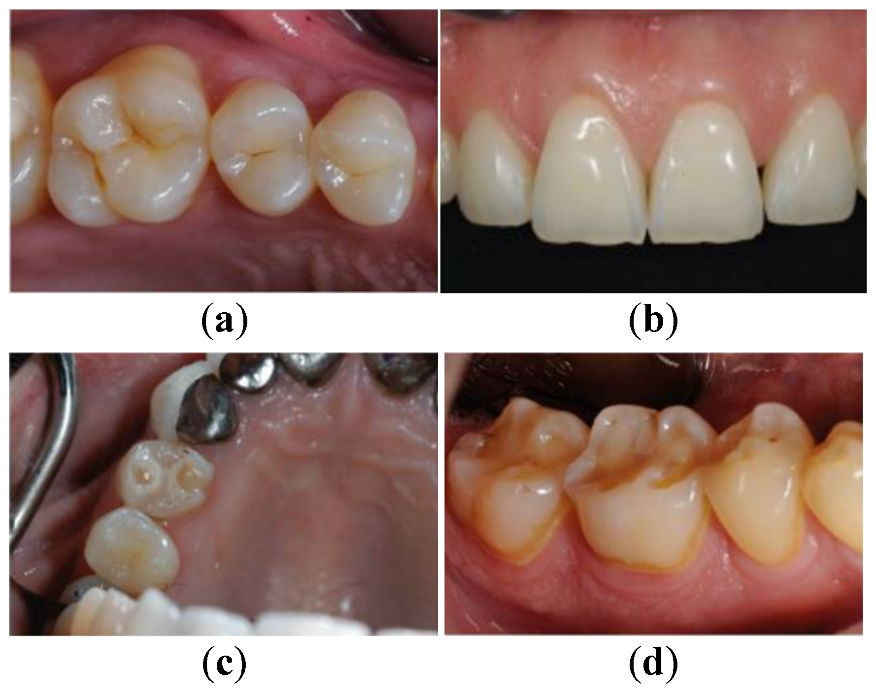
2.2.2. Tooth Wear Index (TWI)
The TWI was proposed by Smith and Knight in 1984 [18] and it is one of the systems capable of measuring the loss of hard tissue in teeth due to a variety of causes. Unlike BEWE, this index independently evaluates the four visible tooth surfaces of all teeth, with a score range of 0–4 [19,20]. Based on these scores, the degree of tooth structure loss in each corresponding region can be evaluated independently [19]. However, TWI is time-consuming and requires high standards from the examiner, which limits its routine clinical use [19]. It is mainly used in clinical practice for detailed evaluation and diagnosis of individual patients, not for screening [19].
2.2.3. Other Common Indices
The Lussi Index and Eccles Index are also commonly used for assessing erosive wear in the anterior region. Wetselaar et al. compared four indices, including BEWE, TWI, Lussi, and Eccles, noting differences in their diagnostic sensitivity and applicable scenarios, and called for the establishment of a unified modular assessment system [21].
2.2.4. Modular System: TWES
The Tooth Wear Evaluation System (TWES) provides a comprehensive framework covering screening, grading, etiological diagnosis, and treatment planning. TWES 2.0 further integrates pathological classification and etiological features, aiming to improve diagnostic consistency and reproducibility [1,3]. Studies indicate it shows acceptable reliability in clinical training and computer-assisted diagnosis, but wider application requires further validation [22].
2.3. Auxiliary Assessment Tools and Techniques
Besides basic examination and indices, intraoral photographs also play an important role in the assessment of tooth wear. Intraoral photographs serve as non-invasive tools to record early signs of wear (e.g., loss of enamel texture and surface gloss changes) and can be combined with indices like BEWE or TWES for longitudinal monitoring [7]. Recently, artificial intelligence (AI)-based automated analysis systems have shown high accuracy and efficiency, promising to become auxiliary tools for future standardized assessment [23].
2.4. Consistency of Assessment Methods and Future Prospects
Despite the coexistence of multiple indices and tools, their diagnostic consistency remains debated. Al-Malik et al. found discrepancies between visual inspection and histological examination in assessing primary tooth wear, suggesting clinical examination might underestimate the actual degree of wear [24]. Inconsistencies in risk stratification and diagnostic thresholds among different indices also affect comparability across studies [21].
In the future, clinical assessment of tooth wear should further develop towards modular, multi-dimensional systems, integrating AI and 3D technology to enhance early diagnostic capability, optimize treatment planning, and enable long-term dynamic monitoring. The synergistic application of standardized indices and digital tools will drive continuous optimization of tooth wear management strategies.
3. Pathophysiology and Etiological Classification
With the clinical picture established, the next step is to interpret these findings in the context of the various etiological factors responsible for tooth wear. The mechanisms of tooth wear are complex. Based on etiology and clinical presentation, tooth wear can be classified into two main categories: mechanical wear (e.g., attrition, abrasion) and chemical wear (e.g., erosion), but in most cases, it results from a combination of factors [25]. Understanding its etiological classification is crucial for diagnosis and treatment strategy formulation. This section systematically elaborates on the pathophysiological mechanisms and etiological classification of tooth wear.
3.1. Pathophysiological Mechanisms
The pathophysiological process of tooth wear primarily involves mechanical wear, chemical wear, biomechanical stress, and material fatigue.
3.1.1. Mechanical Wear
Mechanical wear refers to tissue loss caused by mechanical forces and can be further divided into attrition and abrasion. Attrition is mainly caused by functional or parafunctional tooth-to-tooth contact, commonly seen in bruxism patients. Bruxism is a repetitive masticatory muscle activity that can occur during wakefulness or sleep and is associated with progressive loss of enamel and dentin [26]. Abrasion is typically caused by external mechanical factors, such as excessive toothbrushing, use of abrasive toothpaste leading to cervical and labial wear (“toothpaste abrasion”), or harmful oral habits [11]. Bruxism itself, in most multivariate analyses, shows no significant association with tooth wear, unless combined with cervical abrasion [27].
3.1.2. Chemical Wear
Chemical wear, also known as erosion, is the dissolution of dental hard tissues by intrinsic or extrinsic acids without bacterial involvement. Its main sources include extrinsic acids and intrinsic acids. Extrinsic acids primarily come from dietary acidic beverages (e.g., carbonated drinks, fruit juices), foods, or occupational exposure [28]. Intrinsic acids are related to gastroesophageal reflux disease, eating disorders (e.g., anorexia nervosa, bulimia) causing gastric acid reflux into the oral cavity [29]. The acidic environment demineralizes the enamel surface, reducing its hardness and thereby accelerating the process of mechanical wear [30]. Typical clinical features include loss of enamel surface gloss, a ‘wet clay-like’ appearance, cupping, and restoration ‘overhangs’ [7].
3.1.3. Biomechanics and Material Fatigue
Occlusal forces create stress concentration at the cervical area, particularly in the presence of occlusal interferences or parafunctional movements, leading to microcracks and fractures in the tooth structure, a process termed “abfraction” by some researchers [31]. Dental tissues undergo fatigue fracture under repeated stress, especially at the more vulnerable enamel-dentin junction [32], resulting in tissue loss. Simulation studies of non-carious cervical lesions indicate that the combined action of acid exposure and mechanical loading leads to the most significant tooth tissue loss [32].
3.1.4. Role of Saliva
Saliva plays an important role in protecting teeth from acid erosion. Low stimulated whole saliva pH is significantly associated with tooth wear, while salivary flow rate, buffering capacity, and other properties may also influence the wear process [33,34]. The potential value of monitoring salivary pH for early assessment and prevention of tooth wear has been emphasized [34].
3.2. Etiological Classification
Based on the dominant causative factors, tooth wear can be classified into the following types:
3.2.1. Attrition Type
Attrition type is associated with bruxism, presenting as occlusal flattening, incisal wear, often accompanied by masticatory muscle pain, headache, etc. [26]. Bruxism can be classified into awake and sleep types, and its diagnosis requires combining clinical symptoms, questionnaires, and tools like polysomnography (PSG) [35].
3.2.2. Erosion Type
Extrinsic erosion is common in individuals with frequent intake of acidic beverages (e.g., carbonated drinks, fruit juices), especially adolescents and young adults [36].
Intrinsic erosion is associated with gastroesophageal reflux disease (GERD) and eating disorders (e.g., bulimia nervosa), often presenting as concavities on the palatal surfaces of upper anterior teeth and occlusal surfaces [37]. Patients with eating disorders often exhibit characteristic erosion on the palatal surfaces of the maxillary anterior teeth due to repeated vomiting, and such dental damage may become a “visible scar”, affecting long-term psychological and social function [38].
3.2.3. Mixed Type
Tooth wear is usually not caused by a single factor, and in most patients, mechanical and chemical factors coexist and interact in the process of tooth wear to form mixed forms of tooth wear, such as erosion-attrition and erosion-abrasion [39]. Under acidic conditions, the effects of mechanical factors on teeth are amplified [39]. For example, teeth in bruxism patients are more susceptible to wear in an acidic environment [40].
3.2.4. Other Special Types
Non-carious cervical lesions are a special form of tooth wear, caused by multiple factors including occlusal forces, brushing technique, and acidic environment [41]. Tooth wear may also be associated with systemic diseases such as Prader-Willi syndrome, HIV/AIDS patients, etc., where salivary dysfunction or medication effects make them more susceptible to generalized tooth wear [42,43].
Based on the location, morphology, and number of lesions, different etiological patterns can be identified [25], as summarized in Table 1.
Etiological patterns of tooth wear.
| Etiology | Typical Location | Morphological Features | Behavioral Characteristics |
|---|---|---|---|
| Bruxism | Anterior > Posterior Teeth | Matching occlusal facets, no cupping | Patient often denies, stress-related |
| Toothpaste Abuse | Labial surfaces of mandibular canines/premolars | Slightly rough surface, cupping | Overbrushing, pursuit of whitening |
| Reflux | Palatal surfaces of maxillary anteriors | Even wear from gingival margin, cupping | Common in bulimia, often denied |
| Carbonated Drink Holding | Mandibular first molars | Cupping with sharp enamel edges | Prolonged holding of drinks |
| Carbonated Drink Holding | Posterior teeth, both arches equally | Cupping with worn edges | Health-conscious, often vegetarians |
The pathophysiological mechanisms of tooth wear are complex, involving interactions among chemical, mechanical, behavioral, and physiological aspects. By recognizing typical wear patterns, clinicians can more accurately determine the etiology and formulate targeted prevention and treatment strategies. Future research should focus on developing unified assessment tools, strengthening multivariate analysis and long-term follow-up to further clarify the independent roles of various risk factors and their dynamic changes in disease progression [3].
4. Evidence-Based Strategies for Restorative Management
Based on the underlying etiology and clinical presentation, appropriate restorative strategies can be selected to address tooth wear effectively. This section reviews evidence-based approaches to restorative management.
4.1. Etiological Control and Risk Management
Tooth wear, especially erosive tooth wear, is a multifactorial, progressive disease. Its etiology mainly includes extrinsic erosion (e.g., dietary acids) and intrinsic erosion (e.g., gastric acid reflux). Effective etiological control and risk management are key to preventing disease progression and reducing the need for complex restorative treatment.
First, identifying the source of the etiology is the primary step in management. Based on the location and clinical presentation, a preliminary judgment can be made between intrinsic or extrinsic erosion. Intrinsic erosion is often related to GERD, eating disorders, chronic alcoholism, etc. Extrinsic erosion is associated with acidic diet, occupational exposure, medication use, etc. [7]. Clinicians should conduct systematic assessment through detailed history taking, diet diaries, and clinical examination, combined with the BEWE scoring system.
Regarding dietary management, a hospital-based case-control study by O’Toole et al. (2017) indicated that the frequency of acidic drink intake, consumption of acidic foods between meals, and prolonged intraoral contact time of acidic substances (e.g., swishing juice, slow drinking) are significant risk factors for ETW [44]. Notably, in that study, consuming fruit with meals did not significantly increase the risk, whereas acidic drinks, whether consumed with or between meals, were significantly associated with ETW [44]. Although these findings provide valuable insights, they were derived from a specific population (adults referred to a dental hospital) and require confirmation in longitudinal studies with more diverse samples. From an etiological control perspective, patients should be advised to avoid acidic foods and drinks between meals, reduce the total daily intake of acidic beverages, and modify drinking habits (e.g., using a straw) to reduce contact time with teeth [7,44].
Oral hygiene behaviors also require individualized guidance. Although traditional advice emphasizes delaying brushing after meals to avoid further wear of softened enamel, O’Toole et al. (2017) found no significant independent association between brushing timing and ETW; brushing immediately after meals did not significantly increase tooth wear risk in their study population, suggesting that the traditional recommendation of delaying brushing after meals may need re-evaluation. Therefore, the focus of prevention should lean more towards dietary control rather than simply postponing brushing [44]. Furthermore, using low-abrasiveness toothpaste, soft-bristled toothbrushes, and avoiding immediate brushing after acid exposure—rinsing first with water, fluoride mouthwash, or milk to neutralize the acidic environment—is recommended [7].
For intrinsic causes, such as GERD or eating disorders, multidisciplinary management should be adopted. Patients with suspected GERD should be referred to gastroenterology for further diagnosis and management; patients with bulimia nervosa require psychological counseling; patients with obstructive sleep apnea and nocturnal reflux should undergo sleep studies and consider using CPAP or mandibular advancement devices [7].
In summary, etiological control for tooth wear, especially erosive tooth wear, is a systematic process requiring the integration of etiology identification, dietary intervention, behavior modification, and multidisciplinary collaboration to achieve comprehensive risk control from prevention to management.
4.2. Evidence-Based Evaluation of Restorative Material Performance
In the restorative treatment of tooth wear, material selection should be based on evidence of long-term clinical performance, comprehensively considering aesthetics, function, durability, and individual patient circumstances. Commonly used restorative materials include composite resins, ceramic materials (e.g., lithium disilicate, zirconia), resin nano-ceramics, and CAD/CAM materials. The performance of different materials is systematically evaluated below based on recent clinical studies.
4.2.1. Composite Resin Materials
Composite resins are widely used for direct restoration of moderate to severe tooth wear due to their ease of use, low cost, and minimally invasive nature [45]. The overall survival rate of direct composite restorations is lower than that of resin nanoceramic and indirect ceramic restorations [46], and one of the key factors affecting their long-term survival is the wear behavior of the composite resin [47].
Although composite resins have complications such as wear, staining, or fracture (annual complication rate about 2.16%) [46], their reparability maintains their advantage in early and intermediate restorations [48].
4.2.2. Ceramic Materials
Ceramic materials, known for their excellent mechanical strength, wear resistance, and aesthetic properties, are the preferred choice for restorations in high-load areas [49,50]. Lithium disilicate glass ceramics (e.g., IPS e.max) perform excellently in both anterior and posterior restorations, with high aesthetic match, suitable for veneers, inlays/onlays, and crowns [49,50]. Long-term randomized controlled trials show a success rate exceeding 98% over 6 years, with color match superior to zirconia [49]. Good outcomes have also been achieved in occlusal vertical dimension reconstruction [50]. Zirconia has excellent mechanical properties, suitable for posterior high-load areas, but its high hardness may cause wear of opposing enamel. Studies show that the vertical wear of opposing natural teeth by zirconia crowns (average 46 μm) is significantly higher than controls, suggesting careful occlusal design and consideration for protecting opposing teeth when using it [51]. Systematic reviews indicate that ceramic materials have the lowest annual failure rate (0.04%), significantly lower than composite resin (0.64%) and resin nano-ceramic (0.13%) [46].
4.2.3. CAD/CAM Resin Matrix Composites
The application of CAD/CAM technology provides minimally invasive, personalized solutions for tooth wear restoration, particularly suitable for materials like resin nano-ceramics (e.g., Lava Ultimate) and polymer-infiltrated ceramic network (PICN) [52]. CAD/CAM resin-based composites showed an overall survival rate of 96.8–100% over 5.5 years of follow-up, but failure risk was higher in molar regions, with fracture being the main failure reason [52]. Clinical evidence indicates that one limitation of CAD/CAM resin-matrix composites is their tendency to exhibit more noticeable wear within the first year after placement. To address this, the routine use of an occlusal splint has been proposed [48]. Nevertheless, these materials remain a valuable option for minimally invasive rehabilitation of worn dentition [48].
4.2.4. Emerging Materials and Development Trends
In recent years, new materials such as resin-infiltrated ceramics and polyoxymethylene (POM) have been gradually applied clinically. POM has good mechanical properties and biocompatibility, suitable for removable partial dentures, but its wear resistance still needs improvement [53]. In the future, with advancements in material science and digital technology, restorative materials will become more personalized and functional.
In summary, direct composite resin is suitable for anterior teeth, balancing minimally invasive and repairable advantages [45]; ceramic materials (e.g., lithium disilicate, zirconia) are more suitable for posterior high-load areas, offering excellent mechanical properties and long-term durability [49]; CAD/CAM resin-based composites serve as minimally invasive options with acceptable medium-term survival rates, but require higher maintenance in molar regions [52]. More high-quality, long-cycle randomized controlled trials are still needed in the future to further refine the evidence base for restorative material selection.
4.3. Achieving Long-Term Stable Bonding and Digital Technologies
In the restorative treatment of tooth wear, achieving long-term stable bonding between the restoration and tooth structure is key to success. With the rapid development of material science and digital technology, modern restorative therapy can restore vertical dimension, function, and aesthetics through minimally invasive means, significantly enhancing the longevity and predictability of restorations. This section reviews adhesive techniques and material selection, integration of digital workflows, and long-term stability and clinical outcomes.
4.3.1. Adhesive Techniques and Material Selection
Bonding is the core of long-term survival for minimally invasive restorations. For anterior veneers and posterior inlays/onlays, lithium disilicate ceramics are widely used due to their excellent mechanical properties, aesthetics, and reliable bonding performance [50]. The standard bonding protocol includes hydrofluoric acid etching, silane coupling agent application, selective enamel etching, and the use of universal adhesives and resin cements (e.g., Variolink series) [50]. Studies show that lithium disilicate restorations using this protocol have a high long-term survival rate [50]. For ultra-translucent multilayer zirconia, its chemical inertness must be addressed through specific surface treatment protocols. The “APC zirconia bonding concept” is recommended, including Airborne-particle abrasion (A), ceramic primer containing MDP monomer (B), and composite resin Cement (C), clinically demonstrating reliable bond strength and long-term stability [54].
4.3.2. Digital Workflows
Digital technology provides an efficient and predictable treatment pathway for full-mouth rehabilitation. Acquiring digital models via intraoral scanning, combined with 3D facial photography or stereophotogrammetry systems, allows precise assessment of vertical dimension changes and their impact on facial soft tissues [55]. In virtual design, Digital Smile Design (DSD) and virtual articulators enable clinicians to simulate post-restorative aesthetics and function and communicate expected outcomes with patients [54].
Digital processes have been proven to have certain advantages in multiple studies. Ibrahim et al. reported that by combining facial scanning with CAD/CAM technology, increasing the vertical dimension in full-mouth bonded rehabilitations, and testing the new jaw position using high-performance polycarbonate provisional restorations, the final lithium disilicate restorations showed no failures over 9 years of follow-up [56]. Kreulen et al. also confirmed that CAD/CAM nano-composite resin restorations offer minimally invasive advantages with good functional and aesthetic recovery in severely worn dentitions [57]. Furthermore, Gkantidis et al. developed a digital wear assessment method based on 3D superimposition, providing a high-precision tool for pre-operative planning and post-operative monitoring [58]. The introduction of artificial intelligence further enhances diagnostic efficiency, such as the deep learning-based tooth wear grading system developed by Pang et al., achieving 93% accuracy [23].
4.3.3. Long-Term Stability and Clinical Outcomes
The success of minimally invasive restorations relies on precise bonding and adequate material thickness. Lithium disilicate ceramics demonstrate good fatigue strength even at 0.5 mm thickness [50], while ultra-translucent zirconia offers translucency similar to natural teeth while maintaining strength [54]. The combination of digital design and adhesive techniques not only restores occlusal function and aesthetics but also maintains stability over long-term use [50,54,55]. Clinical studies further support the long-term benefits of integrating adhesion and digitalization. For example, Luna-Dominguez et al. used a fully digital workflow combined with MDP-based bonding protocol to observe restoration edge tightness, color stability, and no shedding or chipping at 2-year follow-up [54].
The combination of adhesive techniques and digital workflows provides minimally invasive, predictable, and long-term stable solutions for tooth wear restoration. Future research should further explore the integration of personalized bonding protocols and digital follow-up systems to achieve more efficient long-term management.
5. Medication
Pharmacological intervention can modulate the pathogenesis of tooth wear from different levels, including inhibiting demineralization, promoting remineralization, desensitization therapy, and improving saliva secretion, which helps to delay disease progression and alleviate symptoms. According to their targets and mechanisms, the agents currently used in the management of tooth wear can be broadly divided into topical remineralizing preparations, proton pump inhibitors (e.g., omeprazole and esomeprazole), salivary function-modulating agents (e.g., pilocarpine and cevimeline), potassium salts, and physical dentinal tubule-occluding agents. Among them, local remineralization preparations are of great significance for the control of early damage and are also an important means of non-restorative management. This section will therefore focus on the mechanisms and clinical evidence of local remineralization preparations in the treatment of tooth wear.
The core pathological process in tooth wear, especially erosive tooth wear, involves demineralization of dental hard tissues in an acidic environment, dissolution of hydroxyapatite crystals, loss of calcium and phosphate ions from the tooth surface [59], and ultimately the formation of tissue defects. Local remineralization preparations are important categories of treatment drugs for tooth wear that combat the pathological process of erosive tooth wear by inhibiting acid dissolution of enamel and dentin (anti-erosion) and promoting remineralization of demineralized tissue. According to the active ingredients, such preparations mainly include fluoride and calcium/phosphate complexes, etc.
5.1. Fluoride Preparations
The core mechanism by which fluoride protects against acid erosion lies in the formation of calcium fluoride (CaF2)-like layers on the tooth surface. On the one hand, this layer releases fluoride ions during the acid etching process, which then form fluorapatite with lower solubility in acid. On the other hand, the CaF2-like layer acts as a physical barrier that reduces direct contact between acid and the tooth surface, while also buffering hydrogen ions, thereby exerting a dual protective effect [60,61].
The anti-erosive effect of fluoride is influenced by both its concentration and dosage form [62]. Commonly used fluoride dosage forms include fluoride toothpaste, high-fluoride toothpaste, fluoride mouthwash, fluoride gel, fluoride varnish and fluoride foam, and their effects and usage methods are different. Clinical evidence supports the effect of fluoride concentration on acid etching. Creeth et al. confirmed through a network meta-analysis that there is a clear dose-effect relationship between toothpaste fluoride concentration (0–1426 ppm F) and enamel remineralization and acid resistance [62]. Another study showed that treatment with 5000 ppm fluoride toothpaste significantly improved tooth enamel’s resistance to orange juice’s aggressive challenge compared to toothpaste with a lower fluoride concentration (1450 ppm) [63].
Different fluorides have varying protective effects against acid erosion. Under the same fluoride concentration, acidic stannous fluoride (SnF2) and amine fluoride (AmF) have better protective effects than sodium fluoride (NaF). Wiegand et al. found that only acidic SnF2 (0.5% and 1%) and 1% AmF solutions could significantly reduce tooth enamel loss, while NaF solutions had limited effects, and the superiority of AmF may be related to its cationic properties [61].
5.2. Calcium/Phosphate Complexes
Calcium/phosphate complexes are another important category of localized remineralization and anti-acid erosion formulations. The mechanism of this type of preparation is to provide calcium and phosphate ions, supplement the mineral substrate for the remineralization process, and promote the remineralization of the demineralized tissue by increasing the local ion concentration gradient [64,65].
Casein phosphopeptide-amorphous calcium phosphate (CPP-ACP) is one of the most studied and widely used of this class of preparations. CPP can stabilize calcium and phosphate ions in solution, thereby promoting remineralization of demineralized tissues [66]. Fluorinated CPP-ACP (CPP-ACPF) is a compound formulation with fluoride ions added to CPP-ACP, the presence of fluoride ions can further stabilize amorphous calcium phosphate and contribute to remineralization [64]. In vitro studies on tooth erosion showed that compared with sodium fluoride varnishes and fluorinated tricalcium phosphate varnishes, the CPP-ACPF varnish group had the lowest surface microhardness loss, indicating that CPP-ACPF varnish had a significant effect in preventing enamel erosion [67]. In addition, a range of other calcium/phosphate-containing preparations, such as amorphous calcium phosphate (ACP), hydroxyapatite (HA) nanoparticles, and the calcium phosphate complex NACP, have been investigated [65,68].
6. Conclusions
Tooth wear is a complex and multifactorial oral condition whose management requires moving beyond restorative intervention alone towards an evidence-based, diagnosis-driven comprehensive strategy. This review systematically summarizes recent progress in this field and leads to several key conclusions.
First, successful clinical management begins with systematic and accurate assessment. This requires the integration of standardized clinical indices, such as BEWE and TWES, with digital tools to achieve early diagnosis, etiological identification, and longitudinal monitoring. Second, a clear understanding of pathophysiology and etiological classification of tooth wear is fundamental to the development of individualized treatment strategies. Third, in the restorative phase, a stepwise, evidence-based approach appears to be effective: priority should first be given to etiological control and risk management, followed by the selection of restorative materials with demonstrated long-term clinical performance according to lesion location and functional demand, and finally by the integration of modern adhesive techniques and digital workflows to achieve minimally invasive, predictable, and long-term stable functional and aesthetic rehabilitation. Finally, pharmacological interventions, particularly topical remineralizing agents, can slow the progression of tooth wear and relieve related symptoms through different mechanisms, and therefore represent an important adjunct in the overall management of this condition.
Author Contributions
T.J.: writing—original draft preparation, writing—reviewing and editing; X.K.: writing—reviewing and editing, conceptualization; Y.L.: supervision, writing—reviewing and editing; Y.Z.: conceptualization, supervision. All authors have read and agreed to the published version of the manuscript.
Funding
This research was funded by National Key R&D Program of China (2025ZD0547100 and 2021YFF0501401 to Y.Z.; 2021YFF0501404 to Y.L.); National Science Foundation of China (82325004 and 92168114 to Y.Z.; 82170422 to Y.L.); Natural Science Foundation of Beijing (7232096 to Y.L.; F251013 to Y.Z.); Research Project of Peking University in State Key Laboratory of Vascular Homeostasis and Remodeling (Peking University) to Y.Z.; National High Level Hospital Clinical Research Funding 2025-PUMCH-A-036 to Y.L.; State Key Laboratory of Respiratory Health and Multimorbidity, State Key Laboratory Special Fund 2060204 (SKLRHM202501205) to Y.L.
Institutional Review Board Statement
Not applicable.
Data Availability Statement
Not applicable.
Conflicts of Interest
The authors declare no conflict of interest.
Use of AI and AI-Assisted Technologies
No AI tools were utilized for this paper.
References
- 1.
Wetselaar, P.; Wetselaar-Glas, M.J.M.; Katzer, L.D.; et al. Diagnosing tooth wear, a new taxonomy based on the revised version of the Tooth Wear Evaluation System (TWES 2.0). J. Oral Rehabil. 2020, 47, 703–712. https://doi.org/10.1111/joor.12972.
- 2.
Wetselaar, P.; Lobbezoo, F.; de Vries, R.; et al. Developing diagnostic criteria for tooth wear, a preliminary beta version based on expert opinion, and a narrative literature review. J. Oral Rehabil. 2023, 50, 1030–1042. https://doi.org/10.1111/joor.13499.
- 3.
Wetselaar, P.; Lobbezoo, F. The tooth wear evaluation system: A modular clinical guideline for the diagnosis and management planning of worn dentitions. J. Oral Rehabil. 2016, 43, 69–80. https://doi.org/10.1111/joor.12340.
- 4.
Papagianni, C.E.; van der Meulen, M.J.; Naeije, M.; Lobbezoo, F. Oral health-related quality of life in patients with tooth wear. J. Oral Rehabil. 2013, 40, 185–190. https://doi.org/10.1111/joor.12025.
- 5.
Lobbezoo, F. Taking up challenges at the interface of wear and tear. J. Dent. Res. 2007, 86, 101–103. https://doi.org/10.1177/154405910708600201.
- 6.
Ganss, C. How valid are current diagnostic criteria for dental erosion? Clin. Oral Investig. 2008, 12, S41–S49. https://doi.org/10.1007/s00784-007-0175-3.
- 7.
Donovan, T.; Nguyen-Ngoc, C.; Abd Alraheam, I.; et al. Contemporary diagnosis and management of dental erosion. J. Esthet. Restor. Dent. 2021, 33, 78–87. https://doi.org/10.1111/jerd.12706.
- 8.
Carvalho, T.S.; Colon, P.; Ganss, C.; et al. Consensus Report of the European Federation of Conservative Dentistry: Erosive tooth wear—Diagnosis and management. Swiss Dent. J. 2016, 126, 342–346. https://doi.org/10.61872/sdj-2016-04-143.
- 9.
Li, M.H.M.; Bernabé, E. Tooth wear and quality of life among adults in the United Kingdom. J. Dent. 2016, 55, 48–53. https://doi.org/10.1016/j.jdent.2016.09.013.
- 10.
van Sambeek, R.M.F.; de Vos, R.; Crins, L.A.M.J.; et al. Perception of oral health related quality of life and orofacial aesthetics following restorative treatment of tooth wear: A five-year follow-up. J. Dent. 2023, 136, 104626. https://doi.org/10.1016/j.jdent.2023.104626.
- 11.
Stănuşi, A.; Stănuşi, A.Ş.; Gîngu, O.; et al. Stereomicroscopic Aspects of Non-Carious Cervical Lesions. Diagnostics 2023, 13, 2590. https://doi.org/10.3390/diagnostics13152590.
- 12.
Urcelay Moreno, N.; Bertuol Gessi, S.F.; Benabdallah M’Rabat, M.; et al. In vivo prospective randomised study of the wear of dental restorations using an intraoral scanner and its correlation with visual assessment. J. Dent. 2025, 153, 105471. https://doi.org/10.1016/j.jdent.2024.105471.
- 13.
O’Toole, S.; Marro, F.; Loomans, B.A.C.; et al. Monitoring of erosive tooth wear: What to use and when to use it. Br. Dent. J. 2023, 234, 463–467. https://doi.org/10.1038/s41415-023-5623-1.
- 14.
Bartlett, D.; Dattani, S.; Mills, I.; et al. Monitoring erosive toothwear: BEWE, a simple tool to protect patients and the profession. Br. Dent. J. 2019, 226, 930–932. https://doi.org/10.1038/s41415-019-0411-7.
- 15.
Aránguiz, V.; Lara, J.S.; Marró, M.L.; et al. Recommendations and guidelines for dentists using the basic erosive wear examination index (BEWE). Br. Dent. J. 2020, 228, 153–157. https://doi.org/10.1038/s41415-020-1246-y.
- 16.
Margaritis, V.; Mamai-Homata, E.; Koletsi-Kounari, H.; et al. Evaluation of three different scoring systems for dental erosion: A comparative study in adolescents. J. Dent. 2011, 39, 88–93. https://doi.org/10.1016/j.jdent.2010.10.014.
- 17.
Martignon, S.; López-Macías, A.M.; Bartlett, D.; et al. The use of index teeth vs. full mouth in erosive tooth wear to assess risk factors in the diet: A cross-sectional epidemiological study. J. Dent. 2019, 88, 103164. https://doi.org/10.1016/j.jdent.2019.07.002.
- 18.
Smith, B.G.; Knight, J.K. An index for measuring the wear of teeth. Br. Dent. J. 1984, 156, 435–438. https://doi.org/10.1038/sj.bdj.4805394.
- 19.
Lorens, M.; Tomaszewska, I. Methods for Assessing and Measuring Tooth Wear-Applications in Clinical Research and a Comparison of the Basic Erosive Wear Examination, Tooth Wear Index and Tooth Wear Evaluation System Version 2.0. J. Oral Rehabil. 2026, 53, 568–578. https://doi.org/10.1111/joor.70104.
- 20.
López-Frías, F.J.; Castellanos-Cosano, L.; Martín-González, J.; et al. Clinical measurement of tooth wear: Tooth wear indices. J. Clin. Exp. Dent. 2012, 4, e48–e53. https://doi.org/10.4317/jced.50592.
- 21.
Wetselaar, P.; Faris, A.; Lobbezoo, F. A plea for the development of an universally accepted modular tooth wear evaluation system. BMC Oral Health 2016, 16, 115. https://doi.org/10.1186/s12903-016-0309-6.
- 22.
Roehl, J.C.; Jakstat, H.A.; Becker, K.; et al. Tooth Wear Evaluation System (TWES) 2.0-Reliability of diagnosis with and without computer-assisted evaluation. J. Oral Rehabil. 2022, 49, 81–91. https://doi.org/10.1111/joor.13277.
- 23.
Pang, Y.N.; Yang, Z.; Zhang, L.X.; et al. Establishment and evaluation of a deep learning-based tooth wear severity grading system using intraoral photographs. J. Dent. Sci. 2025, 20, 477–486. https://doi.org/10.1016/j.jds.2024.05.013.
- 24.
Al-Malik, M.I.; Holt, R.D.; Bedi, R.; et al. Investigation of an index to measure tooth wear in primary teeth. J. Dent. 2001, 29, 103–107. https://doi.org/10.1016/s0300-5712(00)00064-6.
- 25.
Abrahamsen, T.C. The worn dentition--pathognomonic patterns of abrasion and erosion. Int. Dent. J. 2005, 55, 268–276. https://doi.org/10.1111/j.1875-595x.2005.tb00064.x.
- 26.
Manfredini, D.; Ahlberg, J.; Lobbezoo, F. Bruxism definition: Past, present, and future—What should a prosthodontist know? J. Prosthet. Dent. 2022, 128, 905–912. https://doi.org/10.1016/j.prosdent.2021.01.026.
- 27.
Bronkhorst, H.; Kalaykova, S.; Huysmans, M.C.; et al. Tooth wear and bruxism: A scoping review. J. Dent. 2024, 145, 104983. https://doi.org/10.1016/j.jdent.2024.104983.
- 28.
Marschner, F.; Kanzow, P.; Wiegand, A. Anamnestic risk factors for erosive tooth wear: Systematic review, mapping, and meta-analysis. J. Dent. 2024, 144, 104962. https://doi.org/10.1016/j.jdent.2024.104962.
- 29.
Rangé, H.; Colon, P.; Godart, N.; et al. Eating disorders through the periodontal lens. Periodontology 2000 2021, 87, 17–31. https://doi.org/10.1111/prd.12391.
- 30.
West, N.X.; Joiner, A. Enamel mineral loss. J. Dent. 2014, 42, S2–S11. https://doi.org/10.1016/S0300-5712(14)50002-4.
- 31.
Brandini, D.A.; Trevisan, C.L.; Panzarini, S.R.; et al. Clinical evaluation of the association between noncarious cervical lesions and occlusal forces. J. Prosthet. Dent. 2012, 108, 298–303. https://doi.org/10.1016/S0022-3913(12)60180-2.
- 32.
Maayan, E.; Ariel, P.; Waseem, H.; et al. Investigating the etiology of non-carious cervical lesions: Novel µCT analysis. J. Dent. 2023, 136, 104615. https://doi.org/10.1016/j.jdent.2023.104615.
- 33.
Madariaga, V.I.; Pereira-Cenci, T.; Walboomers, X.F.; et al. Association between salivary characteristics and tooth wear: A systematic review and meta-analysis. J. Dent. 2023, 138, 104692. https://doi.org/10.1016/j.jdent.2023.104692.
- 34.
Nobre, M.; Almeida, L.; Magalhães, M.; et al. Tooth Wear and Salivary Factors: Insights from a Cohort of Dental Students. J. Clin. Med. 2025, 14, 1936. https://doi.org/10.3390/jcm14061936.
- 35.
Lobbezoo, F.; Ahlberg, J.; Verhoeff, M.C.; et al. The bruxism screener (BruxScreen): Development, pilot testing and face validity. J. Oral Rehabil. 2024, 51, 59–66. https://doi.org/10.1111/joor.13442.
- 36.
Salas, M.M.; Nascimento, G.G.; Vargas-Ferreira, F.; et al. Diet influenced tooth erosion prevalence in children and adolescents: Results of a meta-analysis and meta-regression. J. Dent. 2015, 43, 865–875. https://doi.org/10.1016/j.jdent.2015.05.012.
- 37.
Valeriani, L.; Frigerio, F.; Piciocchi, C.; et al. Oro-dental manifestations of eating disorders: A systematic review. J. Eat. Disord. 2024, 12, 87. https://doi.org/10.1186/s40337-024-01050-8.
- 38.
Gidlund, U.; Hasselblad, T.; Larsson-Gran, P.; et al. Oral health experience of individuals with eating disorders. J. Eat. Disord. 2024, 12, 158. https://doi.org/10.1186/s40337-024-01117-6.
- 39.
Shellis, R.P.; Addy, M. Attrition, Abrasion and Erosion and Their Interactions in Tooth Wear. Monogr. Oral Sci. 2025, 33, 19–31. https://doi.org/10.1159/000543571.
- 40.
Khan, F.; Young, W.G.; Daley, T.J. Dental erosion and bruxism. A tooth wear analysis from south east Queensland. Aust. Dent. J. 1998, 43, 117–127. https://doi.org/10.1111/j.1834-7819.1998.tb06100.x.
- 41.
Lee, J.C.M.; Burrow, M.F.; Botelho, M.G. A qualitative analysis of dentists’ understanding and management of non-carious cervical lesions (NCCL). J. Dent. 2023, 136, 104640. https://doi.org/10.1016/j.jdent.2023.104640.
- 42.
Sehgal, H.S.; Kohli, R.; Pham, E.; et al. Tooth wear in patients treated with HIV anti-retroviral therapy. BMC Oral Health 2019, 19, 129. https://doi.org/10.1186/s12903-019-0818-1.
- 43.
Olczak-Kowalczyk, D.; Korporowicz, E.; Gozdowski, D.; et al. Oral findings in children and adolescents with Prader-Willi syndrome. Clin. Oral Investig. 2019, 23, 1331–1339. https://doi.org/10.1007/s00784-018-2559-y.
- 44.
O’Toole, S.; Bernabé, E.; Moazzez, R.; et al. Timing of dietary acid intake and erosive tooth wear: A case-control study. J. Dent. 2017, 56, 99–104. https://doi.org/10.1016/j.jdent.2016.11.005.
- 45.
Mehta, S.B.; Lima, V.P.; Bronkhorst, E.M.; et al. Clinical performance of direct composite resin restorations in a full mouth rehabilitation for patients with severe tooth wear: 5.5-year results. J. Dent. 2021, 112, 103743. https://doi.org/10.1016/j.jdent.2021.103743.
- 46.
Fan, J.; Wang, B.; Wang, L.; et al. Clinical performance of minimally invasive full-mouth rehabilitation using different materials and techniques for patients with moderate to severe tooth wear: A systematic review and meta-analysis. Clin. Oral Investig. 2025, 29, 96. https://doi.org/10.1007/s00784-025-06181-z.
- 47.
Ning, K.; Bronkhorst, E.; Crins, L.; et al. Wear behaviour of direct composite restorations in tooth wear patients: A 5-year clinical study. J. Dent. 2022, 127, 104354. https://doi.org/10.1016/j.jdent.2022.104354.
- 48.
Hardan, L.; Mancino, D.; Bourgi, R.; et al. Treatment of Tooth Wear Using Direct or Indirect Restorations: A Systematic Review of Clinical Studies. Bioengineering 2022, 9, 346. https://doi.org/10.3390/bioengineering9080346.
- 49.
Hammoudi, W.; Trulsson, M.; Svensson, P.; et al. Long-term results of a randomized clinical trial of 2 types of ceramic crowns in participants with extensive tooth wear. J. Prosthet. Dent. 2022, 127, 248–257. https://doi.org/10.1016/j.prosdent.2020.08.041.
- 50.
Thorpe-Matthisson, L.; Jäggi, M.; Rohr, N.; et al. Indirect Restorations for Increasing the Vertical Dimension of Occlusion. J. Esthet. Restor. Dent. 2025, 37, 782–791. https://doi.org/10.1111/jerd.13376.
- 51.
Stober, T.; Bermejo, J.L.; Schwindling, F.S.; et al. Clinical assessment of enamel wear caused by monolithic zirconia crowns. J. Oral Rehabil. 2016, 43, 621–629. https://doi.org/10.1111/joor.12409.
- 52.
Maier, E.; Crins, L.; Pereira-Cenci, T.; et al. 5.5-year-survival of CAD/CAM resin-based composite restorations in severe tooth wear patients. Dent. Mater. 2024, 40, 767–776. https://doi.org/10.1016/j.dental.2024.03.001.
- 53.
Schierz, O.; Schmohl, L.; Hahnel, S.; et al. Polyoxymethylene as Material for Removable Partial Dentures-A Literature Review and Illustrating Case Report. J. Clin. Med. 2021, 10, 1458. https://doi.org/10.3390/jcm10071458.
- 54.
Luna-Domínguez, C.R.; Luna-Domínguez, J.H.; Blatz, M. Full-mouth rehabilitation in a completely digital workflow using partially adhesive monolithic zirconia restorations. J. Esthet. Restor. Dent. 2023, 35, 1050–1057. https://doi.org/10.1111/jerd.13048.
- 55.
Benalcazar-Jalkh, E.B.; de Carvalho, L.F.; Zahoui, A.; et al. Digital Workflow for the Restoration of the Vertical Dimension of Occlusion Evaluated by 3D Stereophotogrammetry. J. Esthet. Restor. Dent. 2025, 37, 2493–2509. https://doi.org/10.1111/jerd.70030.
- 56.
Ibrahim, F.; Plaster, U.; Gierthmuehlen, P.C. Digital Approach Provides Predictability in Increasing the VDO in Erosive Wear: Clinical Technique and 9-Year Follow-Up. J. Esthet. Restor. Dent. 2025, 37, 771–781. https://doi.org/10.1111/jerd.13354.
- 57.
Kreulen, C.M.; Crins, L.A.M.J.; Opdam, N.J.M.; et al. Rehabilitation of Worn Dentition with CAD-CAM Restorations: A Case Report. J. Adhes. Dent. 2022, 24, 187–194. https://doi.org/10.3290/j.jad.b2916447.
- 58.
Gkantidis, N.; Dritsas, K.; Ren, Y.; et al. An accurate and efficient method for occlusal tooth wear assessment using 3D digital dental models. Sci. Rep. 2020, 10, 10103. https://doi.org/10.1038/s41598-020-66534-4.
- 59.
Shellis, R.P.; Barbour, M.E.; Jones, S.B.; et al. Effects of pH and acid concentration on erosive dissolution of enamel, dentine, and compressed hydroxyapatite. Eur. J. Oral Sci. 2010, 118, 475–482. https://doi.org/10.1111/j.1600-0722.2010.00763.x.
- 60.
Körner, P.; Georgis, L.; Wiedemeier, D.B.; et al. Potential of different fluoride gels to prevent erosive tooth wear caused by gastroesophageal reflux. BMC Oral Health 2021, 21, 183. https://doi.org/10.1186/s12903-021-01548-6.
- 61.
Wiegand, A.; Bichsel, D.; Magalhães, A.C.; et al. Effect of sodium, amine and stannous fluoride at the same concentration and different pH on in vitro erosion. J. Dent. 2009, 37, 591–595. https://doi.org/10.1016/j.jdent.2009.03.020.
- 62.
Creeth, J.; Smith, G.; Franks, B.; et al. Remineralisation of enamel erosive lesions by daily-use fluoride treatments: Network meta-analysis of an in situ study set. Clin. Oral Investig. 2024, 29, 28. https://doi.org/10.1007/s00784-024-06107-1.
- 63.
Ren, Y.F.; Liu, X.; Fadel, N.; et al. Preventive effects of dentifrice containing 5000ppm fluoride against dental erosion in situ. J. Dent. 2011, 39, 672–678. https://doi.org/10.1016/j.jdent.2011.07.009.
- 64.
Ingle, A.S.; Devadiga, D.; Jain, N.; et al. Biomimetic remineralization of eroded dentin by synergistic effect of calcium phosphate and plant-based biomodifying agents: An in vitro study. J. Conserv. Dent. Endod. 2024, 27, 1221–1227. https://doi.org/10.4103/JCDE.JCDE_611_24.
- 65.
Zhu, W.; Mi, C.; Zhou, R.; et al. Evaluation of caries prevention effect of dental dentin remineralization system constructed by biomimetic peptides and NACP: Molecular properties of calcium phosphate composite material NACP. Int. J. Biol. Macromol. 2025, 315, 144584. https://doi.org/10.1016/j.ijbiomac.2025.144584.
- 66.
Baccolini, V.; da Silva, L.P.; Teixeira, L.; et al. The Role of Casein Phosphopeptide-Amorphous Calcium Phosphate (CPP-ACP) in White Spot Lesion Remineralization-A Systematic Review. J. Funct. Biomater. 2025, 16, 272. https://doi.org/10.3390/jfb16080272.
- 67.
Gokkaya, B.; Ozbek, N.; Guler, Z.; et al. Effect of a Single Application of CPP-ACPF Varnish on the Prevention of Erosive Tooth Wear: An AAS, AFM and SMH Study. Oral Health Prev. Dent. 2020, 18, 311–318. https://doi.org/10.3290/j.ohpd.a43365.
- 68.
Santhosh, V.N.; Ankola, A.V.; Sankeshwari, R.M.; et al. Remineralization Potential of Casein Phosphopeptide-Amorphous Calcium Phosphate, Nanohydroxyapatite Crystals, and Bioactive Glass on Initial Enamel Lesions: A Systematic Review and Meta-analysis. Int. J. Clin. Pediatr. Dent. 2025, 18, 560–572. https://doi.org/10.5005/jp-journals-10005-3129.


This work is licensed under a Creative Commons Attribution 4.0 International License.

 Suite 4002 Level 4, 447 Collins Street, Melbourne, Victoria 3000, Australia
 General Inquiries: info@sciltp.com