
Heart failure with preserved ejection fraction (HFpEF) is a heterogeneous syndrome that accounts for more than half of heart failure cases and disproportionately affects older women. Biological sex is a key determinant of HFpEF, influencing epidemiology, comorbidity profiles, cardiac structure, clinical outcomes, and therapeutic responses. Women more commonly exhibit concentric left ventricular remodeling and diastolic dysfunction, frequently in association with obesity, hypertension, diabetes, and anemia, whereas men more often present with ischemic heart disease, renal dysfunction, and right ventricular involvement. These sex differences extend to prognosis, quality of life, and responses to therapies such as spironolactone and sacubitril/valsartan. At the mechanistic level, women exhibit greater metabolic dysregulation, heightened inflammatory responses, and more pronounced ventricular–arterial stiffening, whereas men show more prominent ischemic and cardiorenal perturbations. Emerging evidence indicates that these sex-specific features arise from distinct but interconnected pathophysiological pathways, including mitochondrial dysfunction, renin–angiotensin–aldosterone system (RAAS) imbalance, microvascular inflammation, impaired NO–cGMP–PKG signaling, and extracellular matrix remodeling. These pathways are differentially regulated by gonadal hormones, sex chromosome dosage, and X-linked gene escape from inactivation, which together shape cardiac stiffness, fibrosis, oxidative stress, and immune activation. Overall, these findings highlight the complex interplay between hormonal, chromosomal, and molecular mechanisms underlying sex differences in HFpEF. A better understanding of these processes may help explain disease heterogeneity, guide the rational identification of sex-dependent therapeutic targets, and support the development of sex-informed prevention and therapeutic strategies.
- Open Access
- Review
Sex Differences in HFpEF: Mechanisms, Manifestations, and Therapeutic Target Identification
- Yue Lu 1,2,
- Dan Hu 1,2,
- Zihao Ren 1,2,
- Yang Cao 1,2,*
Author Information
Received: 09 Feb 2026 | Revised: 20 Apr 2026 | Accepted: 09 May 2026 | Published: 04 Sep 2026
Abstract
Keywords
1. Introduction
Heart failure with preserved ejection fraction (HFpEF) is defined by typical heart-failure symptoms in the presence of a left ventricular ejection fraction (LVEF) ≥50%, together with evidence of diastolic dysfunction or elevated filling pressures, such as abnormal echocardiographic parameters or increased natriuretic peptide levels [1]. Previously described as “diastolic heart failure” or considered a precursor to heart failure with reduced ejection fraction (HFrEF), HFpEF is now recognized as a systemic syndrome. It is characterized by cardiac alterations, including cardiomyocyte hypertrophy, fibrosis, and inflammation, alongside systemic metabolic dysregulation, endothelial dysfunction, and chronic low-grade inflammation [2]. HFpEF now accounts for more than half of all heart-failure cases and continues to increase in prevalence worldwide [3,4,5,6]. Importantly, HFpEF confers risks of hospitalization and mortality comparable to those of HFrEF, underscoring its substantial clinical and public health burden [5,6,7].
Biological sex is an important factor in HFpEF risk. Studies from population cohorts and clinical registries show that women develop HFpEF more often than men, especially in older age groups [3,7,8,9,10]. Compared with men, women with HFpEF tend to have a higher burden of comorbidities such as hypertension, obesity, insulin resistance, anemia, and diabetes, whereas men with HFpEF more commonly present with ischemic heart disease, including prior myocardial infarction, as well as chronic kidney or liver disease [11,12,13,14,15]. Notably, women tend to experience more severe symptoms and exercise intolerance despite comparable or less pronounced cardiac remodeling [16,17,18]. These sex-specific clinical patterns support the existence of distinct biological mechanisms underlying HFpEF development and progression [3].
This review focuses on the influence of sex on HFpEF pathophysiology, emphasizing insights from animal models, the roles of sex hormones and sex chromosomes, and sex-specific molecular pathways, and their translational implications for therapeutic target discovery.
2. Sex-Specific Epidemiology of HFpEF
HFpEF exhibits a clear sex-specific epidemiological pattern, with a higher prevalence in women than in men across diverse populations. Large registry and cohort studies consistently show that women account for the majority of HFpEF cases, often comprising approximately two-thirds of the affected population [4,19,20,21,22].
This sex difference becomes more pronounced with advancing age. HFpEF is relatively uncommon before midlife but increases steeply in older adults, particularly in women. In elderly populations, prevalence in women is nearly double that observed in men, exceeding 8% in those aged ≥80 years [3,7,23].
Lifetime risk analyses further highlight distinct sex-related patterns in heart failure phenotypes. While the overall lifetime risk of HFpEF is comparable between men and women, women are more likely to develop HFpEF than HFrEF, whereas men exhibit a more balanced distribution between the two phenotypes [24].
Together, these findings indicate that the burden of heart failure in women is predominantly driven by HFpEF, whereas men exhibit a more heterogeneous distribution of heart failure subtypes.
3. Sex Differences in Clinical Phenotypes of HFpEF
3.1. Pathophysiological Differences
Echocardiographic studies consistently show that women more frequently exhibit concentric left ventricular (LV) remodeling, characterized by smaller LV cavities and thicker ventricular walls compared with men [25,26,27]. Correspondingly, women demonstrate more pronounced diastolic dysfunction, including impaired LV relaxation, increased diastolic stiffness, and elevated filling pressures at rest [16]. Both end-systolic and end-diastolic LV elastance, as well as arterial stiffness, are higher in women, reflecting a globally stiffer ventricular–arterial system [16].
These sex-specific differences become more evident during physiological stress. In invasive hemodynamic studies combined with exercise echocardiography, women exhibit a reduced diastolic reserve compared with men [16]. Even after adjustment for workload, women show higher pulmonary capillary wedge pressure and greater increases in both systolic and diastolic LV elastance during exercise [28]. In addition, lower systemic and pulmonary compliance and impaired peripheral oxygen kinetics indicate more adverse ventricular–vascular coupling under stress conditions [16].
3.2. Sex-Specific Comorbidity Patterns in HFpEF
Comorbidity patterns in HFpEF display clear sex-specific differences, with additional modulation by age, contributing to distinct clinical phenotypes. Phenotypic analyses from large cohorts suggest that women and men exhibit divergent comorbidity profiles. Women, particularly at younger ages, tend to have milder cardiac remodeling and lower natriuretic peptide levels, whereas older patients—especially men—more often present with chronic kidney disease, pulmonary hypertension, and right ventricular dysfunction, accompanied by more advanced structural remodeling and worse outcomes [29].
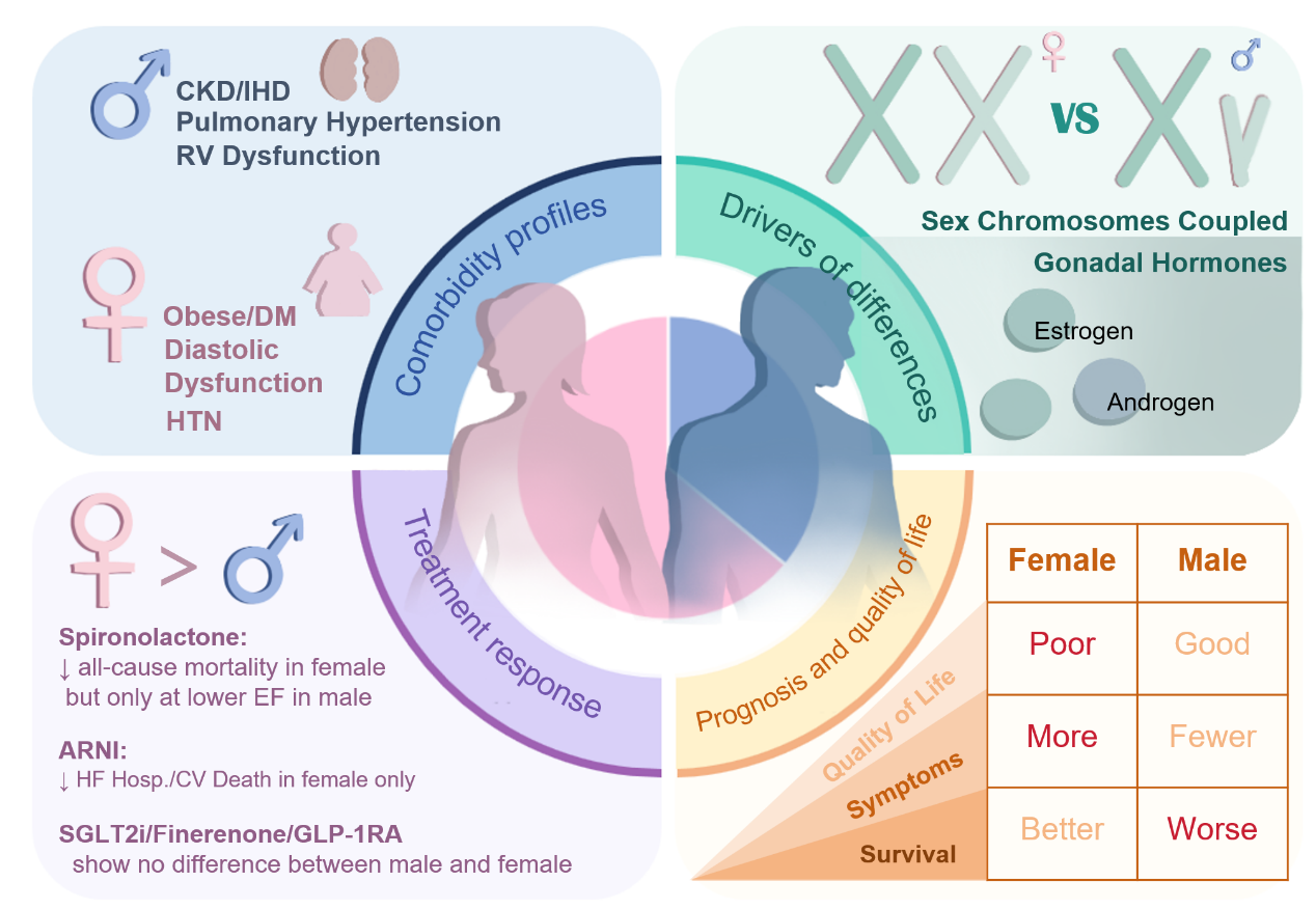
In women with HFpEF, comorbidities more often reflect a cardiometabolic profile and a higher prevalence of hypertension (Figure 1). Hypertension is a major contributor to HFpEF [30] and is more prevalent in women across most age groups, becoming particularly prominent after 75 years of age [24]. In postmenopausal women, it remains a dominant HFpEF risk factor across multiple racial and ethnic groups, including White, African American, and Hispanic women [31]. Obesity is also more prevalent in female HFpEF patients [19]. Notably, body mass index correlates with plasma B-type natriuretic peptide (BNP) levels, a relationship not observed in men [20]. In addition, epicardial fat accumulation is associated with worse outcomes and shows a stronger adverse effect in women [32,33,34]. Diabetes further contributes to this cardiometabolic phenotype, conferring a disproportionately higher risk of HFpEF [35], and more pronounced left ventricular remodeling in women compared with men [36,37].
In contrast, men with HFpEF more frequently exhibit a cardiorenal and ischemic comorbidity profile. Chronic kidney disease and liver dysfunction are more prevalent in male HFpEF patients [19,20], who also show higher rates of ischemic heart disease, prior percutaneous coronary intervention, or coronary artery bypass graft surgery [19,20,38,39] (Figure 1). Furthermore, men are also more prone to a right heart phenotype, characterized by pulmonary vascular disease, elevated diastolic pressure gradients, impaired right ventricular function, and increased cardiac mortality [21].
3.3. Sex-Specific Responses to HFpEF Therapies
Evidence from large clinical trials indicates that women and men with HFpEF can respond differently to pharmacological therapies. The TOPCAT trial (Treatment of Preserved Cardiac Function Heart Failure with an Aldosterone Antagonist) was overall neutral and did not meet its primary endpoint. However, subgroup and exploratory analyses have suggested potential sex-specific differences in treatment response. Spline analyses from sex-specific and phenomapping analyses suggested that women derived benefit from spironolactone across a wide range of LVEF, whereas men showed benefit mainly at lower EF values [29]. Furthermore, additional subgroup analysis indicated that spironolactone was associated with reduced all-cause mortality in women, but not in men [40,41]. Similarly, the PARAGON-HF trial (Prospective Comparison of ARNI with ARB Global Outcomes in HFpEF) demonstrated a significant interaction between sex and treatment, with women appearing to derive greater benefit from sacubitril/valsartan, whereas no clear benefit was observed in men [42,43].
In contrast, large-scale trials of sodium–glucose cotransporter 2 inhibitors, including EMPEROR-Preserved [44] and DELIVER [45], demonstrated consistent benefits in both women and men, without significant sex–treatment interactions. Emerging therapies such as finerenone [46] and glucagon-like peptide-1 receptor [47] agonists have also demonstrated clinical benefit in HFpEF, although data on sex-specific differences remain limited.
Together, these findings suggest that while some therapies may exhibit sex-specific effects, others provide comparable benefit across sexes (Figure 1). These heterogeneous responses may be partly explained by underlying biological differences. Sex-related variation in aldosterone signaling and myocardial fibrosis may contribute to the greater benefit of mineralocorticoid receptor antagonists observed in women, while differences in natriuretic peptide signaling and ventricular–vascular coupling may influence responses to sacubitril/valsartan [48,49]. In addition, women with HFpEF tend to exhibit more concentric remodeling, smaller left ventricular volumes, and higher LVEF than men, such that a similar LVEF may reflect more advanced functional impairment and greater responsiveness to therapies that promote reverse remodeling. Differences in pharmacokinetics and pharmacodynamics may further contribute to variability in treatment response [50].
3.4. Sex Differences in Prognosis and Quality of Life
Sex differences in HFpEF extend beyond pathophysiology and treatment response to include prognosis and quality of life. Women with HFpEF exhibit more congestion-related symptoms and generally report poorer quality of life than men [12,14,51]. Large trials involving over 8000 HFpEF patients from CHARM-Preserve, I-PRESERVE, and TOPCAT-Americas have shown that women experience orthopnea and paroxysmal nocturnal dyspnea more frequently than men [14]. Contemporary studies show that, even after accounting for age and other clinical factors, women score lower on the Kansas City Cardiomyopathy Questionnaire (KCCQ), with median differences exceeding 10 points [14]. In addition, women report higher levels of anxiety and depression [52,53].
Despite reporting lower quality of life, women with HFpEF generally have similar or slightly better long-term survival than men (Figure 1), and their risk of sudden cardiac death is roughly half that of male patients [14]. The I-PRESERVE trial, which included 2500 female HFpEF patients, further confirmed this finding. Even after adjusting for age and other baseline characteristics, women had a 20% lower risk of cardiovascular and non-cardiovascular death or hospitalization compared with men [11]. However, in a recent Chinese cohort of 51,711 patients followed for one year, women had higher adjusted risks of heart failure hospitalization, cardiovascular death, and all-cause hospitalization or death. These associations were consistent across different levels of LVEF [54].
4. Genetic and Hormonal Determinants of Sex Differences in HFpEF
To understand sex differences in HFpEF, it is useful to refer to the conceptual framework proposed by Arnold [55]. According to this framework, sex differences are primarily driven by gonadal hormones and direct effects of sex chromosome genes (Figure 1). Gonadal hormones influence tissue and organ function throughout development and adulthood, while sex chromosome genes can affect phenotype independently of hormones.
4.1. Mouse Models to Dissect Hormonal and Chromosomal Effects
Owing to the natural coupling of chromosomal and gonadal sex, traditional animal models cannot distinguish hormonal from chromosomal effects, so two genetic mouse models—the Four Core Genotypes (FCG) model and the XY* model—were created to overcome this limitation [56] (Figure 1).
The FCG model separates gonadal sex from sex chromosome complement by manipulating the Sry gene, enabling the independent assessment of hormonal and chromosomal influences. Proteomic analyses identified proteins regulated by hormones, sex chromosomes, or both [57].
The XY* model further isolates sex chromosome effects by varying X chromosome number independently of gonadal sex, demonstrating that X chromosome dosage influences susceptibility to cardiac injury [58].
These studies demonstrate that the FCG and XY* mouse models are valuable tools for dissecting the separate and combined effects of sex hormones and sex chromosomes on cardiac structure, function, and molecular pathways. By systematically comparing cardiac phenotypes, molecular pathway activity, and response to interventions between groups with distinct gonadal status and sex chromosome dosage, these models enable researchers to pinpoint critical sex-biased mechanisms, validate their causal roles in HFpEF, and define them as actionable, sex-dependent therapeutic targets for precision drug development.
4.2. Sex Chromosome–Mediated Regulation of Cardiac Function
Growing evidence shows that sex chromosomes exert direct effects on the heart that are independent of gonadal hormones. Studies of cardiac gene expression show clear differences between males and females even before sex hormones rise and before sexual maturation. These differences also appear later in aged females with declining reproductive function, as shown in mouse studies [59]. Proteomic studies also show sex differences which do not depend on gonadal hormones. Analyses of embryonic mouse hearts at E9.5 (embryonic day 9.5, before gonad formation) show that many cardiac proteins already exhibit sex differences. These protein differences are mainly established through post-transcriptional mechanisms [57].
X chromosome inactivation (XCI) ensures dosage compensation in females, but a subset of genes escapes XCI and remains transcriptionally active, contributing to sex-specific gene expression patterns and cardiovascular phenotypes [60,61,62,63].
Research directly addressing HFpEF remains limited, but emerging disease models provide complementary evidence for the role of sex chromosomes beyond specific epigenetic mechanisms. In a recent report using a two-hit HFpEF model (high-fat diet combined with L-NAME), experiments with FCG mice and ovariectomized females showed that both sex hormones and sex chromosomes influence HFpEF development. Estradiol had protective effects, whereas a second X chromosome appeared to exacerbate HFpEF features. Mechanistic studies using RNA sequencing are ongoing [64].
4.3. Estrogen and Estrogen Receptor–Related Phenotypic Modulation in HFpEF
Estrogen, particularly 17β-estradiol (E2), declines markedly after menopause and is closely associated with sex-specific phenotypic features of HFpEF [39,65,66]. Estrogen deficiency has been linked to increased vascular and myocardial stiffness—key pathophysiological features of HFpEF—in women, particularly in older women, and to a diminished cardiac ability to adapt to abnormal hemodynamic load [39,66]. Evidence further shows that estrogen plays an important role in diastolic function by shortening the isovolumic relaxation time (IVRT) and increasing the E/A ratio, thereby improving left ventricular relaxation and early diastolic filling [67]. These alterations provide a potential explanation for the higher prevalence and symptom burden of HFpEF observed in postmenopausal women. Clinical data show that estrogen deficiency is linked to greater left ventricular hypertrophy.
In contrast, estrogen or hormone therapy can reduce vascular resistance, systolic blood pressure, left ventricular mass index, and relative wall thickness in postmenopausal women [68]. However, large clinical trials of hormone therapy in older postmenopausal women have not demonstrated cardiovascular benefit, suggesting that timing and aging may limit efficacy (“critical window” hypothesis) [69,70,71]. However, as these studies were not specifically designed to address HFpEF, the relevance of estrogen signaling to HFpEF therapy remains unclear and warrants further investigation in studies specifically designed for HFpEF.
Estrogen exerts its effects through nuclear receptors estrogen receptor alpha (ERα), estrogen receptor beta (ERβ), and the membrane-bound G-protein-coupled estrogen receptor (GPER/GPR30), which are expressed in multiple cardiac cell types and regulate myocardial remodeling, stiffness, and diastolic function [72,73,74,75,76]. Experimental studies suggest that ERα is associated with preservation of right ventricular structure and function, a key determinant of outcomes in HFpEF [77,78,79], whereas ERβ and GPER signaling are linked to attenuation of hypertrophy, fibrosis, and diastolic dysfunction [72,80,81,82,83,84,85,86,87,88,89,90,91,92,93,94]. Together, estrogen receptor–mediated signaling contributes to sex-specific differences in cardiac structure, function, and clinical presentation in HFpEF.
4.4. Androgens and HFpEF-Related Cardiac Phenotypes
Testosterone is the predominant circulating androgen in men and is present at much lower levels in women [95,96]. Its cardiovascular effects are mainly mediated by the androgen receptor (AR), which is expressed in cardiomyocytes, vascular smooth muscle cells, and endothelial cells [97].
Circulating testosterone declines with age and is associated with increased cardiometabolic risk [97,98,99,100,101,102,103,104,105,106,107]. In men with HFpEF, low testosterone relates to worse clinical status and cardiac dysfunction. In a study of 120 male HFpEF patients, those with low testosterone had more severe symptoms (higher New York Heart Association [NYHA] functional class), higher BNP levels, poorer metabolic and inflammatory profiles, and impaired endothelial function [108]. A pilot study found that about 37% of male HFpEF patients had testosterone deficiency [109].
Mechanistic studies have shown that testosterone can induce vasodilation [110] and that long-term testosterone deficiency leads to diastolic dysfunction and impaired myocardial calcium handling [111]. Given the importance of endothelial function, myocardial relaxation, and calcium handling in HFpEF, testosterone deficiency may represent an underrecognized contributor to HFpEF progression.
5. Key Molecular Pathways Underlying Sex Differences in HFpEF
5.1. Sex-Specific Regulation of Mitochondrial Function in HFpEF
Increasing evidence indicates that mitochondria play a critical role in cardiac function and HFpEF pathophysiology [112,113]. Mitochondrial dysfunction impairs ATP production, disrupts substrate metabolism, increases reactive oxygen species (ROS), and perturbs calcium homeostasis. These alterations compromise cardiomyocyte relaxation and contractile reserve, promote oxidative damage, and exacerbate diastolic dysfunction and ventricular stiffness [114].
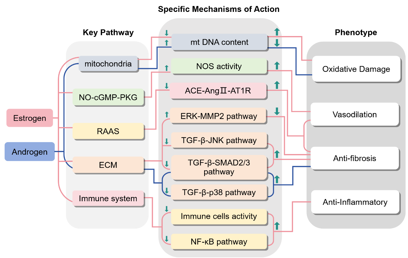
Our laboratory previously investigated sex-dependent mitochondrial regulation in HFpEF using the HMDP mouse panel and “two-hit” HFpEF models [115]. Female mice developed more pronounced diastolic dysfunction than males, accompanied by lower cardiac mitochondrial DNA (mtDNA) content, which negatively correlated with diastolic indices [115]. Gonadectomy and hormone replacement experiments demonstrated that sex hormones modulate mtDNA levels and mitochondrial function, with corresponding effects on diastolic performance (Figure 2): removal of endogenous hormones decreased mtDNA and worsened relaxation in males, whereas ovariectomy in females had the opposite effect [115]. These findings indicate that estrogen and androgens regulate mitochondrial abundance and function, influencing HFpEF susceptibility.
Acsl6 was further identified as a female-biased cardiac gene whose expression correlated with diastolic function [115]. Genetic variation controlling Acsl6 expression associated with HFpEF-relevant traits, highlighting a potential mechanistic link between sex, mitochondrial regulation, and myocardial relaxation.
5.2. Sex-Specific Regulation of the Renin-Angiotensin-Aldosterone System (RAAS)
Persistent activation of the RAAS is a key mechanism underlying left ventricular hypertrophy (LVH) and left ventricular diastolic dysfunction [116,117]. Beyond increasing blood pressure and afterload, RAAS promotes myocardial fibrosis and inflammation, exacerbating ventricular stiffness.
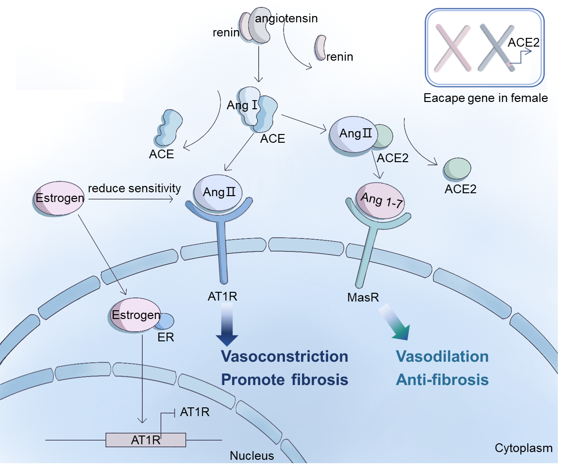
RAAS effects depend on the balance between two opposing axes: a deleterious “classical axis” centered on angiotensin-converting enzyme (ACE)–angiotensin II (Ang II)–angiotensin II type 1 receptor (AT1R) and a protective “counter-regulatory axis” centered on angiotensin-converting enzyme 2 (ACE2)–angiotensin-(1–7) [Ang-(1–7)]–Mas receptor (MasR) [117,118] (Figure 3).
Sex hormones modulate RAAS activity (Figure 2 and Figure 3). Estrogen deficiency enhances vascular and myocardial responses to Ang II, increasing AT1R expression, whereas estrogen replacement reverses this effect [119]. Consequently, premenopausal women exhibit a blunted ACE–Ang II–AT1R response, partially limiting RAAS-mediated cardiac injury. Beyond hormones, sex chromosomes may also contribute. ACE2, which encoded on the X chromosome and potentially escaping X inactivation [120,121], converts Ang II to Ang-(1–7), a peptide that counteracts the harmful effects of the classical RAAS axis. Higher ACE2 expression in females may further contribute to sex differences in RAAS activity [122,123,124].
5.3. Sexual Dimorphism in Immune Regulation and Inflammation
In HFpEF, chronic low-grade systemic inflammation and coronary microvascular endothelial inflammation link comorbidities to left ventricular diastolic dysfunction [125]. An illustrative example of how heightened immune responses interact with HFpEF susceptibility comes from autoimmune diseases: these conditions are more prevalent in women and are associated with diastolic dysfunction and an increased risk of HFpEF, as exemplified by impaired left ventricular diastolic function in patients with rheumatoid arthritis [126].
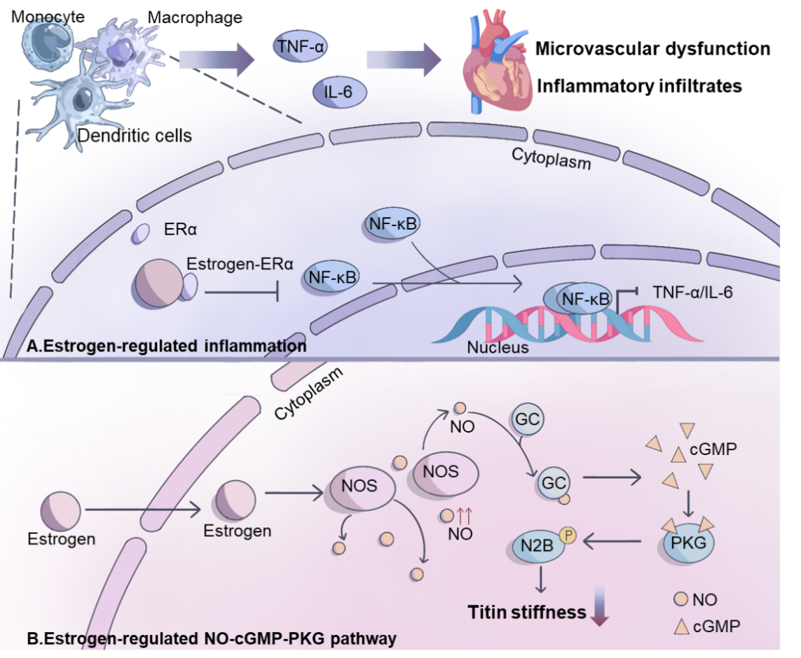
Inflammatory and immune responses display marked sexual dimorphism. Women generally exhibit greater immune responsiveness, characterized by more active transcription of immune-related genes, higher cytokine production, and stronger CD4+/CD8+ T-cell activation [127,128]. This enhanced responsiveness aids pathogen clearance but could predispose to excessive inflammation. In women, estrogen plays a key modulatory role in maintaining immune balance. Specifically, estrogen regulates the activity of T lymphocytes, B cells, and monocyte–macrophage populations through ERα, and suppresses nuclear factor kappa-light-chain-enhancer of activated B cells (NF-κB)–dependent transcription of pro-inflammatory genes, lowering the expression of cytokines such as tumor necrosis factor alpha (TNF-α) and interleukin-6 (IL-6) [129,130] (Figure 2 and Figure 4).
Sex differences in inflammation are also evident at the cardiac tissue level. Transcriptomic analyses show sex-dimorphic gene expression, with female hearts expressing higher levels of genes enriched in inflammatory pathways, including KLF4 and NF-κB [131].
5.4. Estrogen Modulation of the NO–cGMP–PKG Pathway in HFpEF
Within cardiomyocytes, the nitric oxide (NO)–cyclic guanosine monophosphate (cGMP)–protein kinase G (PKG) axis is central to maintaining diastolic function and limiting pathological remodeling (Figure 2). NO, mainly produced by endothelial (eNOS) and neuronal (nNOS) nitric oxide synthases, activates soluble guanylyl cyclase (sGC) to increase cGMP, which in turn activates PKG. PKG phosphorylates titin, reducing cardiomyocyte stiffness and improving diastolic compliance. In HFpEF patients, attenuated NO–cGMP–PKG signaling leads to titin hypophosphorylation, increased passive tension, and diastolic dysfunction [132,133,134,135,136].
Experimental studies indicate that estrogen enhances this axis. Estrogen upregulates eNOS and nNOS expression in cardiomyocytes, promoting NO production and maintaining endothelial function [137,138,139,140] (Figure 2 and Figure 4). Through ERβ signaling, estrogen increases eNOS activity in female cardiac myocytes [84]. In addition, Chen et al. have shown higher nNOS expression in female cardiac myocytes [141,142]. Overall, these findings highlight a key role of estrogen in modulating the NO–cGMP–PKG pathway in HFpEF, linking hormonal status to diastolic function and myocardial stiffness.
5.5. Sex Hormone Regulation of Extracellular Matrix Remodeling in HFpEF
Diastolic function is shaped not only by cardiomyocyte properties but also by the extracellular matrix (ECM), composed mainly of type I and III collagens, elastic fibers, and proteoglycans. Alterations in collagen content, isoform ratio, cross-linking, or ECM turnover affect ventricular stiffness and compliance [143,144].
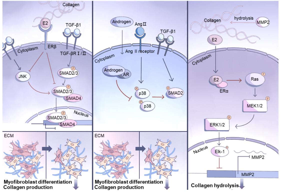
Sex hormones regulate ECM remodeling through distinct pathways in cardiac fibroblasts (Figure 2 and Figure 5). Estrogen signals through nuclear receptors ERα and ERβ as well as the membrane receptor GPER. Activation of ERβ inhibits transforming growth factor beta 1 (TGF-β1)-induced SMAD2/3 activation and nuclear translocation, blocks c-Jun N-terminal kinase (JNK) signaling, prevents fibroblast-to-myofibroblast differentiation, and reduces collagen, vimentin, and fibronectin synthesis [80]. GPER activation suppresses fibroblast proliferation by downregulating cyclins and CDK1 [145]. Androgen–AR signaling also participates in this regulation. Androgens act through AR to inhibit TGF-β1/Ang II-induced fibroblast proliferation, myofibroblast differentiation, and collagen synthesis [146,147]. These effects involve reduced p38 mitogen-activated protein kinase (MAPK) and SMAD2/3 activation [146,147]. AR knockout mice show more severe fibrosis under Ang II stimulation, with increased type I/III collagen, TGF-β1, and SMAD2 activity, highlighting the protective role of androgen–AR signaling [148].
Beyond increased collagen synthesis, impaired ECM degradation also contributes to sex-related fibrosis. ECM turnover is regulated by the balance between matrix metalloproteinases (MMPs) and their tissue inhibitors (TIMPs) [143,144,149]. Tissue inhibitor of metalloproteinases 1 (TIMP1) is an X-linked gene [97,150,151], and elevated TIMP1 inhibits matrix metalloproteinase 2/9 (MMP2/9)-mediated ECM degradation [152,153]. TIMP1 expression is higher in female hearts than in age-matched males in both mice and humans. In human myocardium, this difference is most pronounced in women aged 50–65 years, while younger women (<40 years) show a smaller increase [154,155]. In addition, physiological concentrations of E2 (10−8 M) reduce MMP2 expression in cardiac fibroblasts from adult rats and in HT1080 cells via ERα- and MAPK/extracellular signal-regulated kinase (ERK1/2)-dependent phosphorylation of Elk-1 and subsequent repression of the MMP2 promoter [156] (Figure 5).
In summary, sex hormones and X-linked genes act through distinct signaling mechanisms to regulate the development and progression of myocardial fibrosis in HFpEF.
6. Translational Implications for Therapeutic Target Discovery
The sex-specific pathophysiological mechanisms and preclinical models described in this review provide a robust framework for rational drug discovery in HFpEF. By integrating sex as a fundamental biological variable, researchers can prioritize high-value targets and design more effective clinical trials.
The distinct sex-related pathophysiological patterns observed in HFpEF provide a rational basis for precision therapeutic target discovery. In women, the prominent features of metabolic dysfunction, microvascular inflammation, ventricular-arterial stiffening, and myocardial fibrosis collectively highlight several actionable targets: mitochondrial energy metabolism, NO–cGMP–PKG signaling, inflammatory microvascular injury, and extracellular matrix remodeling. These pathways are not only more severely dysregulated in female patients but also underpin the observed sex-specific therapeutic advantages of spironolactone and sacubitril/valsartan.
In men, the predominant ischemic cardiopathy, cardiorenal involvement, and right ventricular dysfunction shift therapeutic priorities toward cardiorenal protection, anti-ischemic signaling, and right ventricular functional preservation. Although SGLT2 inhibitors and finerenone exhibit consistent cardioprotection across sexes, male patients may derive additional benefit from combined anti-ischemic strategies. Sex differences in pharmacokinetics and pharmacodynamics further warrant sex-stratified dose optimization during early drug development to maximize efficacy and safety.
7. Conclusions
HFpEF is highly prevalent in women and is associated with worse clinical outcomes and reduced quality of life. Women with HFpEF often present with distinct cardiac phenotypes, including concentric left ventricular remodeling, smaller ventricular cavities, pronounced diastolic dysfunction, and common comorbidities such as hypertension, obesity, and diabetes. Systemic factors—such as heightened inflammation, endothelial dysfunction, and metabolic disturbances—further contribute to disease progression.
Accumulating evidence suggests that these sex-specific patterns arise not only from traditional risk factors but also from the interplay of gonadal hormones, sex chromosomes, and downstream molecular pathways. Sex hormones—particularly estradiol and testosterone—their receptors, and X- or Y-linked genes modulate key mechanisms, including mitochondrial function, RAAS signaling, immune activation, nitric oxide–cGMP–PKG signaling, and extracellular matrix remodeling. Studies using genetic mouse models, such as the FCG and XY* models, demonstrate that hormonal and chromosomal factors can act independently or synergistically to shape cardiac responses to stress. These insights help explain the higher HFpEF burden in women and the potential for sex-specific therapeutic responses.
To advance HFpEF research and drug discovery, sex must be considered a fundamental biological variable at every stage of the translational pipeline. Preclinical studies should move beyond the exclusive use of young male animals and incorporate the sex-specific models described herein to identify and validate sex-dependent therapeutic targets. Clinically, many previous heart failure trials underrepresented women or lacked pre-specified sex-stratified analyses, leading to missed opportunities for effective therapies. Future clinical trials should ensure adequate female representation, incorporate pre-specified sex-based subgroup analyses, and evaluate sex-specific dosing regimens to develop truly personalized, sex-informed therapies for HFpEF.
Author Contributions
Y.L.: conceptualization, writing—original draft preparation; D.H.: visualization; Z.R.: data curation; Y.C.: supervision, writing—reviewing and editing. All authors have read and agreed to the published version of the manuscript.
Funding
This research received no external funding.
Institutional Review Board Statement
Not applicable.
Informed Consent Statement
Not applicable.
Data Availability Statement
Not applicable.
Conflicts of Interest
The authors declare no conflict of interest.
Use of AI and AI-Assisted Technologies
No AI tools were utilized for this paper.
List of Abbreviations
| Abbreviation | Full Term |
|---|---|
| HFpEF | heart failure with preserved ejection fraction |
| LVEF | left ventricular ejection fraction |
| HFrEF | heart failure with reduced ejection fraction |
| RAAS | renin–angiotensin–aldosterone system |
| LV | left ventricular |
| BNP | B-type natriuretic peptide |
| TOPCAT | Treatment of Preserved Cardiac Function Heart Failure with an Aldosterone Antagonist |
| PARAGON-HF | Prospective Comparison of ARNI with ARB Global Outcomes in HFpEF |
| ARNI | angiotensin receptor–neprilysin inhibitors |
| KCCQ | Kansas City Cardiomyopathy Questionnaire |
| DM | diabetes mellitus |
| HTN | hypertension |
| CKD | chronic kidney disease |
| IHD | ischemic heart disease |
| RV | right ventricular |
| CV | cardiovascular |
| FCG | four core genotypes |
| Xic | X-inactivation left |
| XIST | X-inactive specific transcript |
| PRC2 | polycomb repressive complex 2 |
| H3K27 | histone H3 lysine 27 |
| XCI | X chromosome inactivation |
| E2 | 17β-estradiol |
| IVRT | isovolumic relaxation time |
| ERα/ERβ | estrogen receptor alpha / beta |
| GPER/GPR30 | G-protein-coupled estrogen receptor |
| AR | androgen receptor |
| NYHA | New York Heart Association |
| ROS | reactive oxygen species |
| mtDNA | mitochondrial DNA |
| LVH | left ventricular hypertrophy |
| ACE | angiotensin-converting enzyme |
| ACE2 | angiotensin-converting enzyme 2 |
| Ang II | angiotensin II |
| AT1R | angiotensin II type 1 receptor |
| MasR | Mas receptor |
| TNF-α | tumor necrosis factor alpha |
| IL-6 | interleukin-6 |
| NF-κB | nuclear factor kappa-light-chain-enhancer of activated B cells |
| NO | nitric oxide |
| eNOS | endothelial nitric oxide synthase |
| nNOS | neuronal nitric oxide synthase |
| sGC | soluble guanylyl cyclase |
| cGMP | cyclic guanosine monophosphate |
| PKG | protein kinase G |
| ECM | extracellular matrix |
| TGF-β | transforming growth factor beta |
| MMPs | matrix metalloproteinases |
| MMP2 | matrix metalloproteinase 2 |
| MMP9 | matrix metalloproteinase 9 |
| TIMPs | tissue inhibitors of metalloproteinases |
| TIMP1 | tissue inhibitor of metalloproteinases 1 |
| TGF-βRII | transforming growth factor-β type II receptor |
| TGF-βRI | transforming growth factor-β type I receptor |
| JNK | c-Jun N-terminal kinase |
| MAPK | mitogen-activated protein kinase |
| ERK | extracellular signal-regulated kinase |
References
- 1.
Bozkurt, B.; Coats, A.J.S.; Tsutsui, H.; et al. Universal definition and classification of heart failure: A report of the Heart Failure Society of America, Heart Failure Association of the European Society of Cardiology, Japanese Heart Failure Society and Writing Committee of the Universal Definition of Heart Failure: Endorsed by the Canadian Heart Failure Society, Heart Failure Association of India, Cardiac Society of Australia and New Zealand, and Chinese Heart Failure Association. Eur. J. Heart Fail. 2021, 23, 352–380. https://doi.org/10.1002/ejhf.2115.
- 2.
Mishra, S.; Kass, D.A. Cellular and molecular pathobiology of heart failure with preserved ejection fraction. Nat. Rev. Cardiol. 2021, 18, 400–423. https://doi.org/10.1038/s41569-020-00480-6.
- 3.
Dunlay, S.M.; Roger, V.L.; Redfield, M.M. Epidemiology of heart failure with preserved ejection fraction. Nat. Rev. Cardiol. 2017, 14, 591–602. https://doi.org/10.1038/nrcardio.2017.65.
- 4.
Benjamin, E.J.; Virani, S.S.; Callaway, C.W.; et al. Heart Disease and Stroke Statistics-2018 Update: A Report from the American Heart Association. Circulation 2018, 137, e67–e492. https://doi.org/10.1161/CIR.0000000000000558.
- 5.
Steinberg, B.A. Trends in patients hospitalized with heart failure and preserved left ventricular ejection fraction: Prevalence, therapies, and outcomes. Circulation 2012, 126, 65–75. https://doi.org/10.1161/circulationaha.111.080770.
- 6.
Oktay, A.A.; Rich, J.D.; Shah, S.J. The emerging epidemic of heart failure with preserved ejection fraction. Curr. Heart Fail. Rep. 2013, 10, 401–410. https://doi.org/10.1007/s11897-013-0155-7.
- 7.
Ceia, F.; Fonseca, C.N.; Mota, T.; et al. Prevalence of chronic heart failure in Southwestern Europe: The EPICA study. Eur. J. Heart Fail. 2002, 4, 531–539. https://doi.org/10.1016/s1388-9842(02)00034-x.
- 8.
Owan, T.E. Trends in prevalence and outcome of heart failure with preserved ejection fraction. N. Engl. J. Med. 2006, 355, 251–259. https://doi.org/10.1056/NEJMoa052256.
- 9.
Guo, L.; Guo, X.; Chang, Y.; et al. Prevalence and Risk Factors of Heart Failure with Preserved Ejection Fraction: A Population-Based Study in Northeast China. Int. J. Environ. Res. Public. Health 2016, 13, 770. https://doi.org/10.3390/ijerph13080770.
- 10.
Sotomi, Y. Sex Differences in Heart Failure with Preserved Ejection Fraction. J. Am. Heart Assoc. 2021, 10, e018574. https://doi.org/10.1161/JAHA.120.018574.
- 11.
Lam, C.S.P.; Carson, P.E.; Anand, I.S.; et al. Sex Differences in Clinical Characteristics and Outcomes in Elderly Patients with Heart Failure and Preserved Ejection Fraction. Circ. Heart Fail. 2012, 5, 571–578. https://doi.org/10.1161/circheartfailure.112.970061.
- 12.
Tromp, J.; MacDonald, M.R.; Tay, W.T.; et al. Heart Failure with Preserved Ejection Fraction in the Young. Circulation 2018, 138, 2763–2773. https://doi.org/10.1161/circulationaha.118.034720.
- 13.
Tromp, J.; Shen, L.; Jhund, P.; et al. Age-Related Characteristics and Outcomes of Patients with Heart Failure with Preserved Ejection Fraction. JACC 2019, 74, 601–612. https://doi.org/10.1016/j.jacc.2019.05.052.
- 14.
Dewan, P.; Rørth, R.; Raparelli, V.; et al. Sex-Related Differences in Heart Failure with Preserved Ejection Fraction. Circ. Heart Fail. 2019, 12, e006539. https://doi.org/10.1161/circheartfailure.119.006539.
- 15.
Tadic, M. Sex and Heart Failure with Preserved Ejection Fraction: From Pathophysiology to Clinical Studies. J. Clin. Med. 2019, 8, 792. https://doi.org/10.3390/jcm8060792.
- 16.
Beale, A.L.; Nanayakkara, S.; Segan, L.; et al. Sex Differences in Heart Failure with Preserved Ejection Fraction Pathophysiology: A Detailed Invasive Hemodynamic and Echocardiographic Analysis. JACC Heart Fail. 2019, 7, 239–249. https://doi.org/10.1016/j.jchf.2019.01.004.
- 17.
Regitz-Zagrosek, V. Sex and Gender Differences in Heart Failure. Int. J. Heart Fail. 2020, 2, 157–181. https://doi.org/10.36628/ijhf.2020.0004.
- 18.
Honigberg, M.C. Sex Differences in Exercise Capacity and Quality of Life in Heart Failure with Preserved Ejection Fraction: A Secondary Analysis of the RELAX and NEAT-HFpEF Trials. J. Card. Fail. 2020, 26, 276–280. https://doi.org/10.1016/j.cardfail.2020.01.001.
- 19.
Goyal, P. Sex- and Race-Related Differences in Characteristics and Outcomes of Hospitalizations for Heart Failure with Preserved Ejection Fraction. J. Am. Heart Assoc. 2017, 6, e003330. https://doi.org/10.1161/jaha.116.003330.
- 20.
Harada, E. Sex Differences in Heart Failure with Preserved Ejection Fraction Reflected by B-type Natriuretic Peptide Level. Am. J. Med. Sci. 2018, 356, 335–343. https://doi.org/10.1016/j.amjms.2018.06.009.
- 21.
Duca, F. Gender-related differences in heart failure with preserved ejection fraction. Sci. Rep. 2018, 8, 1080. https://doi.org/10.1038/s41598-018-19507-7.
- 22.
Gerber, Y.; Weston, S.A.; Redfield, M.M.; et al. A contemporary appraisal of the heart failure epidemic in Olmsted County, Minnesota, 2000 to 2010. JAMA Intern. Med. 2015, 175, 996–1004. https://doi.org/10.1001/jamainternmed.2015.0924.
- 23.
Kitzman, D.W.; O’Neill, T.J.; Brubaker, P.H. Unraveling the Relationship Between Aging and Heart Failure with Preserved Ejection Fraction: The Importance of Exercise and Normative Reference Standards. JACC Heart Fail. 2017, 5, 356–358. https://doi.org/10.1016/j.jchf.2017.01.009.
- 24.
Pandey, A.; Omar, W.; Ayers, C.; et al. Sex and Race Differences in Lifetime Risk of Heart Failure with Preserved Ejection Fraction and Heart Failure with Reduced Ejection Fraction. Circulation 2018, 137, 1814–1823. https://doi.org/10.1161/circulationaha.117.031622.
- 25.
Lundorff, I.J.; Sengeløv, M.; Jørgensen, P.G.; et al. Echocardiographic Predictors of Mortality in Women with Heart Failure with Reduced Ejection Fraction. Circ. Cardiovasc. Imaging 2018, 11, e008031. https://doi.org/10.1161/circimaging.118.008031.
- 26.
Kuch, B.; Muscholl, M.; Luchner, A.; et al. Gender specific differences in left ventricular adaptation to obesity and hypertension. J. Hum. Hypertens. 1998, 12, 685–691. https://doi.org/10.1038/sj.jhh.1000689.
- 27.
Krumholz, H.M.; Larson, M.; Levy, D. Sex differences in cardiac adaptation to isolated systolic hypertension. Am. J. Cardiol. 1993, 72, 310–313. https://doi.org/10.1016/0002-9149(93)90678-6.
- 28.
Lam, C.S.P.; Arnott, C.; Beale, A.L.; et al. Sex differences in heart failure. Eur. Heart J. 2019, 40, 3859–3868c. https://doi.org/10.1093/eurheartj/ehz835.
- 29.
Shah, S.J.; Katz, D.H.; Selvaraj, S.; et al. Phenomapping for novel classification of heart failure with preserved ejection fraction. Circulation 2015, 131, 269–279. https://doi.org/10.1161/circulationaha.114.010637.
- 30.
Tadic, M.; Cuspidi, C.; Frydas, A.; et al. The role of arterial hypertension in development heart failure with preserved ejection fraction: Just a risk factor or something more? Heart Fail. Rev. 2018, 23, 631–639. https://doi.org/10.1007/s10741-018-9698-8.
- 31.
Eaton, C.B.; Pettinger, M.; Rossouw, J.; et al. Risk Factors for Incident Hospitalized Heart Failure with Preserved Versus Reduced Ejection Fraction in a Multiracial Cohort of Postmenopausal Women. Circ. Heart Fail. 2016, 9, e002883. https://doi.org/10.1161/circheartfailure.115.002883.
- 32.
van Woerden, G.; van Veldhuisen, D.J.; Manintveld, O.C.; et al. Epicardial Adipose Tissue and Outcome in Heart Failure with Mid-Range and Preserved Ejection Fraction. Circ. Heart Fail. 2022, 15, e009238. https://doi.org/10.1161/circheartfailure.121.009238.
- 33.
Kim, S.A.; Kim, M.N.; Shim, W.J.; et al. Epicardial adipose tissue is related to cardiac function in elderly women, but not in men. Nutr. Metab. Cardiovasc. Dis. 2017, 27, 41–47. https://doi.org/10.1016/j.numecd.2016.11.001.
- 34.
Kenchaiah, S. Pericardial Fat and the Risk of Heart Failure. JACC 2021, 77, 2638–2652. https://doi.org/10.1016/j.jacc.2021.04.003.
- 35.
Kannel, W.B.; Hjortland, M.; Castelli, W.P. Role of diabetes in congestive heart failure: The Framingham study. Am. J. Cardiol. 1974, 34, 29–34. https://doi.org/10.1016/0002-9149(74)90089-7.
- 36.
Galderisi, M.; Anderson, K.M.; Wilson, P.W.; et al. Echocardiographic evidence for the existence of a distinct diabetic cardiomyopathy (the Framingham Heart Study). Am. J. Cardiol. 1991, 68, 85–89. https://doi.org/10.1016/0002-9149(91)90716-x.
- 37.
Kajimoto, K.; Minami, Y.; Sato, N.; et al. Gender Differences in Anemia and Survival in Patients Hospitalized for Acute Decompensated Heart Failure with Preserved or Reduced Ejection Fraction. Am. J. Cardiol. 2017, 120, 435–442. https://doi.org/10.1016/j.amjcard.2017.04.043.
- 38.
Nakada, Y.; Kawakami, R.; Nakano, T.; et al. Sex differences in clinical characteristics and long-term outcome in acute decompensated heart failure patients with preserved and reduced ejection fraction. Am. J. Physiol. Heart Circ. Physiol. 2016, 310, H813–H820. https://doi.org/10.1152/ajpheart.00602.2015.
- 39.
Gori, M.; Lam, C.S.P.; Gupta, D.K.; et al. Sex-specific cardiovascular structure and function in heart failure with preserved ejection fraction. Eur. J. Heart Fail. 2014, 16, 535–542. https://doi.org/10.1002/ejhf.67.
- 40.
Merrill, M.; Sweitzer, N.K.; Lindenfeld, J. Sex Differences in Outcomes and Responses to Spironolactone in Heart Failure with Preserved Ejection Fraction: A Secondary Analysis of TOPCAT Trial. JACC Heart Fail. 2019, 7, 228–238. https://doi.org/10.1016/j.jchf.2019.01.003.
- 41.
Kanashiro-Takeuchi, R.M.; Heidecker, B.; Lamirault, G. Sex-specific impact of aldosterone receptor antagonism on ventricular remodeling and gene expression after myocardial infarction. Clin. Transl. Sci. 2009, 2, 134–142. https://doi.org/10.1111/j.1752-8062.2009.00094.x.
- 42.
Solomon, S.D.; McMurray, J.J.V.; Anand, I.S.; et al. Angiotensin-Neprilysin Inhibition in Heart Failure with Preserved Ejection Fraction. N. Engl. J. Med. 2019, 381, 1609–1620. https://doi.org/10.1056/NEJMoa1908655.
- 43.
McMurray, J.J.V.; Jackson, A.M.; Lam, C.S.P. Effects of Sacubitril-Valsartan Versus Valsartan in Women Compared with Men with Heart Failure and Preserved Ejection Fraction: Insights From PARAGON-HF. Circulation 2020, 141, 338–351. https://doi.org/10.1161/circulationaha.119.044491.
- 44.
Butler, J.; Filippatos, G.; Siddiqi, T.J.; et al. Effects of Empagliflozin in Women and Men with Heart Failure and Preserved Ejection Fraction. Circulation 2022, 146, 1046–1055. https://doi.org/10.1161/circulationaha.122.059755.
- 45.
Pabón, M.A.; Wang, X.; Lam, C.S.P. Sex Differences in Heart Failure with Improved Ejection Fraction: The DELIVER Trial. JACC Heart Fail. 2024, 12, 1119–1122. https://doi.org/10.1016/j.jchf.2024.03.011.
- 46.
Chimura, M.; Wang, X.; Jhund, P.S. Finerenone in Women and Men with Heart Failure with Mildly Reduced or Preserved Ejection Fraction: A Secondary Analysis of the FINEARTS-HF Randomized Clinical Trial. JAMA Cardiol. 2025, 10, 59–70. https://doi.org/10.1001/jamacardio.2024.4613.
- 47.
Verma, S.; Butler, J.; Borlaug, B.A. Efficacy of Semaglutide by Sex in Obesity-Related Heart Failure with Preserved Ejection Fraction: STEP-HFpEF Trials. J. Am. Coll. Cardiol. 2024, 84, 773–785. https://doi.org/10.1016/j.jacc.2024.06.001.
- 48.
Davel, A.P.; Jaffe, I.Z.; Tostes, R.C.; et al. New roles of aldosterone and mineralocorticoid receptors in cardiovascular disease: Translational and sex-specific effects. Am. J. Physiol. Heart Circ. Physiol. 2018, 315, H989–H999. https://doi.org/10.1152/ajpheart.00073.2018.
- 49.
Dewan, P.; Jackson, A.; Lam, C.S.P.; et al. Interactions between left ventricular ejection fraction, sex and effect of neurohumoral modulators in heart failure. Eur. J. Heart Fail. 2020, 22, 898–901. https://doi.org/10.1002/ejhf.1776.
- 50.
Chyou, J.Y.; Qin, H.; Butler, J.; et al. Sex-related similarities and differences in responses to heart failure therapies. Nat. Rev. Cardiol. 2024, 21, 498–516. https://doi.org/10.1038/s41569-024-00996-1.
- 51.
Chandra, A.; Vaduganathan, M.; Lewis, E.F.; et al. Health-Related Quality of Life in Heart Failure with Preserved Ejection Fraction. JACC Heart Fail. 2019, 7, 862–874. https://doi.org/10.1016/j.jchf.2019.05.015.
- 52.
Lewis, E.F.; Lamas, G.A.; O’Meara, E.; et al. Characterization of health-related quality of life in heart failure patients with preserved versus low ejection fraction in CHARM. Eur. J. Heart Fail. 2007, 9, 83–91. https://doi.org/10.1016/j.ejheart.2006.10.012.
- 53.
Yang, X.; Wen, Y.; Peng, H.; et al. Gender Differences in Anxiety, Depression, Insomnia, and Quality of Life in Heart Failure with Preserved Ejection Fraction: A Multicenter, Cross-sectional Study. J. Cardiovasc. Nurs. 2023, 38, 425–432. https://doi.org/10.1097/jcn.0000000000000951.
- 54.
Cai, A.; Qiu, W.; Xia, S.; et al. Sex-specific characteristics and outcomes in hospitalized heart failure with preserved ejection fraction: The China Cardiovascular Association Database-Heart Failure Center Registry. Eur. Heart J. 2023, 44, 4715–4718. https://doi.org/10.1093/eurheartj/ehad619.
- 55.
Arnold, A.P. Promoting the understanding of sex differences to enhance equity and excellence in biomedical science. Biol. Sex. Differ. 2010, 1, 1. https://doi.org/10.1186/2042-6410-1-1.
- 56.
Arnold, A.P.; Cassis, L.A.; Eghbali, M.; et al. Sex Hormones and Sex Chromosomes Cause Sex Differences in the Development of Cardiovascular Diseases. Arterioscler. Thromb. Vasc. Biol. 2017, 37, 746–756. https://doi.org/10.1161/atvbaha.116.307301.
- 57.
Shi, W.; Sheng, X.; Dorr, K.M.; et al. Cardiac proteomics reveals sex chromosome-dependent differences between males and females that arise prior to gonad formation. Dev. Cell 2021, 56, 3019–3034.e7. https://doi.org/10.1016/j.devcel.2021.09.022.
- 58.
Li, J.; Chen, X.; McClusky, R.; et al. The number of X chromosomes influences protection from cardiac ischaemia/reperfusion injury in mice: One X is better than two. Cardiovasc. Res. 2014, 102, 375–384. https://doi.org/10.1093/cvr/cvu064.
- 59.
Yusifov, A.; Chhatre, V.E.; Koplin, E.K.; et al. Transcriptomic analysis of cardiac gene expression across the life course in male and female mice. Physiol. Rep. 2021, 9, e14940. https://doi.org/10.14814/phy2.14940.
- 60.
Augui, S.; Nora, E.P.; Heard, E. Regulation of X-chromosome inactivation by the X-inactivation centre. Nat. Rev. Genet. 2011, 12, 429–442. https://doi.org/10.1038/nrg2987.
- 61.
Brown, C.J.; Ballabio, A.; Rupert, J.L.; et al. A gene from the region of the human X inactivation centre is expressed exclusively from the inactive X chromosome. Nature 1991, 349, 38–44. https://doi.org/10.1038/349038a0.
- 62.
Rocha, S.T. Jarid2 Is Implicated in the Initial Xist-Induced Targeting of PRC2 to the Inactive X Chromosome. Mol. Cell 2014, 53, 301–316. https://doi.org/10.1016/j.molcel.2014.01.002.
- 63.
Zhao, J.; Sun, B.K.; Erwin, J.A.; et al. Polycomb Proteins Targeted by a Short Repeat RNA to the Mouse X Chromosome. Science 2008, 322, 750–756. https://doi.org/10.1126/science.1163045.
- 64.
Zhou, Q.; Quan, M.; Szczesniak, D.; et al. Abstract Thu162: Sex Chromosomes and Sex Hormones Independently Contribute to Heart Failure with Preserved Ejection Fraction. Circ. Res. 2025, 137. https://doi.org/10.1161/res.137.suppl_1.thu162.
- 65.
Flurkey, K.; Randall, P.K.; Sinha, Y.N.; et al. Transient shortening of estrous cycles in aging C57BL/6J mice: Effects of spontaneous pseudopregnancy, progesterone, L-dihydroxyphenylalanine, and hydergine. Biol. Reprod. 1987, 36, 949–959. https://doi.org/10.1095/biolreprod36.4.949.
- 66.
Mancusi, C.; Gerdts, E.; De Simone, G.; et al. Impact of isolated systolic hypertension on normalization of left ventricular structure during antihypertensive treatment (the LIFE study). Blood Press. 2014, 23, 206–212. https://doi.org/10.3109/08037051.2013.858482.
- 67.
Deng, S.; Lin, J.; Zhao, Z.; et al. Sex differences and related estrogenic effects in heart failure with preserved ejection fraction. Heart Fail. Rev. 2023, 28, 937–948. https://doi.org/10.1007/s10741-022-10274-2.
- 68.
Light, K.C.; Hinderliter, A.L.; West, S.G.; et al. Hormone replacement improves hemodynamic profile and left ventricular geometry in hypertensive and normotensive postmenopausal women. J. Hypertens. 2001, 19, 269–278. https://doi.org/10.1097/00004872-200102000-00014.
- 69.
Rossouw, J.E.; Anderson, G.L.; Prentice, R.L. Risks and benefits of estrogen plus progestin in healthy postmenopausal women: Principal results from the Women’s Health Initiative randomized controlled trial. Jama 2002, 288, 321–333. https://doi.org/10.1001/jama.288.3.321.
- 70.
Grady, D.; Applegate, W.; Bush, T. Heart and Estrogen/progestin Replacement Study (HERS): Design, methods, and baseline characteristics. Control. Clin. Trials 1998, 19, 314–335. https://doi.org/10.1016/s0197-2456(98)00010-5.
- 71.
Hodis, H.N.; Mack, W.J.; Henderson, V.W. Vascular Effects of Early versus Late Postmenopausal Treatment with Estradiol. N. Engl. J. Med. 2016, 374, 1221–1231. https://doi.org/10.1056/NEJMoa1505241.
- 72.
Qian, C.; Liu, J.; Liu, H. Targeting estrogen receptor signaling for treating heart failure. Heart Fail. Rev. 2024, 29, 125–131. https://doi.org/10.1007/s10741-023-10356-9.
- 73.
Nevzati, E.; Shafighi, M.; Bakhtian, K.D.; et al. Estrogen induces nitric oxide production via nitric oxide synthase activation in endothelial cells. Acta Neurochir. Suppl. 2015, 120, 141–145. https://doi.org/10.1007/978-3-319-04981-6_24.
- 74.
Grohé, C.; Kahlert, S.; Löbbert, K.; et al. Cardiac myocytes and fibroblasts contain functional estrogen receptors. FEBS Lett. 1997, 416, 107–112. https://doi.org/10.1016/s0014-5793(97)01179-4.
- 75.
Menazza, S.; Murphy, E. The Expanding Complexity of Estrogen Receptor Signaling in the Cardiovascular System. Circ. Res. 2016, 118, 994–1007. https://doi.org/10.1161/circresaha.115.305376.
- 76.
Aryan, L.; Younessi, D.; Zargari, M. The Role of Estrogen Receptors in Cardiovascular Disease. Int. J. Mol. Sci. 2020, 21, 4314. https://doi.org/10.3390/ijms21124314.
- 77.
Chatterjee, N.A.; Steiner, J.; Lewis, G.D. It is time to look at heart failure with preserved ejection fraction from the right side. Circulation 2014, 130, 2272–2277. https://doi.org/10.1161/circulationaha.114.013536.
- 78.
Frump, A.L.; Goss, K.N.; Vayl, A.; et al. Estradiol improves right ventricular function in rats with severe angioproliferative pulmonary hypertension: Effects of endogenous and exogenous sex hormones. Am. J. Physiol. Lung Cell Mol. Physiol. 2015, 308, L873–L890. https://doi.org/10.1152/ajplung.00006.2015.
- 79.
Cheng, T.C.; Philip, J.L.; Tabima, D.M.; et al. Estrogen receptor-α prevents right ventricular diastolic dysfunction and fibrosis in female rats. Am. J. Physiol. Heart Circ. Physiol. 2020, 319, H1459–H1473. https://doi.org/10.1152/ajpheart.00247.2020.
- 80.
Pedram, A.; Razandi, M.; O’Mahony, F.; et al. Estrogen receptor-beta prevents cardiac fibrosis. Mol. Endocrinol. 2010, 24, 2152–2165. https://doi.org/10.1210/me.2010-0154.
- 81.
Umar, S.; Iorga, A.; Matori, H. Estrogen rescues preexisting severe pulmonary hypertension in rats. Am. J. Respir. Crit. Care Med. 2011, 184, 715–723. https://doi.org/10.1164/rccm.201101-0078OC.
- 82.
Gürgen, D.; Hegner, B.; Kusch, A.; et al. Estrogen Receptor-β Signals Left Ventricular Hypertrophy Sex Differences in Normotensive Deoxycorticosterone Acetate-Salt Mice. Hypertension 2011, 57, 648–654. https://doi.org/10.1161/HYPERTENSIONAHA.110.166157.
- 83.
Pedram, A.; Razandi, M.; Lubahn, D.; et al. Estrogen inhibits cardiac hypertrophy: Role of estrogen receptor-beta to inhibit calcineurin. Endocrinology 2008, 149, 3361–3369. https://doi.org/10.1210/en.2008-0133.
- 84.
Nuedling, S.; Karas, R.H.; Mendelsohn, M.E.; et al. Activation of estrogen receptor beta is a prerequisite for estrogen-dependent upregulation of nitric oxide synthases in neonatal rat cardiac myocytes. FEBS Lett. 2001, 502, 103–108. https://doi.org/10.1016/s0014-5793(01)02675-8.
- 85.
Jessup, J.A.; Zhang, L.; Chen, A.F.; et al. Neuronal nitric oxide synthase inhibition improves diastolic function and reduces oxidative stress in ovariectomized mRen2.Lewis rats. Menopause 2011, 18, 698–708. https://doi.org/10.1097/gme.0b013e31820390a2.
- 86.
Xu, Y.; Arenas, I.A.; Armstrong, S.J. Estrogen modulation of left ventricular remodeling in the aged heart. Cardiovasc. Res. 2003, 57, 388–394. https://doi.org/10.1016/s0008-6363(02)00705-8.
- 87.
Zhang, X.; Li, T.; Cheng, H.J.; et al. Chronic GPR30 agonist therapy causes restoration of normal cardiac functional performance in a male mouse model of progressive heart failure: Insights into cellular mechanisms. Life Sci. 2021, 285, 119955. https://doi.org/10.1016/j.lfs.2021.119955.
- 88.
Olde, B.; Leeb-Lundberg, L.M. GPR30/GPER1: Searching for a role in estrogen physiology. Trends Endocrinol. Metab. 2009, 20, 409–416. https://doi.org/10.1016/j.tem.2009.04.006.
- 89.
Morselli, E.; Santos, R.S.; Criollo, A.; et al. The effects of oestrogens and their receptors on cardiometabolic health. Nat. Rev. Endocrinol. 2017, 13, 352–364. https://doi.org/10.1038/nrendo.2017.12.
- 90.
Pei, H.; Wang, W.; Zhao, D. G Protein-Coupled Estrogen Receptor 1 Inhibits Angiotensin II-Induced Cardiomyocyte Hypertrophy via the Regulation of PI3K-Akt-mTOR Signalling and Autophagy. Int. J. Biol. Sci. 2019, 15, 81–92. https://doi.org/10.7150/ijbs.28304.
- 91.
Wang, Y.; Xing, Y.; Liu, X.; et al. G-protein coupled receptor 30 attenuates myocardial hypertrophy by reducing oxidative stress and apoptosis in Ang II-treated mice. Peptides 2022, 157, 170878. https://doi.org/10.1016/j.peptides.2022.170878.
- 92.
Wang, H.; Sun, X.; Chou, J.; et al. Cardiomyocyte-specific deletion of the G protein-coupled estrogen receptor (GPER) leads to left ventricular dysfunction and adverse remodeling: A sex-specific gene profiling analysis. Biochim. Biophys. Acta Mol. Basis Dis. 2017, 1863, 1870–1882. https://doi.org/10.1016/j.bbadis.2016.10.003.
- 93.
Groban, L.; Tran, Q.K.; Ferrario, C.M. Female Heart Health: Is GPER the Missing Link? Front. Endocrinol. 2019, 10, 919. https://doi.org/10.3389/fendo.2019.00919.
- 94.
Alencar, A.K.; Silva, J.S.; Lin, M. Effect of Age, Estrogen Status, and Late-Life GPER Activation on Cardiac Structure and Function in the Fischer344×Brown Norway Female Rat. J. Gerontol. A Biol. Sci. Med. Sci. 2017, 72, 152–162. https://doi.org/10.1093/gerona/glw045.
- 95.
Lucas-Herald, A.K.; Touyz, R.M. Androgens and Androgen Receptors as Determinants of Vascular Sex Differences Across the Lifespan. Can. J. Cardiol. 2022, 38, 1854–1864. https://doi.org/10.1016/j.cjca.2022.09.018.
- 96.
Naamneh Elzenaty, R.; Toit, T.; Flück, C.E. Basics of androgen synthesis and action. Best. Pract. Res. Clin. Endocrinol. Metab. 2022, 36, 101665. https://doi.org/10.1016/j.beem.2022.101665.
- 97.
Ferreira, C.; Trindade, F.; Ferreira, R. Sexual dimorphism in cardiac remodeling: The molecular mechanisms ruled by sex hormones in the heart. J. Mol. Med. 2022, 100, 245–267. https://doi.org/10.1007/s00109-021-02169-w.
- 98.
Feldman, H.A.; Longcope, C.; Derby, C.A. Age trends in the level of serum testosterone and other hormones in middle-aged men: Longitudinal results from the Massachusetts male aging study. J. Clin. Endocrinol. Metab. 2002, 87, 589–598. https://doi.org/10.1210/jcem.87.2.8201.
- 99.
Zumoff, B.; Strain, G.W.; Miller, L.K. Twenty-four-hour mean plasma testosterone concentration declines with age in normal premenopausal women. J. Clin. Endocrinol. Metab. 1995, 80, 1429–1430. https://doi.org/10.1210/jcem.80.4.7714119.
- 100.
Fujita, S.; Chiba, M.; Ohta, M. Alteration of plasma sex hormone levels associated with old age and its effect on hepatic drug metabolism in rats. J. Pharmacol. Exp. Ther. 1990, 253, 369–374. https://doi.org/10.1016/s0022-3565(25)12470-1.
- 101.
Zhao, D.; Guallar, E.; Ouyang, P. Endogenous Sex Hormones and Incident Cardiovascular Disease in Post-Menopausal Women. J. Am. Coll. Cardiol. 2018, 71, 2555–2566. https://doi.org/10.1016/j.jacc.2018.01.083.
- 102.
Zhao, S.P.; Li, X.P. The association of low plasma testosterone level with coronary artery disease in Chinese men. Int. J. Cardiol. 1998, 63, 161–164. https://doi.org/10.1016/s0167-5273(97)00295-7.
- 103.
Elagizi, A.; Köhler, T.S.; Lavie, C.J. Testosterone and Cardiovascular Health. Mayo Clin. Proc. 2018, 93, 83–100. https://doi.org/10.1016/j.mayocp.2017.11.006.
- 104.
Barrientos, G.; Llanos, P.; Basualto-Alarcón, C. Androgen-Regulated Cardiac Metabolism in Aging Men. Front. Endocrinol. 2020, 11, 316. https://doi.org/10.3389/fendo.2020.00316.
- 105.
Rhoden, E.L.; Ribeiro, E.P.; Teloken, C. Diabetes mellitus is associated with subnormal serum levels of free testosterone in men. BJU Int. 2005, 96, 867–870. https://doi.org/10.1111/j.1464-410X.2005.05728.x.
- 106.
Laughlin, G.A.; Barrett-Connor, E.; Bergstrom, J. Low serum testosterone and mortality in older men. J. Clin. Endocrinol. Metab. 2008, 93, 68–75. https://doi.org/10.1210/jc.2007-1792.
- 107.
Malkin, C.J.; Pugh, P.J.; Morris, P.D.; et al. Low serum testosterone and increased mortality in men with coronary heart disease. Heart 2010, 96, 1821–1825. https://doi.org/10.1136/hrt.2010.195412.
- 108.
Hamam, A.; Abou-Omar, M.; Rabah, H.; et al. Worsening effect of testosterone deficiency on males with heart failure with preserved ejection fraction. BMC Endocr. Disord. 2022, 22, 321. https://doi.org/10.1186/s12902-022-01249-3.
- 109.
Favuzzi, A.M.R.; Venuti, A.; Bruno, C. Hormonal deficiencies in heart failure with preserved ejection fraction: Prevalence and impact on diastolic dysfunction: A pilot study. Eur. Rev. Med. Pharmacol. Sci. 2020, 24, 352–361. https://doi.org/10.26355/eurrev_202001_19933.
- 110.
Jones, R.D.; English, K.M.; Pugh, P.J.; et al. Pulmonary vasodilatory action of testosterone: Evidence of a calcium antagonistic action. J. Cardiovasc. Pharmacol. 2002, 39, 814–823. https://doi.org/10.1097/00005344-200206000-00006.
- 111.
Ayaz, O.; Banga, S.; Heinze-Milne, S.; et al. Long-term testosterone deficiency modifies myofilament and calcium-handling proteins and promotes diastolic dysfunction in the aging mouse heart. Am. J. Physiol. Heart Circ. Physiol. 2019, 316, H768–H780. https://doi.org/10.1152/ajpheart.00471.2018.
- 112.
Brown, D.A.; Perry, J.B.; Allen, M.E.; et al. Expert consensus document: Mitochondrial function as a therapeutic target in heart failure. Nat. Rev. Cardiol. 2017, 14, 238–250. https://doi.org/10.1038/nrcardio.2016.203.
- 113.
Kumar, A.A.; Kelly, D.P.; Chirinos, J.A. Mitochondrial Dysfunction in Heart Failure with Preserved Ejection Fraction. Circulation 2019, 139, 1435–1450. https://doi.org/10.1161/CIRCULATIONAHA.118.036259.
- 114.
Yang, J.; Guo, Q.; Feng, X.; et al. Mitochondrial Dysfunction in Cardiovascular Diseases: Potential Targets for Treatment. Front. Cell Dev. Biol. 2022, 10, 841523. https://doi.org/10.3389/fcell.2022.841523.
- 115.
Cao, Y.; Vergnes, L.; Wang, Y.C.; et al. Sex differences in heart mitochondria regulate diastolic dysfunction. Nat. Commun. 2022, 13, 3850. https://doi.org/10.1038/s41467-022-31544-5.
- 116.
Chinnaiyan, K.M.; Alexander, D.; McCullough, P.A. Role of angiotensin II in the evolution of diastolic heart failure. J. Clin. Hypertens. 2005, 7, 740–747. https://doi.org/10.1111/j.1524-6175.2005.04889.x.
- 117.
Gregori, M.; Tocci, G.; Marra, A.; et al. Inadequate RAAS suppression is associated with excessive left ventricular mass and systo-diastolic dysfunction. Clin. Res. Cardiol. 2013, 102, 725–733. https://doi.org/10.1007/s00392-013-0585-y.
- 118.
Sweeney, M.; Corden, B.; Cook, S.A. Targeting cardiac fibrosis in heart failure with preserved ejection fraction: Mirage or miracle? EMBO Mol. Med. 2020, 12, EMMM201910865. https://doi.org/10.15252/emmm.201910865.
- 119.
Nickenig, G.; Bäumer, A.T.; Grohè, C.; et al. Estrogen modulates AT1 receptor gene expression in vitro and in vivo. Circulation 1998, 97, 2197–2201. https://doi.org/10.1161/01.cir.97.22.2197.
- 120.
Mangs, A.H.; Morris, B.J. The Human Pseudoautosomal Region (PAR): Origin, Function and Future. Curr. Genom. 2007, 8, 129–136. https://doi.org/10.2174/138920207780368141.
- 121.
Gagliardi, M.C.; Tieri, P.; Ortona, E.; et al. ACE2 expression and sex disparity in COVID-19. Cell Death Discov. 2020, 6, 37. https://doi.org/10.1038/s41420-020-0276-1.
- 122.
Chappell, M.C. Nonclassical renin-angiotensin system and renal function. Compr. Physiol. 2012, 2, 2733–2752. https://doi.org/10.1002/cphy.c120002.
- 123.
McKinney, C.A.; Fattah, C.; Loughrey, C.M. Angiotensin-(1-7) and angiotensin-(1-9): Function in cardiac and vascular remodelling. Clin. Sci. 2014, 126, 815–827. https://doi.org/10.1042/cs20130436.
- 124.
Liang, B.; Li, Y.; Han, Z. ACE2-Ang (1-7) axis is induced in pressure overloaded rat model. Int. J. Clin. Exp. Pathol. 2015, 8, 1443–1450.
- 125.
Paulus, W.J.; Tschöpe, C. A novel paradigm for heart failure with preserved ejection fraction: Comorbidities drive myocardial dysfunction and remodeling through coronary microvascular endothelial inflammation. J. Am. Coll. Cardiol. 2013, 62, 263–271. https://doi.org/10.1016/j.jacc.2013.02.092.
- 126.
Aslam, F.; Bandeali, S.J.; Khan, N.A.; et al. Diastolic dysfunction in rheumatoid arthritis: A meta-analysis and systematic review. Arthritis Care Res. 2013, 65, 534–543. https://doi.org/10.1002/acr.21861.
- 127.
Beale, A.L.; Meyer, P.; Marwick, T.H. Sex Differences in Cardiovascular Pathophysiology: Why Women Are Overrepresented in Heart Failure with Preserved Ejection Fraction. Circulation 2018, 138, 198–205. https://doi.org/10.1161/circulationaha.118.034271.
- 128.
Klein, S.L.; Flanagan, K.L. Sex differences in immune responses. Nat. Rev. Immunol. 2016, 16, 626–638. https://doi.org/10.1038/nri.2016.90.
- 129.
Straub, R.H. The complex role of estrogens in inflammation. Endocr. Rev. 2007, 28, 521–574. https://doi.org/10.1210/er.2007-0001.
- 130.
Ghisletti, S.; Meda, C.; Maggi, A. 17beta-estradiol inhibits inflammatory gene expression by controlling NF-kappaB intracellular localization. Mol. Cell Biol. 2005, 25, 2957–2968. https://doi.org/10.1128/mcb.25.8.2957-2968.2005.
- 131.
InanlooRahatloo, K.; Liang, G.; Vo, D.; et al. Sex-based differences in myocardial gene expression in recently deceased organ donors with no prior cardiovascular disease. PLoS ONE 2017, 12, e0183874. https://doi.org/10.1371/journal.pone.0183874.
- 132.
Kötter, S.; Gout, L.; Frieling-Salewsky, M.; et al. Differential changes in titin domain phosphorylation increase myofilament stiffness in failing human hearts. Cardiovasc. Res. 2013, 99, 648–656. https://doi.org/10.1093/cvr/cvt144.
- 133.
Hidalgo, C.; Hudson, B.; Bogomolovas, J. PKC phosphorylation of titin’s PEVK element: A novel and conserved pathway for modulating myocardial stiffness. Circ. Res. 2009, 105, 631–638. https://doi.org/10.1161/circresaha.109.198465.
- 134.
Heerebeek, L.; Hamdani, N.; Falcão-Pires, I. Low myocardial protein kinase G activity in heart failure with preserved ejection fraction. Circulation 2012, 126, 830–839. https://doi.org/10.1161/circulationaha.111.076075.
- 135.
Heerebeek, L.; Hamdani, N.; Handoko, M.L. Diastolic stiffness of the failing diabetic heart: Importance of fibrosis, advanced glycation end products, and myocyte resting tension. Circulation 2008, 117, 43–51. https://doi.org/10.1161/circulationaha.107.728550.
- 136.
Borbély, A.; Velden, J.; Papp, Z.; et al. Cardiomyocyte stiffness in diastolic heart failure. Circulation 2005, 111, 774–781. https://doi.org/10.1161/01.Cir.0000155257.33485.6d.
- 137.
Nuedling, S.; Kahlert, S.; Loebbert, K. 17 Beta-estradiol stimulates expression of endothelial and inducible NO synthase in rat myocardium in-vitro and in-vivo. Cardiovasc. Res. 1999, 43, 666–674. https://doi.org/10.1016/s0008-6363(99)00093-0.
- 138.
Murphy, E.; Steenbergen, C. Cardioprotection in females: A role for nitric oxide and altered gene expression. Heart Fail. Rev. 2007, 12, 293–300. https://doi.org/10.1007/s10741-007-9035-0.
- 139.
Mendelsohn, M.E.; Karas, R.H. The protective effects of estrogen on the cardiovascular system. N. Engl. J. Med. 1999, 340, 1801–1811. https://doi.org/10.1056/nejm199906103402306.
- 140.
Mendelsohn, M.E. Nongenomic, ER-mediated activation of endothelial nitric oxide synthase: How does it work? What does it mean? Circ. Res. 2000, 87, 956–960. https://doi.org/10.1161/01.res.87.11.956.
- 141.
Chen, J.; Petranka, J.; Yamamura, K.; et al. Gender differences in sarcoplasmic reticulum calcium loading after isoproterenol. Am. J. Physiol. Heart Circ. Physiol. 2003, 285, H2657–H2662. https://doi.org/10.1152/ajpheart.00557.2003.
- 142.
Sun, J.; Picht, E.; Ginsburg, K.S. Hypercontractile female hearts exhibit increased S-nitrosylation of the L-type Ca2+ channel alpha1 subunit and reduced ischemia/reperfusion injury. Circ. Res. 2006, 98, 403–411. https://doi.org/10.1161/01.RES.0000202707.79018.0a.
- 143.
González, A.; López, B.; Ravassa, S.; et al. The complex dynamics of myocardial interstitial fibrosis in heart failure. Focus on collagen cross-linking. Biochim. Biophys. Acta Mol. Cell Res. 2019, 1866, 1421–1432. https://doi.org/10.1016/j.bbamcr.2019.06.001.
- 144.
Severino, P.; D’Amato, A.; Prosperi, S. Myocardial Tissue Characterization in Heart Failure with Preserved Ejection Fraction: From Histopathology and Cardiac Magnetic Resonance Findings to Therapeutic Targets. Int. J. Mol. Sci. 2021, 22, 7650. https://doi.org/10.3390/ijms22147650.
- 145.
Wang, H.; Zhao, Z.; Lin, M.; et al. Activation of GPR30 inhibits cardiac fibroblast proliferation. Mol. Cell Biochem. 2015, 405, 135–148. https://doi.org/10.1007/s11010-015-2405-3.
- 146.
Chung, C.C.; Hsu, R.C.; Kao, Y.H. Androgen attenuates cardiac fibroblasts activations through modulations of transforming growth factor-β and angiotensin II signaling. Int. J. Cardiol. 2014, 176, 386–393. https://doi.org/10.1016/j.ijcard.2014.07.077.
- 147.
Iwasaki, T.; Mukasa, K.; Yoneda, M.; et al. Marked attenuation of production of collagen type I from cardiac fibroblasts by dehydroepiandrosterone. Am. J. Physiol. Endocrinol. Metab. 2005, 288, E1222–E1228. https://doi.org/10.1152/ajpendo.00370.2004.
- 148.
Ikeda, Y.; Aihara, K.; Sato, T. Androgen receptor gene knockout male mice exhibit impaired cardiac growth and exacerbation of angiotensin II-induced cardiac fibrosis. J. Biol. Chem. 2005, 280, 29661–29666. https://doi.org/10.1074/jbc.M411694200.
- 149.
Krebber, M.M.; Dijk, C.G.M.; Vernooij, R.W.M. Matrix Metalloproteinases and Tissue Inhibitors of Metalloproteinases in Extracellular Matrix Remodeling during Left Ventricular Diastolic Dysfunction and Heart Failure with Preserved Ejection Fraction: A Systematic Review and Meta-Analysis. Int. J. Mol. Sci. 2020, 21, 6742. https://doi.org/10.3390/ijms21186742.
- 150.
Anderson, C.L.; Brown, C.J. Epigenetic predisposition to expression of TIMP1 from the human inactive X chromosome. BMC Genet. 2005, 6, 48. https://doi.org/10.1186/1471-2156-6-48.
- 151.
Anderson, C.L.; Brown, C.J. Variability of X chromosome inactivation: Effect on levels of TIMP1 RNA and role of DNA methylation. Hum. Genet. 2002, 110, 271–278. https://doi.org/10.1007/s00439-002-0676-8.
- 152.
Korzeń, D.; Sierka, O.; Dąbek, J. Transcriptional Activity of Metalloproteinase 9 (MMP-9) and Tissue Metalloproteinase 1 (TIMP-1) Genes as a Diagnostic and Prognostic Marker of Heart Failure Due to Ischemic Heart Disease. Biomedicines 2023, 11, 2776. https://doi.org/10.3390/biomedicines11102776.
- 153.
Takawale, A.; Zhang, P.; Patel, V.B.; et al. Tissue Inhibitor of Matrix Metalloproteinase-1 Promotes Myocardial Fibrosis by Mediating CD63-Integrin β1 Interaction. Hypertension 2017, 69, 1092–1103. https://doi.org/10.1161/hypertensionaha.117.09045.
- 154.
Isensee, J.; Witt, H.; Pregla, R. Sexually dimorphic gene expression in the heart of mice and men. J. Mol. Med. 2008, 86, 61–74. https://doi.org/10.1007/s00109-007-0240-z.
- 155.
Almuntashiri, S.; Dutta, S.; Zhu, Y.; et al. Estrogen-dependent gene regulation: Molecular basis of TIMP-1 as a sex-specific biomarker for acute lung injury. Physiol. Rep. 2024, 12, e70047. https://doi.org/10.14814/phy2.70047.
- 156.
Mahmoodzadeh, S.; Dworatzek, E.; Fritschka, S.; et al. 17beta-Estradiol inhibits matrix metalloproteinase-2 transcription via MAP kinase in fibroblasts. Cardiovasc. Res. 2010, 85, 719–728. https://doi.org/10.1093/cvr/cvp350.


This work is licensed under a Creative Commons Attribution 4.0 International License.

 Suite 4002 Level 4, 447 Collins Street, Melbourne, Victoria 3000, Australia
 General Inquiries: info@sciltp.com