
Cardiovascular diseases (CVDs) are the leading cause of death globally, propelling the urgent need for more effective drug development strategies. Traditional methods, such as two-dimensional (2D) cell cultures and animal models, fall short due to their limited structural complexity, lack of physiological relevance, and variations across species. These shortcomings hinder their ability to accurately predict human cardiac responses to therapeutic agents. Recently, advanced three-dimensional (3D) cardiac models, including cardiac organoids, multicellular microtissues, engineered heart tissues, and organ-on-chip platforms, have emerged as promising tools to address these limitations. This review summarizes recent advances in 3D cardiac in vitro modeling technologies, outlines major cardiac model construction strategies and their technical characteristics, and discusses their applications in drug toxicity evaluation, high-throughput drug screening, disease modeling, and personalized medicine. In addition, current challenges associated with these systems are discussed, along with future perspectives for improving their physiological relevance, scalability, and translational potential in cardiovascular drug development and precision medicine.
- Open Access
- Review
Advances and Prospects of Cardiac Organoids in Drug Development
- Xiaoying Liu 1,
- Yingjuan Liu 1,
- Yutong Yan 2,
- Fay Pu 1,
- Sam Butterworth 3,
- Hongyuan Zhang 1,*
Author Information
Received: 24 Dec 2025 | Revised: 20 Mar 2026 | Accepted: 09 Apr 2026 | Published: 15 Sep 2026
Abstract
Keywords
3D in vitro cardiac models | cardiac organoids | cardiotoxicity | drug screening | disease modeling
1. Introduction
Cardiovascular diseases (CVDs) rank as the foremost cause of death across the globe. Recent Global Burden of Disease estimates indicate that CVDs remained the leading cause of death worldwide in 2023, accounting for approximately 19.2 million deaths, or about one in three global deaths [1]. Although pharmacological interventions are considered the primary prevention and treatments for CVDs, the development of new drugs continues to face significant challenges, such as prolonged timelines, high costs, and elevated failure rates [2]. Cardiotoxicity, in particular, is a major cause for the discontinuation or withdrawal of drug candidates during safety evaluation. Notable examples include cisapride [3] and astemizole [4], which were withdrawn from the market due to the risk of life-threatening arrhythmias. Other medications have encountered usage limitations or warnings due to comparable issues. Traditional drug screening processes predominantly utilize two-dimensional (2D) cardiomyocyte models and animal experiments. However, these models have several limitations in replicating human cardiac responses due to deficiencies in tissue architecture, multicellular interactions, cellular maturity [5], and interspecies physiological differences such as heart rate [6]. Consequently, these models may not fully capture human-specific cardiac responses.
In recent years, the emergence of organoid technology has provided new avenues for in vitro disease modeling. Organoids can be generated from embryonic stem cells (ESCs), induced pluripotent stem cells (iPSCs), or adult stem cells (ASCs). Considering their developmental plasticity and self-renewal, these stem cells can differentiate into diverse lineages and organize into three-dimensional (3D) multicellular assemblies under appropriate culture conditions [7]. Such organoids carry several hallmarks of early human organs, including the coexistence of multiple cell populations, basic tissue organization, and partial organ-specific functions [8]. To date, organoid culture methods have been proposed to model several organs, including vasculature [9], heart [10], gastrointestinal tract [11,12], brain [13], liver [14] and retina [15]. In contrast to traditional 2D systems, organoids offer a spatially organized environment that more accurately mimics in vivo signaling interactions. This capability allows for the reconstruction of microenvironments typical of both physiological and pathological states. Given that the cellular microenvironment is essential for controlling processes such as proliferation, differentiation, and morphogenesis, organoids enhance the reliability and relevance of in vitro studies. Additionally, they present a promising alternative for minimizing the dependence on animal experiments [16].
Cardiac organoids, a significant subset of organoid technology, consist of essential cardiac cell types, including cardiomyocytes, endothelial cells, and fibroblasts. These organoids are capable of displaying key functional characteristics, such as spontaneous contractions and electrophysiological activity [10]. Cardiac organoids provide a more accurate representation of human physiological conditions than traditional models, enhancing their biological relevance. Additionally, other advanced 3D in vitro cardiac models, including engineered heart tissues (EHTs) and organ-on-chip platforms, have emerged. Together, these models significantly advance preclinical drug testing and disease research. They have shown broad application potential in studies of cardiac development, cardiotoxicity screening [17], drug testing [18], disease modeling [19] and personalized medicine.
Cardiac organoids represent an important subtype of advanced 3D in vitro cardiac models. In addition, other engineered systems, including multicellular microtissues, engineered heart tissues (EHTs), and organ-on-chip platforms, have emerged as complementary approaches for modeling cardiac physiology and pathology. This review highlights recent advances in 3D cardiac modeling strategies and their applications in drug development, with particular emphasis on cardiotoxicity screening, disease modeling, and personalized medicine. It also discusses current limitations and future perspectives, providing insights for both basic research and clinical translation.
2. Three-Dimensional In Vitro Modeling Systems
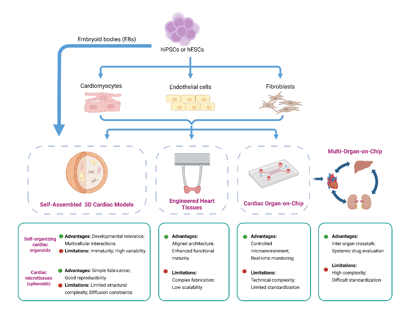
Recent advances in bioengineering and stem cell technologies have led to the development of multiple types of 3D in vitro cardiac models. These systems aim to better recapitulate the cellular organization, mechanical properties, and functional features of human heart tissue compared with traditional 2D cultures. In this review, 3D in vitro cardiac models are discussed in three major groups: self-assembled 3D cardiac models, EHTs, and organ-on-chip platforms. This classification is based on their distinct construction strategies, levels of structural control, and typical experimental applications in cardiovascular research. Although they share the advantage of providing more physiologically relevant microenvironments, each model type differs in its construction principles, level of structural complexity, and suitability for specific applications (Figure 1). The following subsections outline these major 3D cardiac model systems and highlight the unique characteristics of each.
2.1. Self-Assembled 3D Cardiac Models
Self-assembled 3D cardiac models refer to cardiac constructs generated without predefined external scaffolds or rigid geometric templates. Instead, tissue formation arises through cellular aggregation and cell–cell interactions within a three-dimensional environment [20].
Within this category, two major types of models can be distinguished based on their underlying biological mechanisms. The first includes self-organizing cardiac organoids derived from pluripotent stem cells, in which morphogen-driven developmental programs guide tissue patterning and spatial organization. The second includes multicellular cardiac microtissues or spheroids formed by assembling pre-differentiated cardiac cell types in defined ratios.
Although both systems rely on three-dimensional cell aggregation, they differ substantially in their developmental origin, structural autonomy, and experimental applications.
2.1.1. Self-Organizing Cardiac Organoids
Self-organizing cardiac organoids are PSC-derived 3D aggregates that recapitulate aspects of early cardiac morphogenesis through intrinsic developmental programs. These models rely on the coordinated differentiation, lineage specification, and spatial patterning of pluripotent stem cells under temporally controlled signaling modulation. Typically, PSCs are first aggregated into embryoid bodies (EBs) and subsequently guided toward cardiac mesoderm and cardiomyocyte lineages through stage-specific regulation of key developmental pathways, most commonly the WNT/β-catenin signaling pathway [21].
Unlike engineered constructs assembled from pre-specified components, self-organizing organoids undergo endogenous morphogen-driven pattern formation. Through cell–cell interactions and self-sorting processes, multiple cardiac-relevant cell types, including cardiomyocytes, endothelial cells, and epicardial-like cells, emerge and organize into spatially structured 3D tissues.
Several groups have recently developed stem-cell-derived cardiac organoid systems that reproduce key aspects of early heart development. To model chamber-specific morphogenesis, Hofbauer et al. developed human pluripotent stem cell–derived cardioids that self-organize into chamber-like cardiac structures and reproduce fundamental morphogenetic events of early cardiogenesis [22]. Building on this work, Schmidt et al. further established multi-chamber cardioid models capable of generating distinct cardiac compartments, thereby enabling the study of inter-chamber interactions and congenital heart defects in vitro [23]. Other organoid models have focused on reproducing early developmental tissue interactions. Drakhlis et al. reported human heart-forming organoids that recapitulate early cardiac morphogenesis and heart–foregut interactions through the coordinated emergence of myocardial and endocardial-like tissues [24]. Extending this platform, Dardano et al. developed blood-generating heart-forming organoids that model the co-development of cardiac and hematopoietic systems during early embryogenesis [25]. Additional studies highlight the importance of multi-lineage patterning in cardiac development. For instance, Rossi et al. demonstrated that mouse ESC-derived gastruloids can reproduce early cardiogenic patterning events, including the formation of cardiac crescent-like structures and beating cardiac tissues during early embryonic development [26]. Similarly, Silva et al. generated multilineage human iPSC-derived organoids in which cardiac and gut tissues co-emerge from mesendoderm progenitors. In this model, endoderm-derived tissues promote structural and functional maturation of cardiomyocytes through paracrine signaling interactions, including enhanced sarcomere organization and enrichment of atrial/nodal-like cardiomyocytes [27]. Lewis-Israeli et al. further demonstrated the generation of self-organizing human heart organoids derived from pluripotent stem cells and showed their capacity to model aspects of cardiac development and congenital heart disease under altered metabolic conditions [28,29]. These organoids exhibited spontaneous contractions, multicellular composition, and structural features reminiscent of early heart tissue. Such systems provide valuable platforms for studying developmental processes, congenital abnormalities, and early-stage drug-induced developmental toxicity.
Despite their biological relevance, self-organizing cardiac organoids remain subject to several limitations. Variability in organoid size, structural heterogeneity, incomplete maturation of cardiomyocytes, and batch-to-batch inconsistency continue to challenge reproducibility across laboratories. Moreover, the predominantly fetal-like phenotype of PSC-derived cardiomyocytes restricts the modeling of adult-onset cardiac diseases and mature electrophysiological disorders.
2.1.2. Multicellular Cardiac Microtissues
Multicellular cardiac microtissues, also referred to as cardiac spheroids, represent another important class of self-assembled 3D cardiac models. In contrast to self-organizing cardiac organoids, these systems are typically generated by assembling pre-differentiated cardiac cell types rather than relying on intrinsic developmental self-organization.
In many cardiac microtissue models, pluripotent stem cells are first directed to differentiate into multiple cardiac-relevant lineages, including cardiomyocytes, endothelial cells, smooth muscle cells, and cardiac fibroblasts. These pre-differentiated cells are subsequently combined in defined ratios and cultured within a three-dimensional environment, where they spontaneously aggregate to form compact spheroids through cell–cell adhesion and extracellular matrix interactions [30,31]. This “pre-differentiation + co-culture” strategy allows researchers to reconstruct aspects of the multicellular cardiac microenvironment in a controlled manner.
Importantly, multicellular cardiac microtissues are not restricted to systems composed entirely of iPSC-derived cell types. In some studies, iPSC-derived cardiomyocytes have been combined with primary supporting cells, such as endothelial cells and fibroblasts, to better reproduce the native myocardial microenvironment. For example, Richards et al. [32] developed a 3D cardiac microtissue model using iPSC-derived cardiomyocytes together with HUVECs and primary ventricular fibroblasts, highlighting the flexibility of this platform in incorporating mixed cell sources.
In other studies, cardiac spheroids composed of cardiomyocytes together with endothelial cells, smooth muscle cells, and cardiac fibroblasts derived from human induced pluripotent stem cells have been successfully generated and shown to exhibit coordinated contractile activity and multicellular interactions [33]. Similar engineered cardiac spheroid systems have also been proposed as platforms for modeling cardiovascular diseases and evaluating potential therapeutic strategies [34].
Compared with self-organizing cardiac organoids, multicellular cardiac microtissues offer several practical advantages. Because the cellular composition and cell ratios can be precisely controlled, these models generally demonstrate improved experimental reproducibility and reduced batch-to-batch variability. In addition, their relatively simple architecture makes them compatible with high-throughput experimental formats, which is particularly advantageous for drug screening and cardiotoxicity assessment [35].
However, because cardiac microtissues are formed through the aggregation of pre-specified cell populations, they typically lack the morphogen-driven developmental patterning observed in self-organizing cardiac organoids. As a result, these systems are less suitable for studying early cardiac morphogenesis but remain valuable for functional studies and pharmacological testing.
2.2. Engineered Heart Tissues
EHTs, as function-oriented, artificially constructed models, serve as a complementary 3D model system to self-assembling cardiac organoids. Unlike self-organizing cardiac organoids, which arise through intrinsic developmental programs, and multicellular cardiac microtissues, which are formed by aggregation of pre-differentiated cells, EHTs are engineered constructs assembled within defined biomaterial scaffolds, allowing precise control over tissue architecture and mechanical properties. Therefore, 3D bioprinting is often considered as an advanced fabrication technique within the broader spectrum of engineered heart tissues, although it is sometimes discussed as a distinct subcategory due to its unique structural controllability. It has been reported that 3D-based EHT construction strategies provide a supportive platform for the maturation of Human pluripotent stem cell–derived cardiomyocytes (hPSC-CMs).
Several EHT models, including strip- [36,37], ring- [38], and patch-type constructs [39], are established by embedding hPSC-CMs (with or without supporting cardiac cells) into hydrogels such as collagen, Matrigel, fibronectin, or fibrin [40]. During this process, a variety of physiological parameters—including long-term culture [41], substrate stiffness [42], cell patterning and alignment [43,44], electrical and mechanical stimulation [45], mechanical loading [46] and the interaction with other cell types [47], have all been shown to significantly enhance hPSC-CMs maturation [48].
EHTs are recognized as optimal models for investigating cardiac contractility and electrophysiological characteristics because they can replicate specific features of adult human myocardium. Nevertheless, EHTs have limitations in mimicking the intricate 3D structure and the early developmental processes of native cardiac tissue. Additionally, the construction of EHTs is both technically challenging and expensive, which limits their scalability and effectiveness in high-throughput screening applications [20]. Under strict quality control standards, EHTs exhibit relatively high reproducibility in drug-induced response studies [49]. In the future, efforts to improve maturity and throughput of EHT systems are advocated to improve therapeutics and facilitate the establishment of more granular approaches to disease modeling [50].
2.3. Cardiac Organ-on-Chip Platforms
Organ-on-chip systems represent an advanced class of in vitro models. Utilizing microfluidic technology, these models facilitate precise control over fluid dynamics and nutrient supply. This capability enables the incorporation of external stimuli, such as electrical and mechanical signals, while also permitting real-time monitoring of crucial physiological metrics, including pH, oxygen levels, and metabolite concentrations. As a result, organ-on-chip systems effectively mimic the intricate in vivo microenvironment [51]. For example, Marsano et al. developed a chip-based platform that reproduces mechanical conditions relevant to cardiomyocytes and may facilitate the study of hypertrophic responses induced by combined mechanical and biochemical stimulation [52]. Zhang et al. [53] constructed a multifunctional microfluidic chip integrating both electrical stimulation and electrophysiological signal monitoring. This system not only promotes cardiomyocyte maturation but also enables real-time assessment of drug responses, highlighting its broad potential in cardiovascular toxicity testing and drug screening.
Organ-on-chip platforms can also be integrated with models of various organs, including the kidney, liver, and pancreas, to develop “multi-organ-on-chip” systems. These innovative platforms have the potential to accurately replicate the intricate interactions between the cardiovascular system and metabolic or excretory organs. This capability allows for a more realistic assessment of drug-induced systemic effects and the potential for cardiotoxicity. For instance, Cody Juguilon et al. [54] pointed out that the cardiorenal-metabolic syndrome is a progressive condition driven by disrupted signaling among adipose tissue, liver, kidney, and heart, which can eventually lead to multi-organ dysfunction. Multi-organ chip platforms offer a cutting-edge in vitro model that facilitates the systematic investigation of inter-organ communication at different stages of the syndrome. These platforms have the potential to enhance drug discovery processes and improve the translation of findings into clinical applications.
These features highlight the potential of organ-on-chip systems for studying cardiac physiology, drug responses, and inter-organ interactions in a more integrated manner. To facilitate a clearer conceptual comparison of the major 3D cardiac model systems discussed above, their key features, advantages, and limitations are summarized in Table 1.
Comparative features of major 3D cardiac models in drug development, based on representative studies discussed in this review.
| Model Type | Construction Strategy | Structural Organization | Maturity | Throughput | Advantages | Limitations | Applications |
|---|---|---|---|---|---|---|---|
| Self-organizing cardiac organoids | PSC self-organization guided by developmental signaling | Moderate–high (multi-lineage, partial spatial organization) | Low (fetal-like) | Moderate | Recapitulate cardiac development; multicellular interactions | High variability; immature phenotype; limited reproducibility | Developmental studies; congenital disease modeling; early toxicity assessment |
| Cardiac microtissues (spheroids) | Aggregation of pre-differentiated cells | Low–moderate | Low–moderate | High | Good reproducibility; cost-effective; suitable for high-throughput screening | Limited structural complexity; diffusion limitations in larger constructs [55] | Drug screening; cardiotoxicity assessment |
| Engineered heart tissues (EHTs) | Cells embedded in biomaterials with mechanical/electrical stimulation | High (aligned structure) | High | Low–moderate | Enhanced functional maturity; direct measurement of contractility and electrophysiology | Complex fabrication; high cell demand; limited scalability | Functional pharmacology; contractility and electrophysiological analysis |
| Cardiac organ-on-chip platforms | Microfluidic systems with controlled microenvironment | Moderate–high (dynamic and perfusable system) | Moderate–high | Low–moderate | Real-time monitoring; integration of physical cues; potential for multi-organ systems | Technical complexity; limited standardization; potential drug absorption (e.g., polydimethylsiloxane (PDMS)) | Mechanistic studies; system-level drug response; multi-organ interaction studies |
3. Applications of 3D Cardiac Models in Drug Development
Drug-induced cardiotoxicity has emerged as a critical issue in both drug development and clinical practice. While most pharmaceuticals are not specifically designed to affect the heart, many can produce a range of cardiac adverse events, some of which pose significant risks to patient safety. Despite the adoption of various safety screening protocols during preclinical trials, new cardiotoxic effects continue to surface after drugs are brought to market, highlighting the shortcomings of current detection methodologies. It is estimated that approximately 10% of drug withdrawals are attributable to cardiovascular adverse drug reactions (ADRs). The consequences of drug-induced cardiotoxicity, especially in relation to heart failure, are frequently underestimated, with incidence rates reported between 11.0% and over 20.0% [56]. This not only poses a direct threat to patient safety but also significantly increases the cost of drug development.
Historically, numerous drugs have been restricted or withdrawn, either globally or in specific regions, due to cardiac safety concerns. Many of these, including cisapride [3], astemizole [4,57], gatifloxacin [58,59,60], grepafloxacin [58,61,62], bepridil [58,63], levomethadyl acetate [58,64], mesoridazine [58,65], probucol [58,66], sparfloxacin [58,61,62], and terfenadine [57,58,67], were linked to QT interval prolongation or life-threatening arrhythmias such as Torsades de Pointes (TdP). Ezogabine (retigabine) [58,68,69] was also withdrawn globally, with a possible risk of TdP and rare cases of QT prolongation and arrhythmias. Droperidol [70,71], though not withdrawn, carries an FDA black box warning (2001) for QT prolongation and TdP risk. Notably, some drugs with known cardiotoxic potential are applied in clinical use due to their substantial therapeutic benefits, provided that dosing and cardiac monitoring are carefully managed. For example, doxorubicin, an anthracycline anticancer agent, is currently used although it is associated with dose-dependent cardiotoxicity—approximately 4% of patients develop cardiomyopathy at cumulative doses of 500–550 mg/m2, increasing to 36% when the dose exceeds 600 mg/m2 [72].
Traditional cardiac safety assessment relies on multiple approaches, which include in vitro ion channel assays (such as the hERG test [73]), human cardiomyocyte models (e.g., hiPSC-derived cardiomyocytes) [74], animal studies, and clinical ECG monitoring [75,76]. However, both 2D hiPSC-CM models and animal models face significant limitations in structural complexity, functional maturity, and interspecies physiological differences, which compromise their predictive accuracy.
Although 2D cell culture systems are cost-effective and easy to handle, they do not capture the 3D tissue microenvironment in vivo, resulting in inaccurate drug responses [77] and limited predictive power. Animal models are commonly used to assess systemic toxicity, but significant interspecies differences can undermine their relevance to human health. A retrospective analysis of 108 anticancer drugs revealed that the median positive predictive value (PPV) of animal studies in predicting human toxicity was merely 0.65. In contrast, the negative predictive value (NPV) was approximately 0.50—indicating a nearly equal likelihood of encountering false positives and false negatives [78].
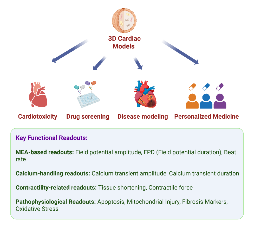
These limitations underscore the pressing necessity for in vitro cardiac models that more accurately replicate human cardiac physiology while providing better predictive accuracy for cardiotoxicity. Among these systems, cardiac organoids and other advanced 3D cardiac models have attracted considerable interest owing to their superior structural and functional biomimicry (Figure 2).
3.1. Cardiotoxicity Evaluation and Drug Screening
Advanced 3D cardiac models, particularly cardiac organoids, have emerged as promising in vitro platforms for evaluating drug-induced cardiotoxicity, owing to their ability to recapitulate multicellular architecture, contractile behavior, and electrophysiological properties that are difficult to achieve in conventional 2D cultures. Existing studies have applied cardiac organoids to assess multiple manifestations of drug-induced cardiotoxicity, particularly structural and cellular injury, electrophysiological disturbances, and functional impairment.
To examine structural and cellular manifestations of cardiotoxicity, Chen et al. [17] used a previously established human ESC-derived cardiac organoid model to investigate doxorubicin-induced toxicity. The model reproduced several clinically relevant hallmarks of cardiotoxicity, including impaired cardiac function, apoptosis, inflammation, and mitochondrial damage, supporting its utility as a physiologically relevant platform for toxicity assessment. Likewise, integrated single-organ and multi-organoid systems have been used to evaluate FDA-recalled compounds associated with human toxicity, and these 3D tissue models, including cardiac organoids, showed sensitivity to toxic agents [79]. Together, these studies support the utility of cardiac organoids for detecting diverse cardiotoxic phenotypes. Nevertheless, most evidence remains feasibility-based, and rigorous demonstrations of superior predictive performance over conventional animal or 2D models are still scarce.
For electrophysiological safety assessment and arrhythmogenic risk prediction, Lee et al. [80] used cardiac organoids to examine the cardiac safety of Echinochrome A (EchA). The organoids expressed characteristic cardiac markers and displayed electrophysiological activity, with no significant adverse effects across multiple functional parameters at concentrations ranging from 0.1 to 30 μM. This study highlights the value of cardiac organoids not only for identifying toxic compounds but also for supporting the preclinical evaluation of candidates with minimal detectable cardiotoxicity. More recently, Choi et al. developed a deformable shell-shaped microelectrode array (shell MEA) that conforms to the organoid surface and enables real-time measurement of global electrical activity [81]. In combination with calcium imaging, this platform detected electrophysiological responses to isoproterenol and the hERG blocker E-4031 [81], demonstrating its potential for early detection of drug-induced arrhythmias. Compared with standard 2D electrophysiological assays, these organoid-based systems may better preserve three-dimensional tissue-level responses. However, the extent to which this translates into improved prediction of clinical arrhythmia liability remains unclear, given the limited benchmarking across diverse drug classes and the still immature phenotype of many current cardiac organoid models.
For functional drug screening and contractile assessment, engineered cardiac microtissues and organoid-related platforms have also shown utility in detecting drug-induced mechanical impairment. Richard J. Mills et al. [18] established a bioengineered human cardiac tissue platform and performed high-throughput screening of 105 small molecules, revealing screening outcomes that differed from those obtained in traditional 2D systems. This study identified two compounds capable of promoting cardiomyocyte proliferation while preserving functional performance, illustrating the advantage of 3D cardiac models for functional screening and mechanistic studies. In another study, an engineered heart tissue (EHT) platform composed of hiPSC-derived cardiomyocytes was coupled with an automated image-based contraction analysis workflow to quantify tissue shortening and PDMS post deflection under standardized conditions [82], thereby enabling indirect assessment of contractile force. Using this system, exposure to the hERG inhibitor E-4031 produced detectable alterations in contractile dynamics [82], supporting its sensitivity for cardiac safety screening. These studies indicate that 3D cardiac models can capture integrated mechanical outputs that are difficult to resolve in simpler monolayer cultures, although differences in platform design and analysis pipelines currently limit cross-study comparability. These studies also reflect an important trend in the field, namely the adaptation of cardiac organoids to higher-throughput and multi-parametric screening formats. However, most available reports still emphasize technical feasibility and platform responsiveness, whereas rigorous benchmarking against conventional models across broader compound sets remains limited.
Importantly, several studies also point to practical limitations and inconclusive aspects that must be addressed before wider adoption. Standardization remains a major challenge, as parameters such as cell source, differentiation protocol, generation time, and organoid size can substantially influence beating behavior, calcium handling, and drug responsiveness. Lee et al. systematically evaluated the beating regularity and calcium signaling of cardiac organoids generated under different conditions and emphasized the need to define parameters such as generation time and size to improve the reproducibility and reliability of toxicity assessments [83]. Similarly, Hoang et al. [84] identified cardiac organoids with a diameter of approximately 600 μm as the most suitable for drug screening and used this optimized model to assess the embryotoxicity of nine FDA-approved pregnancy-related drugs with different teratogenic risks. These findings underscore that organoid performance is highly condition-dependent and that the lack of standardized production criteria may partly explain why results across studies are not always directly comparable.
Overall, cardiac organoids are helping to overcome some of the limitations of traditional models by more closely reproducing human cardiac structure and function. Their particular strengths lie in enabling integrated assessment of structural injury, electrophysiological disturbances, and contractile dysfunction within a more biomimetic context. However, the field is still transitioning from promising proof-of-principle studies to robust validation. Future progress will depend not only on advances in bioengineering and automation, but also on systematic benchmarking, standardization, and transparent reporting of both positive and negative findings. With these improvements, cardiac organoids are likely to become increasingly valuable tools for drug screening and cardiotoxicity risk assessment.
3.2. Disease Modeling
Although 3D cardiac models were initially developed for drug safety assessment and screening, they are increasingly being used as platforms for studying human cardiac diseases. Recent research has demonstrated the successful application of cardiac organoids to simulate various cardiac conditions in vitro, such as ischemic heart disease, metabolic cardiomyopathy, congenital heart defects, and heart failure. These models facilitate targeted drug screening and play a crucial role in advancing more tailored therapeutic approaches for cardiovascular disorders. Beyond phenotypic recapitulation, these systems also enable the investigation of disease-relevant mechanisms, including hypoxia-related injury, metabolic stress, fibrosis-associated remodeling, and abnormal calcium handling.
In the context of ischemic heart disease modeling, Richards et al. [32] developed a 3D cardiac microtissue-based model incorporating an internal oxygen gradient and norepinephrine stimulation to mimic the structural and metabolic characteristics of different myocardial infarction zones (infarct, border, and remote areas). This model effectively replicated infarction characteristics across transcriptomic, morphological, and functional dimensions, while also enabling the study of hypoxia-driven injury responses and regional heterogeneity across infarct, border, and remote zones. It was also used to explore how hypoxic conditions amplify doxorubicin-induced cardiotoxicity, underscoring the potential of 3D cardiac models for modeling non-genetic cardiovascular diseases. Similarly, Song et al. [85] induced ischemia–reperfusion injury in cardiac organoids via hypoxic treatment, thereby enhancing apoptosis, impairing cardiac function, disrupting calcium handling, and elevating fibrosis markers—effectively simulating the pathological processes of acute myocardial infarction and cardiac fibrosis. Together, these studies suggest that 3D cardiac models are particularly useful for investigating ischemia-associated mechanisms, including hypoxia-induced stress, apoptosis, calcium dysregulation, and fibrotic remodeling, within a multicellular, spatially organized tissue context.
For metabolic cardiomyopathy, Wang et al. [86] established a diabetic cardiomyopathy (DCM) model by exposing cardiac organoids to a high-glucose, high-lipid environment. They observed that the treatment induced cellular apoptosis, oxidative stress, mitochondrial dysfunction, and upregulation of cardiac injury markers, inflammatory mediators, and fibrosis-related factors. Subsequent treatment with metformin significantly improved these pathological features, highlighting the model’s effectiveness in assessing drug efficacy and exploring mechanisms associated with diabetic heart disease. These findings indicate that cardiac organoids can capture key mechanisms of diabetic cardiomyopathy, particularly the interplay among metabolic stress, mitochondrial dysfunction, inflammatory signaling, and fibrosis-related remodeling.
In congenital disease modeling, Lewis-Israeli et al. [29] simulated pregestational diabetes conditions (high glucose and high insulin) during organoid culture, successfully recapitulating the morphological and functional abnormalities of malformed fetal hearts. The associated electrophysiological abnormalities, metabolic disturbances, and myocardial disorganization provide a useful platform for investigating developmental mechanisms underlying congenital cardiac defects. However, because these models predominantly reflect early developmental stages, their ability to reproduce later-stage or adult electrophysiology-driven disease phenotypes remains limited.
In addition, researchers have developed 3D cardiac models for heart failure with preserved ejection fraction (HFpEF) that integrate various comorbid conditions, including hypertension, diabetes, and obesity. These models effectively replicate essential pathological features of HFpEF, such as cellular and tissue hypertrophy, fibrosis, increased passive stiffness of cardiac tissue, diastolic dysfunction, abnormal calcium handling, and impaired energy metabolism [19]. This application highlights the potential of advanced 3D cardiac models to integrate multiple disease-relevant stressors and reproduce complex pathological features of adult heart disease. At the same time, whether these in vitro phenotypes fully capture the long-term progression and patient-to-patient heterogeneity of HFpEF remains to be established.
Collectively, these studies show that 3D cardiac models are not only useful for reproducing disease-associated phenotypes, but also for probing tissue-level pathological processes that depend on multicellular crosstalk and three-dimensional microenvironmental context. However, important limitations remain. Many current cardiac organoid models retain fetal-like characteristics, which constrains their ability to faithfully model adult-onset disorders and mature electrophysiology-driven diseases. In addition, although several reported phenotypes are consistent with known human pathological features, systematic validation against clinical patient data is still limited. Further refinement, benchmarking, and clinical correlation will therefore be essential to enhance the mechanistic and translational value of these models.
3.3. Personalized Medicine
Patient-specific iPSC-derived cardiac models provide a powerful platform for disease modeling and drug testing, particularly for developing models that accurately represent individual genetic backgrounds. Unlike traditional animal models, which often face challenges due to interspecies differences, iPSC-derived organoids effectively reflect the genetic and phenotypic diversity among individuals. This capability makes them especially useful for creating and personalizing treatment plans that cater to the specific needs of each patient [10,87].
In studies of genetic cardiac diseases, patient-specific 3D cardiac models derived from iPSCs have been used to reproduce pathological changes in cell alignment and calcium signaling associated with dilated cardiomyopathy [88]. Advances in gene editing have revealed that correcting disease-causing mutations, such as the F508del mutation in the CFTR gene using CRISPR to produce functional lung organoids, could inspire similar applications in cardiac organoid systems [16,89].
With continuous advancements in differentiation efficiency, disease modeling, and phenotypic analysis techniques, iPSC-derived cardiac organoids are poised to significantly enhance drug target discovery, optimize individualized dosing, and inform precision therapeutic decisions. This progress opens new avenues for cardiovascular precision medicine.
4. Perspectives and Challenges
Advanced 3D cardiac models, particularly cardiac organoids, have shown remarkable potential in recent years across drug toxicity assessment, disease modeling, and personalized medicine. Compared with conventional 2D cardiomyocyte models and animal experiments, these systems provide enhanced biomimicry by more accurately reflecting tissue architecture and cellular composition. They can partially recapitulate complex human cardiac functions, including rhythmic contractions, electrophysiological activity, and drug responses, and have been widely used to model cardiotoxicity, screen therapeutic compounds, and simulate disease conditions. This progression is gradually shifting these models from fundamental research toward preclinical applications, positioning them as promising platforms for drug development and precision medicine.
As summarized in Table 1, different 3D cardiac model systems exhibit distinct structural, functional, and practical characteristics, leading to specific advantages and limitations that influence their translational potential. Despite their rapid development, 3D cardiac models still face several challenges. First, there is currently no standardized protocol for organoid generation, leading to significant inter-laboratory variability and reduced reproducibility and comparability [83]. Such variability complicates cross-study comparisons and may reduce the reliability of organoid-based drug response predictions. Furthermore, cardiomyocytes within cardiac organoids often present with a fetal-like phenotype and lack the functional characteristics of mature cardiac tissue [90]. This immature phenotype limits the ability of current organoid systems to faithfully model adult-onset cardiovascular diseases, including arrhythmias, hypertrophic cardiomyopathy, and heart failure. Although physiological interventions such as electrical stimulation have been reported to enhance the maturation of cardiac organoids/EHTs to some extent [91,92], modeling adult cardiac diseases, such as arrhythmias, remains difficult [93]. Moreover, accurate replication of the coordinated regulation of the cardiac microenvironment using systems such as the nervous and immune systems remains to be a critical challenge, although recent advances have partially addressed this limitation [94]. Finally, significant technological barriers to scalability and high-throughput compatibility limit the broader implementation of cardiac organoids in industrial-scale drug screening, particularly given the relatively high cost and time required for organoid generation. In addition, material-related limitations in certain organoid-on-chip or microfluidic platforms, such as small-molecule absorption by PDMS, may affect drug exposure accuracy and compromise pharmacological assessments [95].
Another major challenge lies in the regulatory acceptance of 3D cardiac model-based assays for drug safety evaluation, as standardized validation frameworks and benchmarking datasets are still under development [96,97]. While technical guidelines for cardiac organoid manufacturing have recently been established to address quality control and basic assay requirements [98], these currently serve as preliminary frameworks and highlight the need for further comparative studies to support regulatory qualification [98]. Regulatory agencies, including the FDA, are actively working toward incorporating new approach methodologies (NAMs), but formal acceptance pathways specifically for complex 3D cardiac models have not yet been established [97,99].
The protocol standardization, functional maturation, microenvironmental integration, and regulatory acceptance, represent major bottlenecks to the broader adoption of 3D cardiac model-based platforms [96]. These limitations highlight the current lack of clear pathways for integrating 3D cardiac models into established safety pharmacology frameworks, such as the comprehensive in vitro proarrhythmia assay (CiPA) [97,99]. Recent efforts have focused on qualifying human iPSC-derived cardiomyocytes within the CiPA paradigm, but these have predominantly utilized 2D monolayers rather than 3D configurations [100]. This suggests that 3D cardiac models may serve as complementary platforms to existing CiPA components by providing higher-order tissue-level readouts. While 3D cardiac models offer enhanced structural complexity, achieving the throughput and reproducibility required for regulatory standardization remains a significant challenge [97].
To address these challenges, future research is proposed to promote the optimization and application of cardiac organoids in several key directions. For example, integrating multi-organ-on-chip systems [54] may expand the capacity to simulate systemic drug effects and toxicity. The application of interventions, such as electrical stimulation, mechanical loading, and metabolic modulation, has been reported to facilitate cardiomyocyte maturation toward an adult-like phenotype [101,102]. Artificial intelligence is progressively being utilized in the quality control of organoids, image analysis, and the evaluation of 3D structures. These advances may facilitate the integration of cardiac organoid systems into automated drug discovery pipelines and improve the predictive accuracy of preclinical cardiotoxicity assessment. This advancement significantly enhances the automation and accuracy of high-throughput screening methods that involve cardiac organoids [103]. Ultimately, the integration of iPSC-derived cardiac organoids with patient-specific clinical data may offer powerful tools for individualized drug therapy, disease prediction, and precision cardiovascular medicine [104]. In parallel, collaborative efforts among academia, industry, and regulatory agencies will be essential to generate robust benchmarking datasets and standardized validation frameworks, thereby enabling the regulatory qualification and broader adoption of 3D cardiac model-based assays in drug safety evaluation.
In summary, cardiac organoids and related 3D cardiac models represent a promising advance in drug screening and cardiotoxicity assessment. Although challenges related to cost, preparation time, maturation, standardization, and scalability remain, continued progress in stem cell engineering, tissue biomimicry, and digital technologies is expected to enhance their value in cardiovascular drug development and precision medicine. With further optimization, these systems may increasingly help bridge the gap between conventional preclinical models and human clinical responses.
Author Contributions
X.L.: investigation, writing—original draft preparation, visualization; Y.L.: writing—review and editing; Y.Y.: writing—review and editing; S.B.: supervision; H.Z.: supervision, writing—review and editing. All authors have read and agreed to the published version of the manuscript.
Funding
This work was funded by British Heart Foundation Manchester Centre of Research Excellence grant number RE/24/130017.
Data Availability Statement
Not applicable.
Conflicts of Interest
The authors declare no conflict of interest.
Use of AI and AI-Assisted Technologies
No AI tools were utilized for this paper.
Abbreviations
| Abbreviation | Full Term |
| 2D | Two-dimensional |
| 3D | Three-dimensional |
| ASC | Adult stem cell |
| CiPA | Comprehensive in vitro Proarrhythmia Assay |
| CVD | Cardiovascular disease |
| EHT | Engineered heart tissue |
| ESC | Embryonic stem cell |
| FDA | Food and Drug Administration |
| HFpEF | Heart failure with preserved ejection fraction |
| hPSC-CM | Human pluripotent stem cell–derived cardiomyocyte |
| iPSC | Induced pluripotent stem cell |
| NAM | New Approach Methodology |
| NPV | Negative predictive value |
| PDMS | Polydimethylsiloxane |
| PPV | Positive predictive value |
| TdP | Torsades de Pointes |
References
- 1.
Global Burden of Cardiovascular Diseases and Risks 2023 Collaborators. Global, Regional, and National Burden of Cardiovascular Diseases and Risk Factors in 204 Countries and Territories, 1990–2023. J. Am. Coll. Cardiol. 2025, 86, 2167–2243. https://doi.org/10.1016/j.jacc.2025.08.015.
- 2.
Fordyce, C.B.; Roe, M.T.; Ahmad, T.; et al. Cardiovascular drug development: Is it dead or just hibernating? J. Am. Coll. Cardiol. 2015, 65, 1567–1582. https://doi.org/10.1016/j.jacc.2015.03.016.
- 3.
Layton, D.; Key, C.; Shakir, S.A. Prolongation of the QT interval and cardiac arrhythmias associated with cisapride: Limitations of the pharmacoepidemiological studies conducted and proposals for the future. Pharmacoepidemiol. Drug Saf. 2003, 12, 31–40. https://doi.org/10.1002/pds.781.
- 4.
Vorperian, V.R.; Zhou, Z.; Mohammad, S.; et al. Torsade de pointes with an antihistamine metabolite: Potassium channel blockade with desmethylastemizole. J. Am. Coll. Cardiol. 1996, 28, 1556–1561. https://doi.org/10.1016/s0735-1097(96)00352-x.
- 5.
Liu, C.; Feng, X.; Li, G.; et al. Generating 3D human cardiac constructs from pluripotent stem cells. eBioMedicine 2022, 76, 103813. https://doi.org/10.1016/j.ebiom.2022.103813.
- 6.
Cesarovic, N.; Lipiski, M.; Falk, V.; et al. Animals in cardiovascular research: Clinical relevance and translational limitations of animal models in cardiovascular medicine. Eur. Heart J. 2020, 41, 200–203. https://doi.org/10.1093/eurheartj/ehz933.
- 7.
Brassard, J.A.; Lutolf, M.P. Engineering Stem Cell Self-organization to Build Better Organoids. Cell Stem Cell 2019, 24, 860–876. https://doi.org/10.1016/j.stem.2019.05.005.
- 8.
Fatehullah, A.; Tan, S.H.; Barker, N. Organoids as an in vitro model of human development and disease. Nat. Cell Biol. 2016, 18, 246–254. https://doi.org/10.1038/ncb3312.
- 9.
Wimmer, R.A.; Leopoldi, A.; Aichinger, M.; et al. Generation of blood vessel organoids from human pluripotent stem cells. Nat. Protoc. 2019, 14, 3082–3100. https://doi.org/10.1038/s41596-019-0213-z.
- 10.
Kim, H.; Kamm, R.D.; Vunjak-Novakovic, G.; et al. Progress in multicellular human cardiac organoids for clinical applications. Cell Stem Cell 2022, 29, 503–514. https://doi.org/10.1016/j.stem.2022.03.012.
- 11.
Pang, M.J.; Burclaff, J.R.; Jin, R.; et al. Gastric Organoids: Progress and Remaining Challenges. Cell. Mol. Gastroenterol. Hepatol. 2022, 13, 19–33. https://doi.org/10.1016/j.jcmgh.2021.09.005.
- 12.
Min, S.; Kim, S.; Cho, S.W. Gastrointestinal tract modeling using organoids engineered with cellular and microbiota niches. Exp. Mol. Med. 2020, 52, 227–237. https://doi.org/10.1038/s12276-020-0386-0.
- 13.
Lancaster, M.A.; Renner, M.; Martin, C.A.; et al. Cerebral organoids model human brain development and microcephaly. Nature 2013, 501, 373–379. https://doi.org/10.1038/nature12517.
- 14.
Prior, N.; Inacio, P.; Huch, M. Liver organoids: From basic research to therapeutic applications. Gut 2019, 68, 2228–2237. https://doi.org/10.1136/gutjnl-2019-319256.
- 15.
O'Hara-Wright, M.; Gonzalez-Cordero, A. Retinal organoids: A window into human retinal development. Development 2020, 147, dev189746. https://doi.org/10.1242/dev.189746.
- 16.
Li, J.; Yang, J.; Zhao, D.; et al. Promises and challenges of cardiac organoids. Mamm. Genome 2023, 34, 351–356. https://doi.org/10.1007/s00335-023-09987-y.
- 17.
Chen, X.; Lu, N.; Huang, S.; et al. Assessment of doxorubicin toxicity using human cardiac organoids: A novel model for evaluating drug cardiotoxicity. Chem. Biol. Interact. 2023, 386, 110777. https://doi.org/10.1016/j.cbi.2023.110777.
- 18.
Mills, R.J.; Parker, B.L.; Quaife-Ryan, G.A.; et al. Drug Screening in Human PSC-Cardiac Organoids Identifies Pro-proliferative Compounds Acting via the Mevalonate Pathway. Cell Stem Cell 2019, 24, 895–907.e6. https://doi.org/10.1016/j.stem.2019.03.009.
- 19.
Haim, I.R.; Gruber, A.; Kazma, N.; et al. Modeling Heart Failure with Preserved Ejection Fraction Using Human Induced Pluripotent Stem Cell-Derived Cardiac Organoids. Circ. Heart Fail. 2025, 18, e011690. https://doi.org/10.1161/CIRCHEARTFAILURE.124.011690.
- 20.
Liu, Y.; Abraham, S.; Xu, H. Human Cardiac Organoids: Quantification and Qualification in Cardiovascular Studies. Int. J. Drug Discov. Pharmacol. 2024, 3, 100017. https://doi.org/10.53941/ijddp.2024.100017.
- 21.
Lian, X.; Zhang, J.; Azarin, S.M.; et al. Directed cardiomyocyte differentiation from human pluripotent stem cells by modulating Wnt/beta-catenin signaling under fully defined conditions. Nat. Protoc. 2013, 8, 162–175. https://doi.org/10.1038/nprot.2012.150.
- 22.
Hofbauer, P.; Jahnel, S.M.; Papai, N.; et al. Cardioids reveal self-organizing principles of human cardiogenesis. Cell 2021, 184, 3299–3317.e22. https://doi.org/10.1016/j.cell.2021.04.034.
- 23.
Schmidt, C.; Deyett, A.; Ilmer, T.; et al. Multi-chamber cardioids unravel human heart development and cardiac defects. Cell 2023, 186, 5587–5605.e27. https://doi.org/10.1016/j.cell.2023.10.030.
- 24.
Drakhlis, L.; Biswanath, S.; Farr, C.M.; et al. Human heart-forming organoids recapitulate early heart and foregut development. Nat. Biotechnol. 2021, 39, 737–746. https://doi.org/10.1038/s41587-021-00815-9.
- 25.
Dardano, M.; Kleemiss, F.; Kosanke, M.; et al. Blood-generating heart-forming organoids recapitulate co-development of the human haematopoietic system and the embryonic heart. Nat. Cell Biol. 2024, 26, 1984–1996. https://doi.org/10.1038/s41556-024-01526-4.
- 26.
Rossi, G.; Broguiere, N.; Miyamoto, M.; et al. Capturing Cardiogenesis in Gastruloids. Cell Stem Cell 2021, 28, 230–240.e6. https://doi.org/10.1016/j.stem.2020.10.013.
- 27.
Silva, A.C.; Matthys, O.B.; Joy, D.A.; et al. Co-emergence of cardiac and gut tissues promotes cardiomyocyte maturation within human iPSC-derived organoids. Cell Stem Cell 2021, 28, 2137–2152. e6. https://doi.org/10.1016/j.stem.2021.11.007.
- 28.
Lewis-Israeli, Y.R.; Volmert, B.D.; Gabalski, M.A.; et al. Generating Self-Assembling Human Heart Organoids Derived from Pluripotent Stem Cells. J. Vis. Exp. 2021. https://doi.org/10.3791/63097.
- 29.
Lewis-Israeli, Y.R.; Wasserman, A.H.; Gabalski, M.A.; et al. Self-assembling human heart organoids for the modeling of cardiac development and congenital heart disease. Nat. Commun. 2021, 12, 5142. https://doi.org/10.1038/s41467-021-25329-5.
- 30.
Polonchuk, L.; Chabria, M.; Badi, L.; et al. Cardiac spheroids as promising in vitro models to study the human heart microenvironment. Sci. Rep. 2017, 7, 7005. https://doi.org/10.1038/s41598-017-06385-8.
- 31.
Giacomelli, E.; Bellin, M.; Sala, L.; et al. Three-dimensional cardiac microtissues composed of cardiomyocytes and endothelial cells co-differentiated from human pluripotent stem cells. Development 2017, 144, 1008–1017. https://doi.org/10.1242/dev.143438.
- 32.
Richards, D.J.; Li, Y.; Kerr, C.M.; et al. Human cardiac organoids for the modelling of myocardial infarction and drug cardiotoxicity. Nat. Biomed. Eng. 2020, 4, 446–462. https://doi.org/10.1038/s41551-020-0539-4.
- 33.
Kahn-Krell, A.; Pretorius, D.; Guragain, B.; et al. A three-dimensional culture system for generating cardiac spheroids composed of cardiomyocytes, endothelial cells, smooth-muscle cells, and cardiac fibroblasts derived from human induced-pluripotent stem cells. Front. Bioeng. Biotechnol. 2022, 10, 908848. https://doi.org/10.3389/fbioe.2022.908848.
- 34.
Wang, Y.; Hou, Y.; Hao, T.; et al. Model construction and clinical therapeutic potential of engineered cardiac organoids for cardiovascular diseases. Biomater. Transl. 2024, 5, 337–354. https://doi.org/10.12336/biomatertransl.2024.04.002.
- 35.
Archer, C.R.; Sargeant, R.; Basak, J.; et al. Characterization and Validation of a Human 3D Cardiac Microtissue for the Assessment of Changes in Cardiac Pathology. Sci. Rep. 2018, 8, 10160. https://doi.org/10.1038/s41598-018-28393-y.
- 36.
Ronaldson-Bouchard, K.; Ma, S.P.; Yeager, K.; et al. Advanced maturation of human cardiac tissue grown from pluripotent stem cells. Nature 2018, 556, 239–243. https://doi.org/10.1038/s41586-018-0016-3.
- 37.
Zhao, Y.; Rafatian, N.; Feric, N.T.; et al. A Platform for Generation of Chamber-Specific Cardiac Tissues and Disease Modeling. Cell 2019, 176, 913-927.e18. https://doi.org/10.1016/j.cell.2018.11.042.
- 38.
Goldfracht, I.; Protze, S.; Shiti, A.; et al. Generating ring-shaped engineered heart tissues from ventricular and atrial human pluripotent stem cell-derived cardiomyocytes. Nat. Commun. 2020, 11, 75. https://doi.org/10.1038/s41467-019-13868-x.
- 39.
Amano, Y.; Nishiguchi, A.; Matsusaki, M.; et al. Development of vascularized iPSC derived 3D-cardiomyocyte tissues by filtration Layer-by-Layer technique and their application for pharmaceutical assays. Acta Biomater. 2016, 33, 110–121. https://doi.org/10.1016/j.actbio.2016.01.033.
- 40.
Cho, J.; Lee, H.; Rah, W.; et al. From engineered heart tissue to cardiac organoid. Theranostics 2022, 12, 2758–2772. https://doi.org/10.7150/thno.67661.
- 41.
Lundy, S.D.; Zhu, W.Z.; Regnier, M.; et al. Structural and functional maturation of cardiomyocytes derived from human pluripotent stem cells. Stem Cells Dev. 2013, 22, 1991–2002. https://doi.org/10.1089/scd.2012.0490.
- 42.
Jacot, J.G.; McCulloch, A.D.; Omens, J.H. Substrate stiffness affects the functional maturation of neonatal rat ventricular myocytes. Biophys. J. 2008, 95, 3479–3487. https://doi.org/10.1529/biophysj.107.124545.
- 43.
Gerdes, A.M.; Kellerman, S.E.; Moore, J.A.; et al. Structural remodeling of cardiac myocytes in patients with ischemic cardiomyopathy. Circulation 1992, 86, 426–430. https://doi.org/10.1161/01.cir.86.2.426.
- 44.
Kuo, P.L.; Lee, H.; Bray, M.A.; et al. Myocyte shape regulates lateral registry of sarcomeres and contractility. Am. J. Pathol. 2012, 181, 2030–2037. https://doi.org/10.1016/j.ajpath.2012.08.045.
- 45.
Xia, Y.; Buja, L.M.; Scarpulla, R.C.; et al. Electrical stimulation of neonatal cardiomyocytes results in the sequential activation of nuclear genes governing mitochondrial proliferation and differentiation. Proc. Natl. Acad. Sci. USA 1997, 94, 11399–11404. https://doi.org/10.1073/pnas.94.21.11399.
- 46.
Mihic, A.; Li, J.; Miyagi, Y.; et al. The effect of cyclic stretch on maturation and 3D tissue formation of human embryonic stem cell-derived cardiomyocytes. Biomaterials 2014, 35, 2798–2808. https://doi.org/10.1016/j.biomaterials.2013.12.052.
- 47.
Kim, C.; Wong, J.; Wen, J.; et al. Studying arrhythmogenic right ventricular dysplasia with patient-specific iPSCs. Nature 2013, 494, 105–110. https://doi.org/10.1038/nature11799.
- 48.
Feric, N.T.; Radisic, M. Maturing human pluripotent stem cell-derived cardiomyocytes in human engineered cardiac tissues. Adv. Drug Deliv. Rev. 2016, 96, 110–134. https://doi.org/10.1016/j.addr.2015.04.019.
- 49.
Arefin, A.; Mendoza, M.; Dame, K.; et al. Reproducibility of drug-induced effects on the contractility of an engineered heart tissue derived from human pluripotent stem cells. Front. Pharmacol. 2023, 14, 1212092. https://doi.org/10.3389/fphar.2023.1212092.
- 50.
Gokhan, I.; Blum, T.S.; Campbell, S.G. Engineered heart tissue: Design considerations and the state of the art. Biophys. Rev. 2024, 5, 021308. https://doi.org/10.1063/5.0202724.
- 51.
Saorin, G.; Caligiuri, I.; Rizzolio, F. Microfluidic organoids-on-a-chip: The future of human models. Semin. Cell Dev. Biol. 2023, 144, 41–54. https://doi.org/10.1016/j.semcdb.2022.10.001.
- 52.
Marsano, A.; Conficconi, C.; Lemme, M.; et al. Beating heart on a chip: A novel microfluidic platform to generate functional 3D cardiac microtissues. Lab Chip 2016, 16, 599–610. https://doi.org/10.1039/c5lc01356a.
- 53.
Zhang, F.; Qu, K.Y.; Zhou, B.; et al. Design and fabrication of an integrated heart-on-a-chip platform for construction of cardiac tissue from human iPSC-derived cardiomyocytes and in situ evaluation of physiological function. Biosens. Bioelectron. 2021, 179, 113080. https://doi.org/10.1016/j.bios.2021.113080.
- 54.
Juguilon, C.; Khosravi, R.; Radisic, M.; et al. In Vitro Modeling of Interorgan Crosstalk: Multi-Organ-on-a-Chip for Studying Cardiovascular-Kidney-Metabolic Syndrome. Circ. Res. 2025, 136, 1476–1493. https://doi.org/10.1161/CIRCRESAHA.125.325497.
- 55.
Idais, D.; Roche, C.D.; Kalogianni, G.; et al. Generation and applications of cardiac spheroids. npj Biomed. Innov. 2025, 2, 17. https://doi.org/10.1038/s44385-025-00024-y.
- 56.
Destere, A.; Merino, D.; Lavrut, T.; et al. Drug-induced cardiac toxicity and adverse drug reactions, a narrative review. Therapie 2024, 79, 161–172. https://doi.org/10.1016/j.therap.2023.10.008.
- 57.
Paakkari, I. Cardiotoxicity of new antihistamines and cisapride. Toxicol. Lett. 2002, 127, 279–284. https://doi.org/10.1016/s0378-4274(01)00510-0.
- 58.
QTdrugs Lists. Available online: https://www.crediblemeds.org/druglist (accessed on 30 July 2025).
- 59.
Ansari, S.R.; Chopra, N. Gatifloxacin and prolonged QT interval. Am. J. Med. Sci. 2004, 327, 55–56. https://doi.org/10.1097/00000441-200401000-00012.
- 60.
Lexchin, J. Information about a discontinued drug. CMAJ 2008, 178, 730. https://doi.org/10.1503/cmaj.1080001.
- 61.
Ball, P. Future of the quinolones. Semin. Respir. Infect. 2001, 16, 215–224. https://doi.org/10.1053/srin.2001.25628.
- 62.
Freeman, M.Z.; Cannizzaro, D.N.; Naughton, L.F.; et al. Fluoroquinolones-Associated Disability: It Is Not All in Your Head. NeuroSci 2021, 2, 235–253.
- 63.
Matsui, K.; Mukai, Y.; Sakakura, K.; et al. Relationship between serum bepridil concentration and corrected QT interval. Int. J. Clin. Pharmacol. Ther. 2021, 59, 63–70. https://doi.org/10.5414/CP203843.
- 64.
Deamer, R.L.; Wilson, D.R.; Clark, D.S.; et al. Torsades de pointes associated with high dose levomethadyl acetate (ORLAAM). J. Addict. Dis. 2001, 20, 7–14. https://doi.org/10.1300/J069v20n04_02.
- 65.
Su, Z.; Martin, R.; Cox, B.F.; et al. Mesoridazine: An open-channel blocker of human ether-a-go-go-related gene K+ channel. J. Mol. Cell. Cardiol. 2004, 36, 151–160. https://doi.org/10.1016/j.yjmcc.2003.10.017.
- 66.
McCaughan, D. Probucol and the QT interval. Lancet 1982, 2, 161. https://doi.org/10.1016/s0140-6736(82)91132-1.
- 67.
Honig, P.K.; Wortham, D.C.; Zamani, K.; et al. Terfenadine-ketoconazole interaction. Pharmacokinetic and electrocardiographic consequences. JAMA 1993, 269, 1513–1518.
- 68.
Rubi, L.; Kovar, M.; Zebedin-Brandl, E.; et al. Modulation of the heart's electrical properties by the anticonvulsant drug retigabine. Toxicol. Appl. Pharmacol. 2017, 329, 309–317. https://doi.org/10.1016/j.taap.2017.06.018.
- 69.
Hackethal, V. Anticonvulsant Potiga Discontinued in June 2017. Available online: https://www.neurologylive.com/view/anticonvulsant-potiga-discontinued-june-2017 (accessed on 30 July 2025).
- 70.
SoRelle, R. Warnings strengthened on tranquilizer inapsine (Droperidol). Circulation 2001, 104, E9061–E9062.
- 71.
Richards, J.R.; Schneir, A.B. Droperidol in the emergency department: Is it safe? J. Emerg. Med. 2003, 24, 441–447. https://doi.org/10.1016/s0736-4679(03)00044-1.
- 72.
Chatterjee, K.; Zhang, J.; Honbo, N.; et al. Doxorubicin cardiomyopathy. Cardiology 2010, 115, 155–162. https://doi.org/10.1159/000265166.
- 73.
Garrido, A.; Lepailleur, A.; Mignani, S.M.; et al. hERG toxicity assessment: Useful guidelines for drug design. Eur. J. Med. Chem. 2020, 195, 112290. https://doi.org/10.1016/j.ejmech.2020.112290.
- 74.
Stella Stoter, A.M.; Hirt, M.N.; Stenzig, J.; et al. Assessment of Cardiotoxicity With Stem Cell-based Strategies. Clin. Ther. 2020, 42, 1892–1910. https://doi.org/10.1016/j.clinthera.2020.08.012.
- 75.
Malik, M. Errors and misconceptions in ECG measurement used for the detection of drug induced QT interval prolongation. J. Electrocardiol. 2004, 37, 25–33. https://doi.org/10.1016/j.jelectrocard.2004.08.005.
- 76.
Magdy, T.; Schuldt, A.J.T.; Wu, J.C.; et al. Human Induced Pluripotent Stem Cell (hiPSC)-Derived Cells to Assess Drug Cardiotoxicity: Opportunities and Problems. Annu. Rev. Pharmacol. Toxicol. 2018, 58, 83–103. https://doi.org/10.1146/annurev-pharmtox-010617-053110.
- 77.
Anthon, S.G.; Valente, K.P. Vascularization Strategies in 3D Cell Culture Models: From Scaffold-Free Models to 3D Bioprinting. Int. J. Mol. Sci. 2022, 23, 14582. https://doi.org/10.3390/ijms232314582.
- 78.
Atkins, J.T.; George, G.C.; Hess, K.; et al. Pre-clinical animal models are poor predictors of human toxicities in phase 1 oncology clinical trials. Br. J. Cancer 2020, 123, 1496–1501. https://doi.org/10.1038/s41416-020-01033-x.
- 79.
Skardal, A.; Aleman, J.; Forsythe, S.; et al. Drug compound screening in single and integrated multi-organoid body-on-a-chip systems. Biofabrication 2020, 12, 025017. https://doi.org/10.1088/1758-5090/ab6d36.
- 80.
Lee, S.J.; Kim, E.; Jeong, Y.; et al. Evaluation of the cardiotoxicity of Echinochrome A using human induced pluripotent stem cell-derived cardiac organoids. Ecotoxicol. Environ. Saf. 2025, 289, 117489. https://doi.org/10.1016/j.ecoenv.2024.117489.
- 81.
Choi, S.J.; Liu, Z.; Yang, F.; et al. 3D Spatiotemporal Electrophysiology of Cardiac Organoids Using Shell Microelectrode Arrays. Adv. Mater. 2026, 38, e06793. https://doi.org/10.1002/adma.202506793.
- 82.
Mannhardt, I.; Saleem, U.; Benzin, A.; et al. Automated Contraction Analysis of Human Engineered Heart Tissue for Cardiac Drug Safety Screening. J. Vis. Exp. 2017, 55461. https://doi.org/10.3791/55461.
- 83.
Lee, S.W.; Song, M.; Woo, D.H.; et al. Proposal for considerations during human iPSC-derived cardiac organoid generation for cardiotoxicity drug testing. Biomed. Pharmacother. 2024, 174, 116511. https://doi.org/10.1016/j.biopha.2024.116511.
- 84.
Hoang, P.; Kowalczewski, A.; Sun, S.; et al. Engineering spatial-organized cardiac organoids for developmental toxicity testing. Stem Cell Rep. 2021, 16, 1228–1244. https://doi.org/10.1016/j.stemcr.2021.03.013.
- 85.
Song, M.; Choi, D.B.; Im, J.S.; et al. Modeling acute myocardial infarction and cardiac fibrosis using human induced pluripotent stem cell-derived multi-cellular heart organoids. Cell Death Dis. 2024, 15, 308. https://doi.org/10.1038/s41419-024-06703-9.
- 86.
Wang, X.; Tan, X.; Zhang, T.; et al. Modeling diabetic cardiomyopathy using human cardiac organoids: Effects of high glucose and lipid conditions. Chem. Biol. Interact. 2025, 411, 111421. https://doi.org/10.1016/j.cbi.2025.111421.
- 87.
Paik, D.T.; Chandy, M.; Wu, J.C. Patient and Disease-Specific Induced Pluripotent Stem Cells for Discovery of Personalized Cardiovascular Drugs and Therapeutics. Pharmacol. Rev. 2020, 72, 320–342. https://doi.org/10.1124/pr.116.013003.
- 88.
Filippo Buono, M.; von Boehmer, L.; Strang, J.; et al. Human Cardiac Organoids for Modeling Genetic Cardiomyopathy. Cells 2020, 9, 1733.
- 89.
Firth, A.L.; Menon, T.; Parker, G.S.; et al. Functional Gene Correction for Cystic Fibrosis in Lung Epithelial Cells Generated from Patient iPSCs. Cell Rep. 2015, 12, 1385–1390. https://doi.org/10.1016/j.celrep.2015.07.062.
- 90.
Ahmed, R.E.; Anzai, T.; Chanthra, N.; et al. A Brief Review of Current Maturation Methods for Human Induced Pluripotent Stem Cells-Derived Cardiomyocytes. Front. Cell Dev. Biol. 2020, 8, 178. https://doi.org/10.3389/fcell.2020.00178.
- 91.
Sesena-Rubfiaro, A.; Prajapati, N.J.; Paolino, L.; et al. Membrane Remodeling of Human-Engineered Cardiac Tissue by Chronic Electric Stimulation. ACS Biomater. Sci. Eng. 2023, 9, 1644–1655. https://doi.org/10.1021/acsbiomaterials.2c01370.
- 92.
Shen, S.; Sewanan, L.R.; Shao, S.; et al. Physiological calcium combined with electrical pacing accelerates maturation of human engineered heart tissue. Stem Cell Rep. 2022, 17, 2037–2049. https://doi.org/10.1016/j.stemcr.2022.07.006.
- 93.
Sahara, M. Recent Advances in Generation of In Vitro Cardiac Organoids. Int. J. Mol. Sci. 2023, 24, 6244. https://doi.org/10.3390/ijms24076244.
- 94.
Olmsted, Z.T.; Paluh, J.L. Co-development of central and peripheral neurons with trunk mesendoderm in human elongating multi-lineage organized gastruloids. Nat. Commun. 2021, 12, 3020. https://doi.org/10.1038/s41467-021-23294-7.
- 95.
Toepke, M.W.; Beebe, D.J. PDMS absorption of small molecules and consequences in microfluidic applications. Lab Chip 2006, 6, 1484–1486. https://doi.org/10.1039/b612140c.
- 96.
Chen, M.; Zhang, T.; Yang, S.; et al. Human Cardiac Organoids: Advances and Prospects from Construction to Preclinical Drug Evaluation. Cells 2025, 15, 7. https://doi.org/10.3390/cells15010007.
- 97.
Pierson, J.B.; Bahinski, A.; Berridge, B.; et al. Validating and Using Cardiac NAMs for Toxicity Screening and Drug Development. Int. J. Toxicol. 2025, 44, 499–506. https://doi.org/10.1177/10915818251375348.
- 98.
Lee, H.A.; Woo, D.H.; Lim, D.S.; et al. Guidelines for Manufacturing and Application of Organoids: Heart. Int. J. Stem Cells 2024, 17, 130–140. https://doi.org/10.15283/ijsc24046.
- 99.
Zhou, L.; Huang, J.; Li, C.; et al. Organoids and organs-on-chips: Recent advances, applications in drug development, and regulatory challenges. Med 2025, 6, 100667. https://doi.org/10.1016/j.medj.2025.100667.
- 100.
Konala, V.B.R.; Kuhikar, R.; More, S.; et al. CiPA-qualified human iPSC-derived cardiomyocytes: A new frontier in toxicity testing by evaluating drug-induced arrhythmias. Toxicol. In Vitro 2025, 108, 106100. https://doi.org/10.1016/j.tiv.2025.106100.
- 101.
Li, W.; Luo, X.; Strano, A.; et al. Comprehensive promotion of iPSC-CM maturation by integrating metabolic medium with nanopatterning and electrostimulation. Nat. Commun. 2025, 16, 2785. https://doi.org/10.1038/s41467-025-58044-6.
- 102.
Sun, X.; Nunes, S.S. Bioengineering Approaches to Mature Human Pluripotent Stem Cell-Derived Cardiomyocytes. Front. Cell Dev. Biol. 2017, 5, 19. https://doi.org/10.3389/fcell.2017.00019.
- 103.
Maramraju, S.; Kowalczewski, A.; Kaza, A.; et al. AI-organoid integrated systems for biomedical studies and applications. Bioeng. Transl. Med. 2024, 9, e10641. https://doi.org/10.1002/btm2.10641.
- 104.
Wu, X.; Swanson, K.; Yildirim, Z.; et al. Clinical trials in-a-dish for cardiovascular medicine. Eur. Heart J. 2024, 45, 4275–4290. https://doi.org/10.1093/eurheartj/ehae519.


This work is licensed under a Creative Commons Attribution 4.0 International License.

 Suite 4002 Level 4, 447 Collins Street, Melbourne, Victoria 3000, Australia
 General Inquiries: info@sciltp.com