
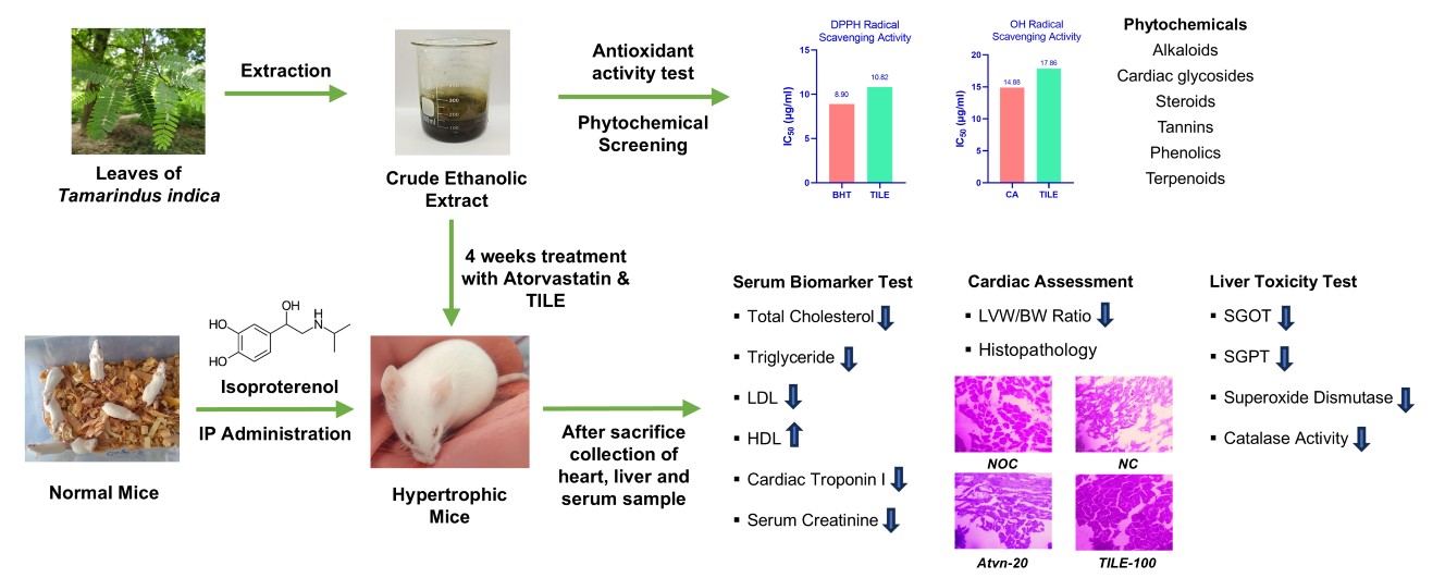
Introduction: Cardiovascular diseases (CVDs) are the leading cause of death globally, with left ventricular hypertrophy (LVH) as a key independent risk factor. Objective: This study evaluates the cardioprotective potential of Tamarindus indica leaf extract (TILE) in mitigating isoproterenol-induced Left Ventricular Hypertrophy (LVH), a critical risk factor for cardiovascular diseases (CVDs). Methods: The crude extract underwent phytochemical screening followed by total phenolic content, total flavonoid content, DPPH, and OH radical scavenging assays. Swiss albino mice with isoproterenol-induced LVH were treated orally with TILE (three doses) over four weeks. Key parameters, including lipid profiles (TC, TG, HDL, LDL), cardiac biomarkers (Troponin I), oxidative stress markers (SOD, CAT), liver and renal function (SGPT, SGOT, Serum Creatinine), LVW/BW ratio, and histopathology, were measured and compared with atorvastatin, which has been taken as a standard drug. Results: TILE showed considerable phenolic (29.69 ± 2.14 mg/g) and flavonoid contents (145.42 ± 7.58 mg/g) and potent antioxidant activity with IC50 values of 10.82 μg/mL (DPPH) and 17.86 μg/mL (OH radical scavenging assay). Isoproterenol elevated TC, TG, LDL cholesterol, Troponin I, SGPT, SGOT, and serum creatinine, while reducing HDL cholesterol, SOD, and catalase levels. TILE treatment reversed these changes significantly, demonstrating antioxidative and antihyperlipidemic properties, and reducing cardiac hypertrophy by improving the LVW/BW ratio. Conclusion: TILE exhibited significant cardioprotective effects against isoproterenol-induced LVH, which were associated with improvements in antioxidant enzyme activities and lipid profile parameters. However, further studies are required to elucidate the precise molecular mechanisms underlying these effects.
- Open Access
- Article
Cardio-Protection by Tamarindus indica Leaves: Mitigating Left Ventricular Hypertrophy in Isoproterenol-Induced Mice
- Farjana Sharmin 1,2,
- Farhad Hossain 2,3,
- Joy Sarker 2,
- Ibrahim Hossain 2,
- Rozina Khatun 2,
- Khadiza Khanam 2,
- A. B. M. Ashraful 2,4,
- Medha Islam 2,
- AHM Khurshid Alam 2,
- Mamunur Rashid 2,*
Author Information
Received: 23 May 2026 | Revised: 22 Jun 2026 | Accepted: 26 Jun 2026 | Published: 30 Jun 2026
Abstract
Graphical Abstract
References
- 1.
Messerli, F.H.; Ketelhut, R. Left ventricular hypertrophy: An independent risk factor. J. Cardiovasc. Pharmacol. 1991, 17 (Suppl. S4), S59–S67.
- 2.
Verdecchia, P.; Porcellati, C.; Reboldi, G.; et al. Left ventricular hypertrophy as an independent predictor of acute cerebrovascular events in essential hypertension. Circulation 2001, 104, 2039–2044. https://doi.org/10.1161/hc4201.097944.
- 3.
Shimizu, I.; Minamino, T. Physiological and pathological cardiac hypertrophy. J. Mol. Cell Cardiol. 2016, 97, 245–262. https://doi.org/10.1016/j.yjmcc.2016.06.001.
- 4.
Bruckschlegel, G.; Holmer, S.R.; Jandeleit, K.; et al. Blockade of the renin-angiotensin system in cardiac pressure-overload hypertrophy in rats. Hypertension 1995, 25, 250–259. https://doi.org/10.1161/01.hyp.25.2.250.
- 5.
Alexander, R.W.; Brock, T.A.; Gimbrone, M.A., Jr.; Rittenhouse, S.E. Angiotensin increases inositol trisphosphate and calcium in vascular smooth muscle. Hypertension 1985, 7, 447–451.
- 6.
Griendling, K.K.; Rittenhouse, S.E.; Brock, T.A.; et al. Sustained diacylglycerol formation from inositol phospholipids in angiotensin II-stimulated vascular smooth muscle cells. J. Biol. Chem. 1986, 261, 5901–5906.
- 7.
Vanderheyden, V.; Devogelaere, B.; Missiaen, L.; et al. Regulation of inositol 1,4,5-trisphosphate-induced Ca2+ release by reversible phosphorylation and dephosphorylation. Biochim. Biophys. Acta 2009, 1793, 959–970. https://doi.org/10.1016/j.bbamcr.2008.12.003.
- 8.
Kolczynska, K.; Loza-Valdes, A.; Hawro, I.; et al. Diacylglycerol-evoked activation of PKC and PKD isoforms in regulation of glucose and lipid metabolism: A review. Lipids Health Dis. 2020, 19, 113. https://doi.org/10.1186/s12944-020-01286-8.
- 9.
Shimokawa, H.; Takeshita, A. Rho-kinase is an important therapeutic target in cardiovascular medicine. Arterioscler. Thromb. Vasc. Biol. 2005, 25, 1767–1775. https://doi.org/10.1161/01.ATV.0000176193.83629.c8.
- 10.
Striessnig, J.; Pinggera, A.; Kaur, G.; et al. L-type Ca(2+) channels in heart and brain. Wiley Interdiscip. Rev. Membr. Transp. Signal 2014, 3, 15–38. https://doi.org/10.1002/wmts.102.
- 11.
Molkentin, J.D.; Dorn, G.W., 2nd. Cytoplasmic signaling pathways that regulate cardiac hypertrophy. Annu. Rev. Physiol. 2001, 63, 391–426. https://doi.org/10.1146/annurev.physiol.63.1.391.
- 12.
Proud, C.G. Ras, PI3-kinase and mTOR signaling in cardiac hypertrophy. Cardiovasc. Res. 2004, 63, 403–413. https://doi.org/10.1016/j.cardiores.2004.02.003.
- 13.
Rashid, M.; Tawara, S.; Fukumoto, Y.; et al. Importance of Rac1 signaling pathway inhibition in the pleiotropic effects of HMG-CoA reductase inhibitors. Circ. J. 2009, 73, 361–370. https://doi.org/10.1253/circj.cj-08-0817.
- 14.
Lezoualc’h, F.; Métrich, M.; Hmitou, I.; et al. Small GTP-binding proteins and their regulators in cardiac hypertrophy. J. Mol. Cell Cardiol. 2008, 44, 623–632. https://doi.org/10.1016/j.yjmcc.2008.01.011.
- 15.
Liu, W.; Zi, M.; Naumann, R.; et al. Pak1 as a novel therapeutic target for antihypertrophic treatment in the heart. Circulation 2011, 124, 2702–2715. https://doi.org/10.1161/circulationaha.111.048785.
- 16.
Kuru, P. Tamarindus indica and its health related effects. Asian Pac. J. Trop. Biomed. 2014, 4, 676–681. https://doi.org/10.12980/APJTB.4.2014APJTB-2014-0173.
- 17.
Devi, B.; Boruah, T. Tamarind (Tamarindus indica); Springer: Singapore, 2020; pp. 317–332.
- 18.
Yogeswari, M.; Baharin, B.; Ganesan, P. Effect of Room Temperature Storage on the Physicochemical and Antioxidant Properties of Oven Dried Young Tamarind Leaves (Tamarindus Indica) Chutney Powder. Indian J. Sci. Technol. 2016, 9, 1–8 https://doi.org/10.17485/ijst/2016/v9i48/91997.
- 19.
Imam, S.; Azhar, I.; Hasan, M.M.; et al. Two triterpenes lupanone and lupeol isolated and identified from Tamarindus indica linn. Pak. J. Pharm. Sci. 2007, 20, 125–127.
- 20.
Pino, J.; Escalona Arranz, J.; Licea, I.; et al. Leaf Oil of Tamarindus indica L. J. Essent. Oil Res. 2002, 14, 187–188. https://doi.org/10.1080/10412905.2002.9699819.
- 21.
Hossain, M.S.; Jami, M.A.B.S.; Ashraful, A.; et al. Unveiling the anti-inflammatory activity of chloroform fraction of curcuma wallichii and its phytoconstituents by in vivo and in silico studies. Sci. Rep. 2026, 16, 1762.
- 22.
Islam, M.T.; Saha, S.; Ashraful, A.; et al. Phytochemical Profile, Antioxidant, Antidiabetic, and Antidiarrheal Potentials of Hylocereus undatus Leaf Extract: GC–MS and In Silico Insights. J. Food Biochem. 2026, 2026, 3039549.
- 23.
Reza, A.A.; Haque, M.A.; Sarker, J.; et al. Antiproliferative and antioxidant potentials of bioactive edible vegetable fraction of Achyranthes ferruginea Roxb. in cancer cell line. Food Sci. Nutr. 2021, 9, 3777–3805.
- 24.
Hossain, M.S.; Ashraful, A.; Rahman, A.A.; et al. Anti-ROS and Anticancer Potential of Rhizomes and a Polyunsaturated Fatty Acid From Chloroform Fraction of Curcuma wallichii as a Bioactive Compound. J. Food Biochem. 2025, 2025, 9517484.
- 25.
Sarker, J.; Reza, A.A.; Ashraful, A.; et al. In Vitro Antioxidant and p53-Targeted Anticancer Activity of Aerva sanguinolenta (L.) Blume Across Different Human Cancer Cell Lines and Ehrlich Ascites Carcinoma Model. J. Chem. Health Risks 2026, 16, 1292–1316
- 26.
Hossain, F.; Zamaly, S.; Ashraful, A.; et al. Exploring the therapeutic potential of Leea rubra (Vitaceae): Antioxidant efficacy, brine shrimp lethality, and in vivo anticancer activity against EAC cells in Swiss albino mice. J. Biol. Act. Prod. Nat. 2025, 15, 181–195.
- 27.
Zhang, Y.; Xu, J.; Long, Z.; et al. Hydrogen (H2) inhibits isoproterenol-induced cardiac hypertrophy via antioxidative pathways. Front. Pharmacol. 2016, 7, 217818.
- 28.
Haroon, H.B.; Ahmed, N.; Sampath, M.K.; et al. Tamarindus indica. Linn leaves ameliorates experimental induced heart failure in Wistar rats. J. Basic. Clin. Physiol. Pharmacol. 2022, 33, 363–371.
- 29.
Tushar, M.A.N.; Rashid, M.M.; Hossain, F.; et al. Investigation of cardioprotective effects of Leea rubra Blume leaves on isoproterenol-induced left ventricular hypertrophy in mice. J. Herbmed Pharmacol. 2026, 15, 27–35.
- 30.
Marklund, S.; Marklund, G. Involvement of the superoxide anion radical in the autoxidation of pyrogallol and a convenient assay for superoxide dismutase. Eur. J. Biochem. 1974, 47, 469–474. https://doi.org/10.1111/j.1432-1033.1974.tb03714.x.
- 31.
Hadwan, M.H. Simple spectrophotometric assay for measuring catalase activity in biological tissues. BMC Biochem. 2018, 19, 7. https://doi.org/10.1186/s12858-018-0097-5.
- 32.
Khatun, R.; Sarker, J.; Sharmin, F.; et al. Attenuation of Left Ventricular Hypertrophy by Psidium guajava Leaf Extract Via Inhibition of Oxidative Stress and Cardiac Troponin I Levels in Adrenaline-Induced Hypertrophic Rats. J. Med. Nat. Prod. 2026, 3, 100011.
- 33.
Slaoui, M.; Fiette, L. Histopathology procedures: From tissue sampling to histopathological evaluation. Methods Mol. Biol. 2011, 691, 69–82. https://doi.org/10.1007/978-1-60761-849-2_4.
- 34.
Sookying, S.; Duangjai, A.; Saokaew, S.; et al. Botanical aspects, phytochemicals, and toxicity of Tamarindus indica leaf and a systematic review of antioxidant capacities of T. indica leaf extracts. Front Nutr. 2022, 9, 977015.
- 35.
Ołdakowski, Ł.; Taylor, J.R. Oxidative damage and antioxidant defense are assay and tissue-dependent both in captive and wild-caught bank voles (Myodes glareolus) before and after reproduction. Ecol. Evol. 2018, 8, 7543–7552.
- 36.
McGill, H.C., Jr.; McMahan, C.A.; Zieske, A.W.; et al. Association of Coronary Heart Disease Risk Factors with microscopic qualities of coronary atherosclerosis in youth. Circulation 2000, 102, 374–379. https://doi.org/10.1161/01.cir.102.4.374.
- 37.
Sharma, S.; Jackson, P.G.; Makan, J. Cardiac troponins. J. Clin. Pathol. 2004, 57, 1025–1026. https://doi.org/10.1136/jcp.2003.015420.
- 38.
Halliwell, B. Antioxidants: The basics--what they are and how to evaluate them. Adv. Pharmacol. 1997, 38, 3–20. https://doi.org/10.1016/s1054-3589(08)60976-x.
- 39.
Gilbert, C.J.; Longenecker, J.Z.; Accornero, F. ERK1/2: An Integrator of Signals That Alters Cardiac Homeostasis and Growth. Biology 2021, 10, 346. https://doi.org/10.3390/biology10040346.
- 40.
Moreau, S. Characterizing Rho Kinase Activity Using a Novel PET Tracer in Hypertrophied Cardiomyocytes; University of Ottawa: Ottawav, Canada, 2012.

This work is licensed under a Creative Commons Attribution 4.0 International License.

 Suite 4002 Level 4, 447 Collins Street, Melbourne, Victoria 3000, Australia
 General Inquiries: info@sciltp.com