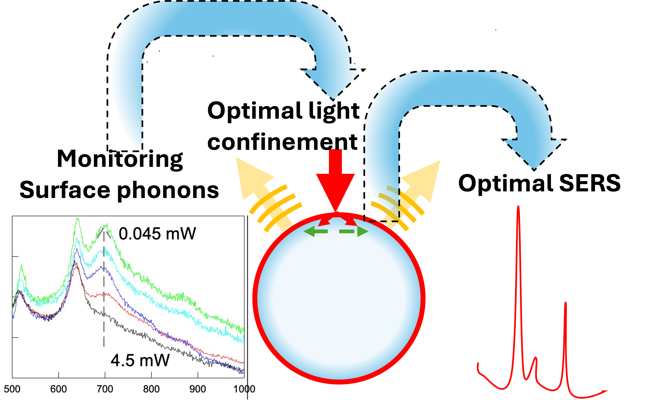
The characterization of surface plasmon–polaritons (SPPs) involved in surface-enhanced Raman scattering (SERS) and plasmon-driven photochemical processes is commonly performed through auxiliary techniques such as optical-, electron energy loss- or scanning probe-spectroscopy that neither reproduce the actual excitation conditions of a microRaman setup nor provide a direct, in situ probe of light confinement in real experiments. Here, we demonstrate an alternative approach based on the intrinsic Raman response of TiO2 hollow-shell cavities coated with ultrathin gold layers (T-horex@Au). The Raman spectra of these hybrid core/shell structures exhibit an additional mode that cannot be assigned to bulk TiO2 phonons. Confocal microRaman analysis reveals a clear dependence of this mode’s intensity on the laser penetration depth, enabling its attribution to surface phonon modes activated at the Au/TiO2 interface. This plasmon-mediated activation takes advantage of interfacial symmetry lowering and efficient electromagnetic field confinement. The simultaneous observation of this mode together with conventional Raman signals provides a direct, non-perturbative diagnostic of SPP excitation quality under realistic experimental conditions. Exploiting this feature, we show that the Raman enhancement induced by identical gold layers is strongly substrate-dependent and that the intensity of the TiO2 surface mode can be used as an internal standard to optimize and tune SERS performance. This strategy offers a practical route for large-scale fabrication and rapid optimization of metal/TiO2-based SERS substrates, opening intriguing perspectives for real-time control and monitoring of plasmon-driven photocatalytic and photochemical reactions at solid interfaces.
- Open Access
- Article
Using Surface Phonons as a Guide for Optimizing SERS and Light-Driven Processes †
Author Information
Received: 15 Dec 2025 | Revised: 03 Mar 2026 | Accepted: 04 Mar 2026 | Published: 07 May 2026
Abstract
Graphical Abstract
Keywords
TiO2 | surface phonons | surface plasmon polaritons | SERS | photocatalysis
References
- 1.
Stiles, P.L.; Dieringer, J.A.; Shah, N.C.; et al. Surface-enhanced Raman spectroscopy. Annu. Rev. Anal. Chem. 2008, 1, 601–626. https://doi.org/10.1146/annurev.anchem.1.031207.112814.
- 2.
Vassalini, I.; Rotunno, E.; Lazzarini, L.; et al. “Stainless” gold nanorods: Preserving shape, optical properties, and SERS activity in oxidative environment. ACS Appl. Mater. Interfaces 2015, 7, 18794–18802.
- 3.
Stefancu, A.; Aizpurua, J.; Alessandri, I.; et al. Impact of surface enhanced Raman spectroscopy in catalysis. ACS Nano 2024, 18, 29337–29379.
- 4.
Liu, X.; Ye, M.; Zhang, S.; et al. Enhanced photocatalytic CO2 valorization over TiO2 hollow microspheres by synergetic surface tailoring and Au decoration. J. Mater. Chem. A 2018, 6, 24245–24255.
- 5.
Xu, T.; Jia, B.; Yan, K.; et al. Boosting visible light photocatalytic oxidation of CO using Au nanocatalysts through synergistic preparation of an Fe-doped TiO2 support and cold plasma treatment. Catal. Sci. Technol. 2025, 15, 2844–2851.
- 6.
Mahdavi-Shakib, A.; Sempel, J.; Hoffman, M.; et al. Au/TiO2-Catalyzed Benzyl Alcohol Oxidation on Morphologically Precise Anatase Nanoparticles. ACS Appl. Mater. Interfaces 2021, 13, 11793–11804. https://doi.org/10.1021/acsami.0c20442.
- 7.
Xu, Q.; Liu, Z. Studies on Photocatalytic Degradation for Organic Pollutants by TiO2/Au Composite and its Antibacterial Properties. Theor. Found. Chem. Eng. 2023, 57, 1610–1617.
- 8.
Reichert, R.; Jusys, Z.; Behm, R.J. Au/TiO2 Photo(electro)catalysis: The Role of the Au Cocatalyst in Photoelectrochemical Water Splitting and Photocatalytic H2 Evolution. J. Phys. Chem. C 2015, 119, 24750–24759.
- 9.
Wang, K.; Lu, J.; Lau, C.H.; et al. Unravelling the C-C coupling in CO2 photocatalytic reduction with H2O on Au/TiO2-x: Combination of plasmonic excitation and oxygen vacancy. Appl. Catal. B Environ. 2021, 292, 120147.
- 10.
Ben-Jaber, S.; Peveler, W.J.; Quesada-Cabrera, R.; et al. Photo-induced enhanced Raman spectroscopy for universal ultra-trace detection of explosives, pollutants and biomolecules. Nat Commun. 2016, 7, 12189. https://doi.org/10.1038/ncomms12189.
- 11.
Alessandri, I. Enhancing Raman scattering without plasmons: Unprecedented sensitivity achieved by TiO2 shell-based resonators. J. Am. Chem. Soc. 2013, 135, 5541–5544.
- 12.
Bontempi, N.; Vassalini, I.; Alessandri, I. All-dielectric core/shell resonators: From plasmon-free SERS to multimodal analysis. J. Raman Spectrosc. 2018, 49, 943–953.
- 13.
Alessandri, I.; Carletti, L.; Ferroni, M.; et al. Bioinspired self-similar all-dielectric antennas: Probing the effect of secondary scattering centres by Raman spectroscopy. Mater. Adv. 2020, 1, 2443–2449.
- 14.
Bontempi, N.; Carletti, L.; De Angelis, C.; et al. Plasmon-free SERS detection of environmental CO2 on TiO2 surfaces. Nanoscale 2016, 8, 3226–3231.
- 15.
Alessandri, I.; Lombardi, J.R. Enhanced Raman scattering with dielectrics. Chem. Rev. 2016, 116, 14921–14981.
- 16.
Boontanom, A.; Moscolari, L.; Kozma, E.; et al. Poly (dopamine)-coated TiO2/SiO2 resonators for the non-plasmonic SERS detection of organic analytes. Appl. Surf. Sci. 2026, 720, 165249. https://doi.org/10.1016/j.apsusc.2025.165249.
- 17.
Salmistraro, M.; Schwartzberg, A.; Bao, W.; et al. Triggering and monitoring plasmon-enhanced reactions by optical nanoantennas coupled to photocatalytic beads. Small 2013, 9, 3301–3307.
- 18.
Kadkhodazadeh, S.; Rosenkrantz de Lasson, J.; Beleggia, M.; et al. Scaling of the Surface Plasmon Resonance in Gold and Silver Dimers Probed by EELS. J. Phys. Chem. C 2014, 118, 5478–5485. https://doi.org/10.1021/jp500288s.
- 19.
Garcia de Abajo, F.J. Optical excitations in electron microscopy. Rev. Mod. Phys. 2010, 82, 209–275.
- 20.
Mooradian, A. Photoluminescence of metals. Phys. Rev. Lett. 1969, 22, 185–187.
- 21.
Boyd, G.T.; Yu, Z.H.; Shen, Y.R. Photoinduced luminescence from the noble metals and its enhancement on roughened surfaces. Phys. Rev. B 1986, 33, 7923–7936.
- 22.
Ngoc, L.L.T.; Wiedemair, J.; van den Berg, A.; et al. Plasmon-modulated photoluminescence from gold nanostructures and its dependence on plasmon resonance, excitation energy, and band structure. Opt. Express 2015, 23, 5547–5564. https://doi.org/10.1364/OE.23.005547.
- 23.
Durinck, G.; Poelman, H.; Clauws, P.; et al. Observation of surface phonons on the (001) and (100) surfaces of anatase minerals. Solid State Commun. 1991, 80, 579–581. https://doi.org/10.1016/0038-1098(91)90155-O.
- 24.
Poelman, H.; Fiermans, L. Surface Phonon Spectra of Mineral TiO2 Anatase (001) and (100). Surf. Sci. Spectra 1998, 5, 252–256. https://doi.org/10.1116/1.1247845.
- 25.
Alessandri, I.; Ferroni, M.; Depero, L.E. Plasmon-Assisted, Spatially Resolved Laser Generation of Transition Metal Oxides from Liquid Precursors. J. Phys. Chem. C 2011, 115, 5174–5180. https://doi.org/10.1021/jp110324y.
- 26.
Alessandri, I. 4-Aminothiophenol Photodimerization without Plasmons. Angew. Chem. Int. Ed. 2022, 61, e202205013. https://doi.org/10.1002/anie.202205013.
- 27.
Alessandri, I.; Vassalini, I. Oxygen-mediated surface photoreactions: Exploring new pathways for sustainable chemistry. ChemPhotoChem. 2023, 7, e202300069. https://doi.org/10.1002/cptc.202300069.

This work is licensed under a Creative Commons Attribution 4.0 International License.
 Suite 4002 Level 4, 447 Collins Street, Melbourne, Victoria 3000, Australia
 General Inquiries: info@sciltp.com