
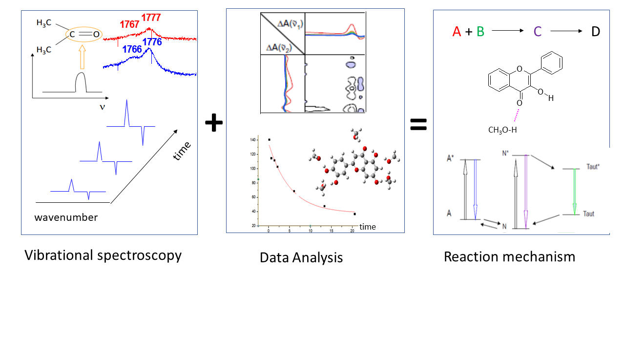
The detailed investigation of reaction mechanisms is nowadays a requirement in nearly all fields of chemistry. Molecular spectroscopy can provide key pieces of information on the reaction scheme. Among all available techniques, vibrational spectroscopy ones (notably mid- IR absorption, Near-IR absorption, non-resonant Raman scattering, Resonance Raman scattering) represent possibly the most appropriate approaches to get valuable insight on reaction mechanism. In this account/perspective, following the research I have developed so far in different fields (ranging from photochemistry/photobiology to surface chemistry), I will describe the most appropriate vibrational spectroscopy techniques needed to get specific pieces of information. Emphasis will be put on the interplay between the different vibrational spectroscopies, and on the synergy with advanced data analysis techniques, quantum chemistry calculations, and other experimental approaches. After general conclusions, the perspectives in the field of vibrational spectroscopy applied to the study of reaction mechanisms—or, more broadly speaking, to the study of time-evolving systems—will be presented.
- Open Access
- Perspective
Vibrational Spectroscopy in the Study of Reaction Mechanism
- Alberto Mezzetti
Author Information
Received: 12 Dec 2025 | Revised: 18 Mar 2026 | Accepted: 19 Mar 2026 | Published: 30 Jul 2026
Abstract
Graphical Abstract
Keywords
Raman | time-resolved FTIR | photosynthetic reaction centers | flavonoids | electron transfer | proton transfer
References
- 1.
Clark, G.L.; Thee, W.C. Ultra-Violet Spectroscopy of Flames of Motor Fuels. Ind. Eng. Chem. 1926, 18, 528–531.
- 2.
Siebert, F.; Hildebrandt, P. Vibrational Spectroscopy in Life Science; John Wiley & Sons: Berlin/Heidelberg, Germany, 2008.
- 3.
Hess, C. New advances in using Raman spectroscopy for the characterization of catalysts and catalytic reactions. Chem. Soc. Rev. 2021, 50, 3519–3564.
- 4.
Ryczkowski, J. IR spectroscopy in catalysis. Catal. Today 2001, 68, 263–381.
- 5.
Mezzetti, A.; Schnee, J.; Lapini, A.; et al. Time-resolved infrared absorption spectroscopy applied to photoinduced reactions: How and why. Photochem. Photobiol. Sci. 2022, 21, 557–584.
- 6.
Burgi, T.; Baiker, A. Attenuated Total Reflection Infrared Spectroscopy of Solid Catalysts Functioning in the Presence of Liquid-Phase Reactants. Adv. Catal. 2006, 50, 227–283.
- 7.
Mezzetti, A.; Nabedryk, E.; Breton, J.; et al. Rapid-scan Fourier transform infrared spectroscopy shows coupling of GLu-L212 protonation and electron transfer to QB in Rhodobacter sphaeroides reaction centers. Biochim. Biophys. Acta BBA Bioenerg. 2002, 1553, 320–330.
- 8.
Braiman, M.S.; Ahl, P.L.; Rothschild, K.J. Millisecond Fourier-transform infrared difference spectra of bacteriorhodopsin’’s’ M412 photoproduct. Proc. Natl. Acad. Sci. USA 1987, 84, 5221–5225.
- 9.
Smith, G.D.; Palmer, R.A. Fast Time-resolved Mid-infrared Spectroscopy Using an Interferometer. In Handbook of Vibrational Spectroscopy; Chalmers, J.M., Griffiths, P.R., Eds.; Wiley: New York, NY, USA, 2002.
- 10.
Uhmann, W.; Becker, A.; Taran, C.; et al. Time-Resolved FT-IR Absorption Spectroscopy Using a Step-Scan Interferometer. Appl. Spectrosc. 1991, 45, 390–397.
- 11.
Mäntele, W. Reaction-induced infrared difference spectroscopy for the study of protein function and reaction mechanisms. Trends Biochem. Sci. 1993, 18, 197–202.
- 12.
Betz, K.C.; Heinz, R.; Tesch, M.; et al. NIR spectroscopy for reaction monitoring and reaction mixture identification of cycloaliphatic epoxides. J. Polym. Res. 2022, 29, 199.
- 13.
Lillhong, T.; Dahlbackaa, J.; Geladib, P. Hyperspectral Near Infrared Image Analysis of a Phenol Formaldehyde Resin Curing Reaction. J. Near Infrared Spectrosc. 2012, 20, 559–572.
- 14.
Czarnecki, M.A.; Morisawa, Y.; Futami, Y.; et al. Advances in Molecular Structure and Interaction Studies Using Near Infrared Spectroscopy. Chem. Rev. 2015, 115, 9707–9744.
- 15.
Jurasekova, Z.; Domingo, C.; Garcia-Ramos, J.V.; et al. Effect of pH on the chemical modification of quercetin and structurally related flavonoids characterized by optical (UV-visible and Raman) spectroscopy. Phys. Chem. Chem. Phys. 2014, 16, 12802–12811.
- 16.
Barras, A.; Mezzetti, A.; Richard, A.; et al. Formulation and characterization of polyphenol-loaded lipid nanocapsules. Int. J. Pharm. 2009, 379, 270–277.
- 17.
Kobayashi, A.; Osawa, K.; Terazima, M.; et al. Solute–solvent hydrogen-bonding in room temperature ionic liquids studied by Raman spectroscopy. Phys. Chem. Chem. Phys. 2012, 14, 13676–13683.
- 18.
Sahoo, S.K.; Umapathy, S.; Parker, A.W. Time-Resolved Resonance Raman Spectroscopy: Exploring Reactive Intermediates. Appl. Spectrosc. 2011, 65, 1087–1115.
- 19.
Batignani, G.; Ferrante, C.; Fumero, G.; et al. Femtosecond stimulated Raman spectroscopy. Nat. Rev. Methods Primers 2024, 4, 34.
- 20.
Lorenz-Fonfria, V.A. Infrared difference spectroscopy of proteins: From bands to bonds. Chem. Rev. 2020, 120, 3466–3576.
- 21.
Noda, I.; Ozaki, Y. Two-Dimensional Correlation Spectroscopy—Applications in Vibrational and Optical Spectroscopy; John Wiley & Sons: Chichester, UK, 2004.
- 22.
Okamura, M.Y.; Paddock, M.L.; Graige, M.S.; et al. Proton and electron transfer in bacterial reaction centers. Biochim. Biophys. Acta BBA Bioenerg. 2000, 1458, 148–163.
- 23.
Malferrari, M.; Mezzetti, A.; Francia, F.; et al. Effects of dehydration on light-induced conformational changes in bacterial photosynthetic reaction centers probed by optical and differential FTIR spectroscopy. Biochim. Biophys. Acta BBA Bioenerg. 2013, 1827, 328–339.
- 24.
Malferrari, M.; Turina, P.; Francia, F.; et al. Dehydration affects the electronic structure of the primary electron donor in bacterial photosynthetic reaction centers: Evidence from visible-NIR and light-induced difference FTIR spectroscopy. Photochem. Photobiol. Sci. 2015, 14, 238–251.
- 25.
Mezzetti, A.; Malferrari, M.; Francia, F.; et al. Effect of Dehydration on Light-Adapted States of Bacterial Reaction Centers Studied by Time-Resolved Rapid-Scan FTIR Difference Spectroscopy. Spectrosc. J. 2025, 3, 5.
- 26.
Mezzetti, A.; Malferrari, M.; Venturoli, G.; et al. Rapid-scan Fourier Transform Infrared Difference Spectroscopy with Two-Dimensional Correlation Analysis to Show the Build-Up of Light-Adapted States in Bacterial Photosynthetic Reaction Centers. Appl. Spectrosc. 2025, 79, 756–766.
- 27.
Sipka, G.; Magyar, M.; Mezzetti, A.; et al. Light-adapted charge-separated state of photosystem II: Structural and functional dynamics of the closed reaction center. Plant Cell 2021, 33, 1286–1302.
- 28.
Malferrari, M.; Venturoli, G.; Francia, F.; et al. A New Method for D2O/H2O Exchange in Infrared Spectroscopy of Proteins. J. Spectrosc. 2012, 27, 337–342.
- 29.
Malferrari, M.; Francia, F.; Mezzetti, A.; et al. Kinetic effects in dehydration, rehydration, and isotopic exchange of bacterial photosynthetic reaction centers. Biomed. Spectr. Imaging 2016, 5, 185–196.
- 30.
Remy, A.; Gerwert, K. Coupling of light-induced electron transfer to proton uptake in photosynthesis. Nat. Struct. Mol. Biol. 2003, 10, 637–644.
- 31.
Mezzetti, A.; Leibl, W. Proton and electron transfer in wild-type and mutant reaction centers from Rhodobacter sphaeroides followed by rapid-scan FTIR spectroscopy. Vib. Spectrosc. 2008, 48, 126–134.
- 32.
Mezzetti, A.; Paul, J.-F.; Leibl, W. Identification of a Ubiquinone–Ubiquinol Quinhydrone Complex in Bacterial Photosynthetic Membranes and Isolated Reaction Centers by Time-Resolved Infrared Spectroscopy. Int. J. Mol. Sci. 2023, 24, 5233.
- 33.
Mezzetti, A.; Leibl, W.; Breton, J.; et al. Photoreduction of the quinone pool in the bacterial photosynthetic membrane: Identification of infrared marker bands for quinol formation. FEBS Lett. 2003, 537, 161–165.
- 34.
Blanchet, L.; Mezzetti, A.; Ruckebusch, C. Multivariate curve resolution of rapid-scan FTIR difference spectra of quinone photoreduction in bacterial photosynthetic membranes. Anal. Bioanal. Chem. 2007, 387, 1863–1873.
- 35.
Blanchet, L.; Ruckebusch, C.; Mezzetti, A.; et al. Monitoring and interpretation of photoinduced biochemical processes by rapid-scan FTIR difference spectroscopy and hybrid hard and soft modeling. J. Phys. Chem. B 2009, 113, 6031–6040.
- 36.
Mezzetti, A.; Leibl, W.; Johnson, J.A.; et al. Monitoring molecular events during photo-driven ubiquinone pool reduction in PufX⁺ and PufX− membranes from Rhodobacter capsulatus by time-resolved FTIR difference spectroscopy. Plant Physiol. Biochem. 2024, 216, 109139.
- 37.
Mezzetti, A.; Leibl, W. Investigation of ubiquinol formation in isolated photosynthetic reaction centers by rapid-scan Fourier transform IR spectroscopy. Eur. Biophys. J. 2005, 34, 921–936.
- 38.
Mezzetti, A.; Blanchet, L.; de Juan, A.; et al. Ubiquinol formation in isolated photosynthetic reaction centres monitored by time-resolved differential FTIR in combination with 2D correlation spectroscopy and multivariate curve resolution. Anal. Bioanal. Chem. 2011, 399, 1999–2014.
- 39.
Mezzetti, A. Rapid-scan FTIR difference spectroscopy applied to ubiquinone reduction in photosynthetic reaction centers: Role of redox mediators. J. Spectrosc. 2010, 24, 79–87.
- 40.
Mezzetti, A.; Seo, D.; Leibl, W.; et al. Time-resolved step-scan FTIR investigation on the primary donor of the reaction center from the green sulfur bacterium Chlorobium tepidum. Photosynth. Res. 2003, 75, 161–166.
- 41.
Mezzetti, A. Light-Induced Infrared Difference Spectroscopy in the Investigation of Light Harvesting Complexes. Molecules 2015, 20, 12229–12249.
- 42.
Mezzetti, A.; Spezia, R. Time-resolved step scan FTIR spectroscopy and DFT investigation on triplet formation in peridinin–chlorophyll a protein from Amphidinium carterae at low temperature. J. Spectrosc. 2008, 22, 235–250.
- 43.
Carbonera, D.; Di Valentin, M.; Spezia, R.; et al. The unique photophysical properties of the Peridinin-Chlorophyll-a-Protein. Curr. Protein Pept. Sci. 2014, 15, 332–350.
- 44.
Bovi, D.; Mezzetti, A.; Vuilleumier, R.; et al. Environmental effects on vibrational properties of carotenoids: Experiments and calculations on peridinin. Phys. Chem. Chem. Phys. 2011, 13, 20954–20964.
- 45.
Kish, E.; Mendes Pinto, M.M.; Bovi, D.; et al. Fermi resonance as a tool for probing peridinin environment. J. Phys. Chem. B 2014, 118, 5873–5881.
- 46.
Mezzetti, A.; Kish, E.; Robert, B.; et al. Assignment of IR bands of isolated and protein-bound Peridinin in its fundamental and triplet state by static FTIR, time-resolved step-scan FTIR and DFT calculations. J. Mol. Struct. 2015, 1090, 58–64.
- 47.
Muzzopappa, F.; Kirilovsky, D. Changing Color for Photoprotection: The Orange Carotenoid Protein. Trends Plant Sci. 2020, 25, 92–104.
- 48.
Leccese, S.; Onfroy, T.; Wilson, A.; et al. Immobilization of Orange Carotenoid Protein on mesoporous silica SBA-15 for the development of photoactivable nanodevices. Microporous Mesoporous Mater. 2022, 340, 112007.
- 49.
Leccese, S.; Calcinoni, A.; Wilson, A.; et al. Orange carotenoid protein in mesoporous silica: A new system towards the development of colorimetric and fluorescent sensors for pH and temperature. Micromachines 2023, 14, 1871.
- 50.
Slomimskiy, Y.B.; Maksimov, E.G.; Lukashev, E.P.; et al. Engineering the photoactive orange carotenoid protein with redox-controllable structural dynamics and photoprotective function. Biochim. Biophys. Acta BBA Bioenerg. 2020, 1861, 148174.
- 51.
Dominguez-Martin, M.A.; Kerfeld, C.A. Engineering the orange carotenoid protein for applications in synthetic biology. Curr. Opin. Struct. Biol. 2019, 57, 110–117.
- 52.
Wilson, A.; Punginelli, C.; Gall, A.; et al. A photoactive carotenoid protein acting as light intensity sensor. Proc. Natl. Acad. Sci. USA 2008, 105, 12075–12080.
- 53.
Wilson, A.; Gwizdala, M.; Mezzetti, A.; et al. The essential role of the N-terminal domain of the orange carotenoid protein in cyanobacterial photoprotection: Importance of a positive charge for phycobilisome binding. Plant Cell 2012, 24, 1972–1983.
- 54.
Mezzetti, A.; Alexandre, M.; Thurotte, A.; et al. Two-step structural changes in orange carotenoid protein photoactivation revealed by time-resolved Fourier transform infrared spectroscopy. J. Phys. Chem. B 2019, 123, 3259–3266.
- 55.
Leccese, S.; Wilson, A.; Kirilovsky, D.; et al. Light-induced infrared difference spectroscopy on three different forms of orange carotenoid protein: Focus on carotenoid vibrations. Photochem. Photobiol. Sci. 2023, 22, 1379–1391.
- 56.
Protti, S.; Mezzetti, A.; Cornard, J.-P.; et al. Hydrogen bonding properties of DMSO in ground-state formation and optical spectra of 3-hydroxyflavone anion. Chem. Phys. Lett. 2008, 467, 88–93.
- 57.
Mezzetti, A.; Protti, S.; Lapouge, C.; et al. Protic equilibria as the key factor of quercetin emission in solution. Relevance to biochemical and analytical studies. Phys. Chem. Chem. Phys. 2011, 13, 6858–6864.
- 58.
Tong, C.; Shi, F.; Tong, X.; et al. Shining natural flavonols in sensing and bioimaging. TrAC Trends Anal. Chem. 2021, 137, 116222.
- 59.
Mezzetti, A.; Protti, S. Flavonols and 3-hydroxychromones: New applications for a class of environment-sensitive fluorescent molecules. Chim. Oggi–Chem. Today 2020, 38, 34–37.
- 60.
Soboleva, T.; Berreau, L.M. 3-Hydroxyflavones and 3-Hydroxy-4-oxoquinolines as Carbon Monoxide-Releasing Molecules. Molecules 2019, 24, 1252.
- 61.
Protti, S.; Mezzetti, A. Any colour you like. Excited state and ground state proton transfer in flavonols and applications. In Photochemistry; Albini, A., Ed.; Royal Society of Chemistry: London, UK, 2012; Volume 40, pp. 295–322.
- 62.
Protti, S.; Mezzetti, A.; Lapouge, C.; et al. Photochemistry of metal complexes of 3-hydroxyflavone: Towards a better understanding of the influence of solar light on the metal-soil organic matter interactions. Photochem. Photobiol. Sci. 2008, 7, 109–119.
- 63.
Yokoe, I.; Higugi, K.; Shirataki, Y.; et al. Photochemistry of Flavonoids. III. Photorearrangement of Flavonols. Chem. Pharm. Bull. 1981, 29, 894–898.
- 64.
Protti, S.; Raulin, K.; Cristini, O.; et al. Wavelength shifting systems based on flavonols and their metal complexes encapsulated by post-doping in porous SiO2 xerogel matrices. J. Mol. Struct. 2011, 993, 485–490.
- 65.
Landström, A.; Leccese, S.; Abadian, H.; et al. Fluorescent silica MCM-41 nanoparticles based on flavonoids: Direct post-doping encapsulation and spectral characterization. Dye. Pigm. 2021, 185, 108870.
- 66.
Landström, A.; Seitsonen, A.P.; Leccese, S.; et al. Electron spectroscopies of 3-hydroxyflavone and 7-hydroxyflavone in MCM-41 silica nanoparticles and in acetonitrile solutions. Experimental data and DFT/TD-DFT calculations. Data Brief 2021, 34, 106630.
- 67.
Seitsonen, A.P.; Idrissi, A.; Protti, S.; et al. Solvent effects on the vibrational spectrum of 3-hydroxyflavone. J. Mol. Liq. 2019, 275, 723–728.
- 68.
Loco, D.; Protti, S.; Mennucci, B.; et al. Critical assessment of solvent effects on absorption and fluorescence of 3HF in acetonitrile in the QM/PCM framework: A synergic computational and experimental study. J. Mol. Struct. 2019, 1182, 283–291.
- 69.
Lazzaroni, S.; Dondi, D.; Mezzetti, A.; et al. Role of solute-solvent hydrogen bonds on the ground state and the excited state proton transfer in 3-hydroxyflavone. A systematic spectrophotometry study. Photochem. Photobiol. Sci. 2018, 17, 923–933.
- 70.
Solomon, G.; Landström, A.; Loria, S.R.; et al. Tunable physics through coordination chemistry: Formation on oxide surface of Ti and Al chelates with 3-hydroxyflavone capable of electron injection and light emission. Dalton Trans. 2022, 51, 18489–18501.
- 71.
Marrassini, C.; Idrissi, A.; De Waele, I.; et al. Organic solvent–luteolin interactions studied by FT-Raman, Vis-Raman, UV-Raman spectroscopy and DFT calculations. J. Mol. Liq. 2015, 205, 2–8.
- 72.
Exarchou, V.; Troganis, A.; Gerothanassis, I.P.; et al. Do strong intramolecular hydrogen bonds persist in aqueous solution? Variable temperature gradient 1H, 1H–13C GE-HSQC and GE-HMBC NMR studies of flavonols and flavones in organic and aqueous mixtures. Tetrahedron 2002, 58, 7423–7429.
- 73.
Smith, G.J.; Thomsen, S.J.; Markham, K.R.; et al. The photostabilities of naturally occurring 5-hydroxyflavones, flavonols, their glycosides and their aluminium complexes. J. Photochem. Photobiol. A Chem. 2000, 136, 87–91.
- 74.
Kaneta, M.; Sugiyama, N. The light resistance of the flavones and the flavonols. Bull. Chem. Soc. Jpn. 1971, 44, 3211.
- 75.
Miano, J.M. Serum Response Factor: Toggling between disparate programs of gene expression. J. Mol. Cell. Cardiol. 2003, 35, 577–593.
- 76.
Mueller, C.; Nordheim, A. A protein domain conserved between yeast MCM1 and human SRF directs ternary complex formation. EMBO J. 1991, 10, 4219–4229.
- 77.
Pellegrini, L.; Tan, S.; Richmond, T.J. Structure of serum response factor core bound to DNA. Nature 1995, 376, 490–498.
- 78.
Mo, Y.; Ho, W.; Johnston, K.; et al. Crystal structure of a ternary SAP-1/SRF/c-fos SRE DNA complex. J. Mol. Biol. 2001, 314, 495–506.
- 79.
Benevides, J.M.; Overman, S.A.; Thomas, G.J., Jr. Raman, polarized Raman and ultraviolet resonance Raman spectroscopy of nucleic acids and their complexes. J. Raman Spectrosc. 2005, 36, 279–299.
- 80.
Stepanek, J.; Kopecky, V., Jr.; Mezzetti, A.; et al. Structural and dynamic changes of the serum response element and the core domain of serum response factor induced by their association. Biochem. Biophys. Res. Commun. 2010, 391, 203–208.
- 81.
De La Rica, R.; Matsui, H. Applications of peptide and protein-based materials in bionanotechnology. Chem. Soc. Rev. 2010, 39, 3499–3509.
- 82.
Pattabiraman, V.R.; Bode, J.W. Rethinking amide bond synthesis. Nature 2011, 480, 471–479.
- 83.
Feyzizarnagh, H.; Yoon, D.-Y.; Goltz, M.; et al. Peptide nanostructures in biomedical technology. WIREs Nanomed. Nanobiotechnol. 2016, 8, 730–743.
- 84.
Fosgerau, K.; Hoffmann, T. Peptide therapeutics: Current status and future directions. Drug Discov. Today 2015, 20, 122–128.
- 85.
Lambert, J.-F. Adsorption and polymerization of amino acids on mineral surfaces: A review. Orig. Life Evol. Biosph. 2008, 38, 211–242.
- 86.
El Samrout, O.; Mezzetti, A.; Berlier, G.; et al. Cyclic or Linear? Parameters Determining the Outcome of Glycine Polymerization in Silica Surface Prebiotic Scenarios. Chem. Eur. J. 2023, 29, e202204010.
- 87.
Sakhno, Y.; Battistella, A.; Mezzetti, A.; et al. One Step up the Ladder of Prebiotic Complexity: Formation of Nonrandom Linear Polypeptides from Binary Systems of Amino Acids on Silica. Chem. Eur. J. 2019, 25, 1275–1285.
- 88.
Abadian, H.; Cornette, P.; Costa, D.; et al. Leucine on Silica: A Combined Experimental and Modeling Study of a System Relevant for Origins of Life, and the Role of Water Coadsorption. Langmuir 2022, 38, 8038–8053.
- 89.
Ter-Ovanessian, L.M.P.; Rigaud, B.; Mezzetti, A.; et al. Carbamoyl phosphate and its substitutes for the uracil synthesis in origins of life scenarios. Sci. Rep. 2021, 11, 19356.
- 90.
Schuh Frantz, A.C.; Mezzetti, A.; Seitsonen, A.P.; et al. Investigating nalidixic acid adsorption onto ferrihydrite and maghemite surfaces: Molecular-level insights via continuous-flow ATR-FTIR spectroscopy. New J. Chem. 2024, 48, 19403–19417.
- 91.
Schuh Frantz, A.C.; Robinson M.R.; Mezzzetti, A.; et al. Heterogeneous Fenton Degradation of Nalidixic Acid on Fe(II)-Sorbed Maghemite, as Probed by In Situ Attenuated Total Reflectance-Fourier Transform Infrared Spectroscopy. ChemPhysChem 2026, 27, e202500857.
- 92.
Wang, I.; Nancollas, G.H. Pathways to biomineralization and biodemineralization of calcium phosphates: The thermodynamic and kinetic controls. Dalton Trans. 2009, 2665–2672. https://doi.org/10.1039/B815887H.
- 93.
Foley, B.; Guibert, C.; Selmane, M.; et al. Tunable Enzyme-Assisted Mineralization of Apatitic Calcium Phosphate by Homogeneous Catalysis. Int. J. Mol. Sci. 2023, 24, 43.
- 94.
Jaabar, I.L.; Foley, B.; Mezzetti, A. Unraveling the Mechanisms of Hypertrophy-Induced Matrix Mineralization and Modifications in Articular Chondrocytes. Calcif. Tissue Int. 2024, 115, 269–282.
- 95.
Foley, B.; Nadaud, F.; Selmane, M.; et al. Seriation of Enzyme-Functionalized Multilayers for the Design of Scalable Biomimetic Mineralized Structures. Small 2024, 20, 2402128.
- 96.
Mezzetti, A.; Leibl, W. Time-resolved infrared spectroscopy in the study of photosynthetic systems. Photosynth. Res. 2017, 131, 121–144.
- 97.
Mezzetti, A. Photobiological systems studied by time-resolved infrared spectroscopy (2021–2022). In Specialist Periodical Reports–Photochemistry; Crespi, S., Protti, S., Eds.; Royal Society of Chemistry: London, UK, 2023; Volume 51, pp. 126–158.
- 98.
Buhrke, D.; Hildebrandt, P. Probing Structure and Reaction Dynamics of Proteins Using Time-Resolved Resonance Raman Spectroscopy. Chem. Rev. 2020, 120, 3577–3630.
- 99.
Hamm, P. Transient 2D IR spectroscopy from micro- to milliseconds. J. Chem. Phys. 2021, 154, 104201.
- 100.
Intze, A.; Temperini, M.E.; Baldassarre, L.; et al. Time-resolved investigation of nanometric cell membrane patches with a mid-infrared laser microscope. Front. Photonics 2023, 4, 117033.
- 101.
Wilcken, R.; Nishida, J.; Triana, J.F.; et al. Antenna-coupled infrared nanospectroscopy of intramolecular vibrational interaction. Proc. Natl. Acad. Sci. USA 2023, 120, e2220852120.
- 102.
Chrimes, A.F.; Khoshmanesh, K.; Stoddart, P.R.; et al. Microfluidics and Raman microscopy: Current applications and future challenges. Chem. Soc. Rev. 2013, 42, 5880.
- 103.
Perro, A.; Lebourdon, G.; Henry, S.; et al. Combining microfluidics and FT-IR spectroscopy: Towards spatially resolved information on chemical processes. React. Chem. Eng. 2016, 1, 577–594.
- 104.
Edwards, M.D.; Pratley, M.T.; Gordon, C.M.; et al. Process Intensification of the Continuous Synthesis of Bio-Derived Monomers for Sustainable Coatings Using a Taylor Vortex Flow Reactor. Org. Process Res. Dev. 2024, 28, 1917–1928.
- 105.
Mezzetti, A.; Schnee, J. Molecular spectroscopy in the study of reaction mechanisms. New J. Chem. 2025, 49, 1566.
- 106.
Kubarych, K.J.; Thielges, M.C.; Tahara, T.; et al. Special issue on time-resolved vibrational spectroscopy. J. Chem. Phys. 2023, 158, 160401.
- 107.
Ferri, D.; Nachtegaal, M. Operando VII. Chimia 2024, 78, 73–74.

This work is licensed under a Creative Commons Attribution 4.0 International License.

 Suite 4002 Level 4, 447 Collins Street, Melbourne, Victoria 3000, Australia
 General Inquiries: info@sciltp.com