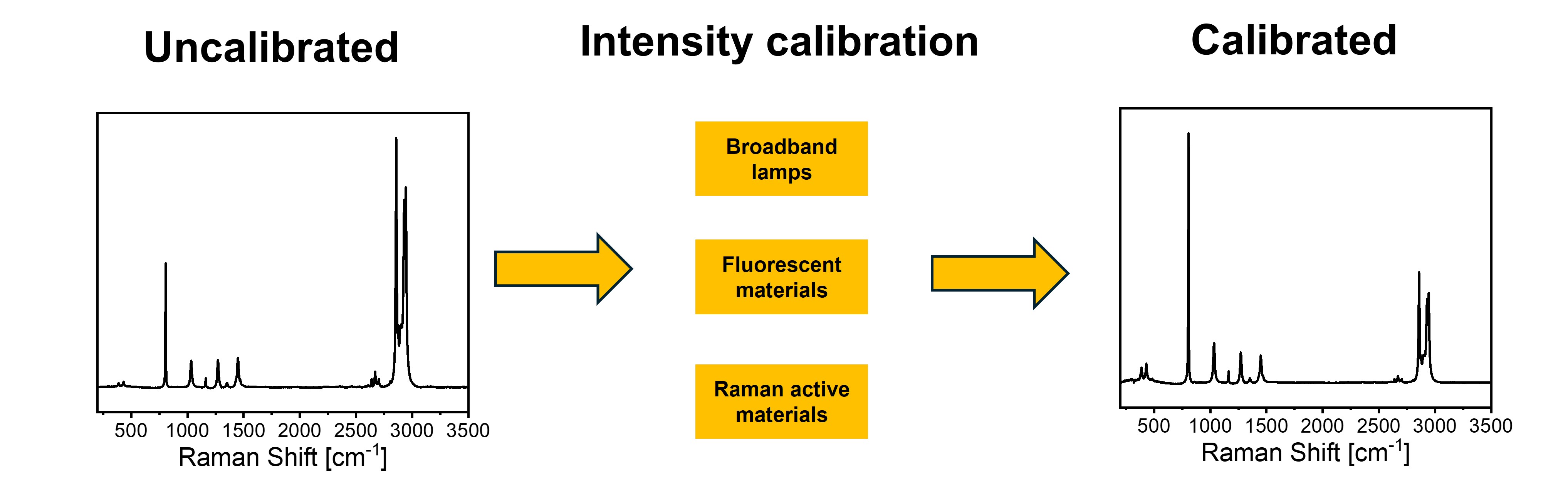
Raman spectroscopy is a powerful analytical technique widely used in many scientific fields due to its molecular fingerprinting capabilities and non-destructive nature. However, the recorded Raman spectrum is strongly dependent on the characteristics of the instrument and on the experimental configuration: in particular, the relative intensities of Raman bands can vary substantially between instruments, limiting reproducibility and accuracy. Intensity-calibrated spectra are therefore increasingly needed, as their availability would facilitate studies based on band intensity ratios, the building of unified spectral databases, and the inter-instrument transferability of chemometric and machine learning models. This review provides an overview of the methods available for the intensity calibration of Raman spectrometers. After introducing the definition of the instrument response function and the required spectral transformations, we examine calibration approaches based on broadband lamps, luminescent materials (certified and non-certified), and Raman scatterers, discussing their experimental implementation, advantages and limitations. Insights from interlaboratory comparison studies are also reviewed, demonstrating that inter-instrument variability remains substantial, and highlighting the importance of intensity calibration. Finally, since inter-instrument transferability can be addressed both by physically calibrating the instrument response and through data-processing strategies known as calibration transfer methods, a brief overview of the latter is also included, as the two approaches are complementary and inherently intertwined.
- Open Access
- Review
Intensity Calibration of Raman Instruments: A Review †
Author Information
Received: 30 Nov 2025 | Revised: 19 Mar 2026 | Accepted: 24 Mar 2026 | Published: 13 May 2026
Abstract
Graphical Abstract
Keywords
Raman | intensity calibration | instrument response function | machine learning | model transfer
References
- 1.
Smith, E.; Dent, G. Modern Raman Spectroscopy: A Practical Approach; John Wiley & Sons, Inc.: Chichester, UK, 2005; ISBN 0-471-49668-5.
- 2.
Handbook of Raman Spectroscopy: From the Research Laboratory to the Process Line; Lewis, I.R., Edwards, H.G.M., Eds.; Practical Spectroscopy; CRC Press: New York, NY, USA, 2001; Volume 28; ISBN 978-0-8247-0557-2.
- 3.
Esmonde-White, K.A.; Cuellar, M.; Uerpmann, C.; et al. Raman Spectroscopy as a Process Analytical Technology for Pharmaceutical Manufacturing and Bioprocessing. Anal. Bioanal. Chem. 2017, 409, 637–649. https://doi.org/10.1007/s00216-016-9824-1.
- 4.
Vankeirsbilck, T.; Vercauteren, A.; Baeyens, W.; et al. Applications of Raman Spectroscopy in Pharmaceutical Analysis. TrAC Trends Anal. Chem. 2002, 21, 869–877. https://doi.org/10.1016/S0165-9936(02)01208-6.
- 5.
Eshbekova, N.; Sowndarya, A.; Thangadurai, T.D.; et al. Recent Advancements in Raman Instrumentation and Capabilities for Pharmaceutical and Biomedical Applications. Appl. Spectrosc. Rev. 2024, 59, 798–849. https://doi.org/10.1080/05704928.2024.2355193.
- 6.
Laplant, F.; De Paepe, A. Raman Spectroscopy for Identifying Polymorphs. In Pharmaceutical Applications of Raman Spectroscopy; Šašić, S., Ed.; John Wiley & Sons, Ltd.: Hoboken, NJ, USA, 2007; pp. 85–115. ISBN 978-0-470-22588-2.
- 7.
Cong, X.; Liu, X.L.; Lin, M.L.; et al. Application of Raman Spectroscopy to Probe Fundamental Properties of Two-Dimensional Materials. 2D Mater. Appl. 2020, 4, 1–12. https://doi.org/10.1038/s41699-020-0140-4.
- 8.
Jorio, A.; Saito, R.; Dresselhaus, G.; et al. Raman Spectroscopy in Graphene Related Systems; Wiley-VCH Verlag GmbH & Co. KGaA: Weinheim, Germany, 2011; ISBN 978-3-527-40811-5.
- 9.
Castiglioni, C.; Mapelli, C.; Negri, F.; et al. Origin of the D Line in the Raman Spectrum of Graphite: A Study Based on Raman Frequencies and Intensities of Polycyclic Aromatic Hydrocarbon Molecules. J. Chem. Phys. 2001, 114, 963–974. https://doi.org/10.1063/1.1329670.
- 10.
Ferrari, A.C.; Robertson, J. Interpretation of Raman Spectra of Disordered and Amorphous Carbon. Phys. Rev. B 2000, 61, 14095–14107. https://doi.org/10.1103/PhysRevB.61.14095.
- 11.
Vibrational Spectroscopy of Polymers. Principles and Practises; Everall, N.J., Chalmers, J.M., Griffiths, P.R., Eds.; John Wiley & Sons, Ltd.: Chichester, UK, 2007; ISBN 978-0-470-01662-6.
- 12.
Modern Polymer Spectroscopy; Zerbi, G., Ed.; Wiley-VCH Verlag GmbH: New York, NY, USA, 2008; ISBN 978-3-527-61393-9.
- 13.
Castiglioni, C.; Tommasini, M.; Zerbi, G. Raman Spectroscopy of Polyconjugated Molecules and Materials: Confinement Effect in One and Two Dimensions. Philos. Trans. R. Soc. Math. Phys. Eng. Sci. 2004, 362, 2425–2459. https://doi.org/10.1098/RSTA.2004.1448.
- 14.
Caggiani, M.C.; Colomban, P. Advanced Procedures in Raman Forensic, Natural, and Cultural Heritage Studies: Mobile Set-up, Optics, and Data Treatment—State of the Art and Perspectives. J. Raman Spectrosc. 2024, 55, 116–124. https://doi.org/10.1002/JRS.6633.
- 15.
Rousaki, A.; Vandenabeele, P. In Situ Raman Spectroscopy for Cultural Heritage Studies. J. Raman Spectrosc. 2021, 52, 2178–2189. https://doi.org/10.1002/JRS.6166.
- 16.
Froment, F.; Tournié, A.; Colomban, P. Raman Identification of Natural Red to Yellow Pigments: Ochre and Iron-Containing Ores. J. Raman Spectrosc. 2008, 39, 560–568. https://doi.org/10.1002/JRS.1858.
- 17.
Angelini, I.; Asscher, Y.; Secco, M.; et al. The Pigments of the Frigidarium in the Sarno Baths, Pompeii: Identification, Stratigraphy and Weathering. J. Cult. Herit. 2019, 40, 309–316. https://doi.org/10.1016/J.CULHER.2019.04.021.
- 18.
Vandenabeele, P.; Edwards, H.G.M.; Jehlička, J. The Role of Mobile Instrumentation in Novel Applications of Raman Spectroscopy: Archaeometry, Geosciences, and Forensics. Chem. Soc. Rev. 2014, 43, 2628–2649. https://doi.org/10.1039/C3CS60263J.
- 19.
Rull, F.; Maurice, S.; Hutchinson, I.; et al. The Raman Laser Spectrometer for the ExoMars Rover Mission to Mars. Astrobiology 2017, 17, 627–654. https://doi.org/10.1089/AST.2016.1567.
- 20.
LeRu, E.C.; Etchegoin, P.G. Principles of Surface Enhanced Raman Spectroscopy; Elsevier: Amsterdam, The Netherlands, 2009; ISBN 978-0-444-52779-0.
- 21.
Pilot, R.; Signorini, R.; Fabris, L. Surface-Enhanced Raman Spectroscopy: Principles, Substrates, and Applications. In Metal Nanoparticles and Clusters: Advances in Synthesis, Properties and Applications; Deepak, F.L., Ed.; Springer: Berlin, Germany, 2017; pp. 89–164. ISBN 978-3-319-68052-1.
- 22.
Pilot, R.; Signorini, R.; Durante, C.; et al. A Review on Surface-Enhanced Raman Scattering. Biosensors 2019, 9, 57. https://doi.org/10.3390/bios9020057.
- 23.
Yi, J.; You, E.M.; Hu, R.; et al. Surface-Enhanced Raman Spectroscopy: A Half-Century Historical Perspective. Chem. Soc. Rev. 2025, 54, 1453–1551. https://doi.org/10.1039/D4CS00883A.
- 24.
Langer, J.; Aberasturi, D.J. de; Aizpurua, J.; et al. Present and Future of Surface-Enhanced Raman Scattering. ACS Nano 2019, 14, 28–117. https://doi.org/10.1021/acsnano.9b04224.
- 25.
Bell, S.E.J.; Charron, G.; Cortés, E.; et al. Towards Reliable and Quantitative Surface-Enhanced Raman Scattering (SERS): From Key Parameters to Good Analytical Practice. Angew. Chem. Int. Ed. 2020, 59, 5454–5462. https://doi.org/10.1002/ANIE.201908154.
- 26.
Bodelón, G.; Pastoriza-Santos, I. Recent Progress in Surface-Enhanced Raman Scattering for the Detection of Chemical Contaminants in Water. Front. Chem. 2020, 8, 518752. https://doi.org/10.3389/fchem.2020.00478.
- 27.
Ong, T.T.X.; Blanch, E.W.; Jones, O.A.H. Surface Enhanced Raman Spectroscopy in Environmental Analysis, Monitoring and Assessment. Sci. Total Environ. 2020, 720, 137601. https://doi.org/10.1016/J.SCITOTENV.2020.137601.
- 28.
Zheng, J.; He, L. Surface-Enhanced Raman Spectroscopy for the Chemical Analysis of Food. Compr. Rev. Food Sci. Food Saf. 2014, 13, 317–328. https://doi.org/10.1111/1541-4337.12062.
- 29.
Cialla-May, D.; Bonifacio, A.; Bocklitz, T.; et al. Biomedical SERS—The Current State and Future Trends. Chem. Soc. Rev. 2024, 53, 8957–8979. https://doi.org/10.1039/D4CS00090K.
- 30.
Fornasaro, S.; Cialla-May, D.; Sergo, V.; et al. The Role of Surface Enhanced Raman Scattering for Therapeutic Drug Monitoring of Antimicrobial Agents. Chemosensors 2022, 10, 128. https://doi.org/10.3390/CHEMOSENSORS10040128.
- 31.
Chisanga, M.; Muhamadali, H.; Ellis, D.I.; et al. Surface-Enhanced Raman Scattering (SERS) in Microbiology: Illumination and Enhancement of the Microbial World. Appl. Spectrosc. 2018, 72, 987–1000. https://doi.org/10.1177/0003702818764672.
- 32.
Stefancu, A.; Aizpurua, J.; Alessandri, I.; et al. Impact of Surface Enhanced Raman Spectroscopy in Catalysis. ACS Nano 2024, 18, 29337–29379. https://doi.org/10.1021/acsnano.4c06192.
- 33.
Pozzi, F.; Leona, M. Surface-Enhanced Raman Spectroscopy in Art and Archaeology. J. Raman Spectrosc. 2016, 47, 67–77. https://doi.org/10.1002/JRS.4827.
- 34.
Nicolson, F.; Kircher, M.F.; Stone, N.; et al. Spatially Offset Raman Spectroscopy for Biomedical Applications. Chem. Soc. Rev. 2021, 50, 556–568. https://doi.org/10.1039/D0CS00855A.
- 35.
Matousek, P. Spatially Offset Raman Spectroscopy for Non-Invasive Analysis of Turbid Samples. TrAC Trends Anal. Chem. 2018, 103, 209–214. https://doi.org/10.1016/J.TRAC.2018.04.002.
- 36.
Eliasson, C.; Matousek, P. Noninvasive Authentication of Pharmaceutical Products through Packaging Using Spatially Offset Raman Spectroscopy. Anal. Chem. 2007, 79, 1696–1701. https://doi.org/10.1021/ac062223z.
- 37.
Eliasson, C.; Macleod, N.A.; Matousek, P. Noninvasive Detection of Concealed Liquid Explosives Using Raman Spectroscopy. Anal. Chem. 2007, 79, 8185–8189. https://doi.org/10.1021/ac071383n.
- 38.
McCreery, R.L. Raman Spectroscopy for Chemical Analysis; Winefordner, J.D., Ed.; Chemical Analysis; John Wiley & Sons, Inc.: Hoboken, NJ, USA, 2000; Volume 157; ISBN 978-0-471-25287-0.
- 39.
Raj, A.; Kato, C.; Witek, H.A.; et al. Toward Standardization of Raman Spectroscopy: Accurate Wavenumber and Intensity Calibration Using Rotational Raman Spectra of H2, HD, D2, and Vibration–Rotation Spectrum of O2. J. Raman Spectrosc. 2020, 51, 2066–2082. https://doi.org/10.1002/jrs.5955.
- 40.
Itoh, N.; Shirono, K. Reliable Estimation of Raman Shift and Its Uncertainty for a Non-Doped Si Substrate (NMIJ CRM 5606-a). J. Raman Spectrosc. 2020, 51, 2496–2504. https://doi.org/10.1002/JRS.6003.
- 41.
Standard Guide for Raman Shift Standards for Spectrometer Calibration; ASTM International: West Conshohocken, PA, USA, 2014.
- 42.
Standard Guide for Testing the Resolution of a Raman Spectrometer; ASTM International: West Conshohocken, PA, USA, 2022.
- 43.
Jakubek, R.S.; Fries, M.D. Calibration of the Temporal Drift in Absolute and Relative Raman Intensities in Large Raman Images Using a Mercury–Argon Lamp. J. Raman Spectrosc. 2022, 53, 137–147. https://doi.org/10.1002/JRS.6259.
- 44.
Jakubek, R.S.; Fries, M.D. Calibration of Raman Wavenumber in Large Raman Images Using a Mercury-Argon Lamp. J. Raman Spectrosc. 2020, 51, 1172–1185. https://doi.org/10.1002/JRS.5887.
- 45.
Jakubek, R.S.; Fries, M.D. Raman Instrument Calibration for Astromaterials and Analysis of Mars Return Samples. Meteorit. Planet. Sci. 2023, 58, 98–110. https://doi.org/10.1111/maps.13940.
- 46.
Gou, L.; Zeng, X.; Du, H.; et al. Sensitive Detection of Trace 4-Methylimidazole Utilizing a Derivatization Reaction-Based Ratiometric Surface-Enhanced Raman Scattering Platform. Talanta 2022, 237, 122925. https://doi.org/10.1016/j.talanta.2021.122925.
- 47.
Li, L.; Zhang, L.; Gou, L.; et al. Au Nanoparticles Decorated CoP Nanowire Array: A Highly Sensitive, Anticorrosive, and Recyclable Surface-Enhanced Raman Scattering Substrate. Anal. Chem. 2023, 95, 11037–11046. https://doi.org/10.1021/acs.analchem.3c01282.
- 48.
Standard Guide for Relative Intensity Correction of Raman Spectrometers; ASTM International: West Conshohocken, PA, USA, 2023.
- 49.
Lussier, F.; Thibault, V.; Charron, B.; et al. Deep Learning and Artificial Intelligence Methods for Raman and Surface-Enhanced Raman Scattering. TrAC Trends Anal. Chem. 2020, 124, 115796. https://doi.org/10.1016/J.TRAC.2019.115796.
- 50.
Guo, S.; Popp, J.; Bocklitz, T. Chemometric Analysis in Raman Spectroscopy from Experimental Design to Machine Learning–Based Modeling. Nat. Protoc. 2021, 16, 5426–5429. https://doi.org/10.1038/s41596-021-00620-3.
- 51.
Blake, N.; Gaifulina, R.; Griffin, L.D.; et al. Machine Learning of Raman Spectroscopy Data for Classifying Cancers: A Review of the Recent Literature. Diagnostics 2022, 12, 1491. https://doi.org/10.3390/diagnostics12061491.
- 52.
Coca-Lopez, N.; Alcolea-Rodriguez, V.; Ares, M.A.B.; et al. Artificial Intelligence-Powered Raman Spectroscopy through Open Science and FAIR Principles. ACS Nano 2025, 19, 38189–38218. https://doi.org/10.1021/ACSNANO.5C09165.
- 53.
Yang, Y.; Xu, B.; Murray, J.; et al. Rapid and Quantitative Detection of Respiratory Viruses Using Surface-Enhanced Raman Spectroscopy and Machine Learning. Biosens. Bioelectron. 2022, 217, 114721. https://doi.org/10.1016/J.BIOS.2022.114721.
- 54.
Botto, C.S.; Orecchio, C.; D’Errico, C.; et al. Rapid Classification of Bacteria by a Portable Raman Spectrometer and Machine Learning. Spectrochim. Acta. A. Mol. Biomol. Spectrosc. 2026, 344, 126701. https://doi.org/10.1016/J.SAA.2025.126701.
- 55.
Zhang, L.; Li, C.; Peng, D.; et al. Raman Spectroscopy and Machine Learning for the Classification of Breast Cancers. Spectrochim. Acta. A. Mol. Biomol. Spectrosc. 2022, 264, 120300. https://doi.org/10.1016/J.SAA.2021.120300.
- 56.
Bocklitz, T.W.; Dörfer, T.; Heinke, R.; et al. Spectrometer Calibration Protocol for Raman Spectra Recorded with Different Excitation Wavelengths. Spectrochim. Acta. A. Mol. Biomol. Spectrosc. 2015, 149, 544–549. https://doi.org/10.1016/j.saa.2015.04.079.
- 57.
Georgiev, G.; Coca-Lopez, N.; Lellinger, D.; et al. Open Source for Raman Spectroscopy Data Harmonization. J. Raman Spectrosc. 2025, 56, 878–881. https://doi.org/10.1002/jrs.6789.
- 58.
Workman, J.J. A Review of Calibration Transfer Practices and Instrument Differences in Spectroscopy. Appl. Spectrosc. 2018, 72, 340–365. https://doi.org/10.1177/0003702817736064.
- 59.
Feudale, R.N.; Woody, N.A.; Tan, H.; et al. Transfer of Multivariate Calibration Models: A Review. Chemom. Intell. Lab. Syst. 2002, 64, 181–192. https://doi.org/10.1016/S0169-7439(02)00085-0.
- 60.
Practical Guide to Chemometrics, 2nd ed.; Gemperline, P., Ed.; CRC Press: Boca Raton, FL, USA, 2006; ISBN 978-0-429-11956-9.
- 61.
Ramadan, A.; Robert, G.; Kersaudy, R.; et al. Calibration Transfer and Maintenance in the Pharmaceutical Industry: A Systematic Review. Eur. J. Pharm. Sci. 2025, 209, 107114. https://doi.org/10.1016/j.ejps.2025.107114.
- 62.
Barton, B.; Thomson, J.; Lozano Diz, E.; et al. Chemometrics for Raman Spectroscopy Harmonization. Appl. Spectrosc. 2022, 76, 1021–1041. https://doi.org/10.1177/00037028221094070.
- 63.
Ntziouni, A.; Thomson, J.; Xiarchos, I.; et al. Review of Existing Standards, Guides, and Practices for Raman Spectroscopy. Appl. Spectrosc. 2022, 76, 747–772. https://doi.org/10.1177/00037028221090988.
- 64.
Guo, S.; Popp, J.; Bocklitz, T. Key Steps in the Workflow to Analyze Raman Spectra. Spectroscopy 2023, 38, 30–33. https://doi.org/10.56530/spectroscopy.fl6984w5.
- 65.
Adar, F. Raman Polarization Measurements: Keeping Track of the Instrumental Components’ Behavior. Spectroscopy 2017, 32, 14–22.
- 66.
Frost, K.J.; McCreery, R.L. Calibration of Raman Spectrometer Instrument Response Function with Luminescence Standards: An Update. Appl. Spectrosc. 1998, 52, 1614–1618.
- 67.
Tuschel, D. Spectral Resolution and Dispersion in Raman Spectroscopy. Spectroscopy 2020, 35, 9–15.
- 68.
Mooney, J.; Kambhampati, P. Get the Basics Right: Jacobian Conversion of Wavelength and Energy Scales for Quantitative Analysis of Emission Spectra. J. Phys. Chem. Lett. 2013, 4, 3316–3318. https://doi.org/10.1021/jz401508t.
- 69.
Malyj, M.; Griffiths, J.E. Stokes/Anti-Stokes Raman Vibrational Temperatures: Reference Materials, Standard Lamps, and Spectrophotometric Calibrations. Appl. Spectrosc. 1983, 37, 315–333. https://doi.org/10.1366/0003702834634325.
- 70.
Choquette, S.J.; Etz, E.S.; Hurst, W.S.; et al. Relative Intensity Correction of Raman Spectrometers: NIST SRMs 2241 through 2243 for 785 nm, 532 nm, and 488 nm/514.5 nm Excitation. Appl. Spectrosc. 2007, 61, 117–129. https://doi.org/10.1366/000370207779947585.
- 71.
Ying, X. An Overview of Overfitting and Its Solutions. J. Phys. Conf. Ser. 2019, 1168, 022022. https://doi.org/10.1088/1742-6596/1168/2/022022.
- 72.
Fang, S.; Wu, S.; Chen, Z.; et al. Recent Progress and Applications of Raman Spectrum Denoising Algorithms in Chemical and Biological Analyses: A Review. TrAC Trends Anal. Chem. 2024, 172, 117578. https://doi.org/10.1016/j.trac.2024.117578.
- 73.
Palchetti, L.; Bianchini, G.; Castagnoli, F. Design and Characterisation of Black-Body Sources for Infrared Wide-Band Fourier Transform Spectroscopy. Infrared Phys. Technol. 2008, 51, 207–215. https://doi.org/10.1016/J.INFRARED.2007.06.001.
- 74.
Petty, C.J.; Warnes, G.M.; Hendra, P.J.; et al. Relative Intensity Calibration of Single-Beam near-Infrared Spectrometers. Spectrochim. Acta Part Mol. Spectrosc. 1991, 47, 1179–1187. https://doi.org/10.1016/0584-8539(91)80205-W.
- 75.
Georgiev, G.T.; Butler, J.J. Long-Term Calibration Monitoring of Spectralon Diffusers BRDF in the Air-Ultraviolet. Appl. Opt. 2007, 46, 7892–7899. https://doi.org/10.1364/AO.46.007892.
- 76.
Hapke, B. Theory of Reflectance and Emittance Spectroscopy; 2nd ed.; Cambridge University Press: Cambridge, UK, 2012; ISBN 978-0-521-88349-8.
- 77.
Fryling, M.; Frank, C.J.; McCreery, R.L. Intensity Calibration and Sensitivity Comparisons for CCD/Raman Spectrometers. Appl. Spectrosc. 1993, 47, 1965–1974. https://doi.org/10.1366/0003702934066226.
- 78.
McConkey, J.W.; Woolsey, J.M. Errors in Absolute Intensity Measurements Using Tungsten Lamp Standards. Appl. Opt. 1969, 8, 205. https://doi.org/10.1364/ao.8.000205.
- 79.
Purcell, F.J.; Kaminski, R.; Russavage, E. Radiometric Correction of Raman Spectra. Appl. Spectrosc. 1980, 34, 323–326.
- 80.
Ray, K.G.; McCreery, R.L. Simplified Calibration of Instrument Response Function for Raman Spectrometers Based on Luminescent Intensity Standards. Appl. Spectrosc. 1997, 51, 108–116. https://doi.org/10.1366/0003702971938849.
- 81.
Standard Reference Material 2242a Relative Intensity Correction Standard for Raman Spectroscopy: 532 nm Excitation; National Institute of Standards and Technology (NIST): Gaithersburg, MD, USA, 2025; pp. 1–5.
- 82.
Standard Reference Material 2246a Relative Intensity Correction Standard for Raman Spectroscopy: 830 nm Excitation; National Institute of Standards and Technology (NIST): Gaithersburg, MD, USA, 2022; pp. 1–7.
- 83.
Standard Reference Material 2244 Relative Intensity Correction Standard for Raman Spectroscopy: 1064 nm Excitation; National Institute of Standards and Technology (NIST): Gaithersburg, MD, USA, 2020; pp. 1–5.
- 84.
Standard Reference Material 2241 Relative Intensity Correction Standard for Raman Spectroscopy: 785 nm Excitation; National Institute of Standards and Technology (NIST): Gaithersburg, MD, USA, 2022; pp. 1–5.
- 85.
Standard Reference Material 2243 Relative Intensity Correction Standard for Raman Spectroscopy: 488 nm and 514.5 nm Excitation; National Institute of Standards and Technology (NIST): Gaithersburg, MD, USA, 2009; pp. 1–8.
- 86.
Standard Reference Material 2245 Relative Intensity Correction Standard for Raman Spectroscopy: 633 nm Excitation; National Institute of Standards and Technology (NIST): Gaithersburg, MD, USA, 2016; pp. 1–6.
- 87.
Lakowicz, J.R. Principles of Fluorescence Spectroscopy; 3rd ed.; Springer New York: New York, NY, USA, 2006; ISBN 978-0-387-31278-1.
- 88.
Iwata, K.; Hamaguchi, H.; Tasumi, M. Sensitivity Calibration of Multichannel Raman Spectrometers Using the Least-Squares-Fitted Fluorescence Spectrum of Quinine. Appl. Spectrosc. 1988, 42, 12–14. https://doi.org/10.1366/0003702884428608.
- 89.
Melhuish, W.H. Modified Technique for Determining the Wavelength-Sensitivity Curve of a Spectrofluorimeter. Appl. Opt. 1975, 14, 26–27. https://doi.org/10.1364/AO.14.000026.
- 90.
Raj, A.; Kato, C.; Witek, H.A.; et al. Accurate Intensity Calibration of Multichannel Spectrometers Using Raman Intensity Ratios. J. Raman Spectrosc. 2021, 52, 2038–2050. https://doi.org/10.1002/jrs.6221.
- 91.
Guo, S.; Kohler, A.; Zimmermann, B.; et al. Extended Multiplicative Signal Correction Based Model Transfer for Raman Spectroscopy in Biological Applications. Anal. Chem. 2018, 90, 9787–9795. https://doi.org/10.1021/acs.analchem.8b01536.
- 92.
Guo, S.; Heinke, R.; Stöckel, S.; et al. Model Transfer for Raman-Spectroscopy-Based Bacterial Classification. J. Raman Spectrosc. 2018, 49, 627–637. https://doi.org/10.1002/jrs.5343.
- 93.
Brouckaert, D.; Uyttersprot, J.-S.; Broeckx, W.; et al. Calibration Transfer of a Raman Spectroscopic Quantification Method for the Assessment of Liquid Detergent Compositions from At-Line Laboratory to in-Line Industrial Scale. Talanta 2018, 179, 386–392. https://doi.org/10.1016/j.talanta.2017.11.025.
- 94.
Myers, N.M.; Gao, B.; Amchin, D.; et al. Calibration Transfer Across Instrument Vendors for Bioprocess Raman Monitoring. AAPS J. 2026, 28, 1–15. https://doi.org/10.1208/s12248-025-01156-0.
- 95.
Lai, J.; Li, M.; Chen, S.; et al. Calibration Transfer of Deep Learning Models among Multiple Raman Spectrometers via Low-Rank Adaptation. Anal. Chem. 2025, 97, 19009–19018. https://doi.org/10.1021/acs.analchem.5c01846.
- 96.
Rusu, E.A.; Baia, M. Moving from Raman Spectroscopy Lab towards Analytical Applications: A Review of Interlaboratory Studies. Instruments 2023, 7, 1–16. https://doi.org/10.3390/instruments7040030.
- 97.
Guo, S.; Beleites, C.; Neugebauer, U.; et al. Comparability of Raman Spectroscopic Configurations: A Large Scale Cross-Laboratory Study. Anal. Chem. 2020, 92, 15745–15756. https://doi.org/10.1021/acs.analchem.0c02696.
- 98.
Turner, P.; Paton, K.R.; Legge, E.J.; et al. International Interlaboratory Comparison of Raman Spectroscopic Analysis of CVD-Grown Graphene. 2D Mater. 2022, 9, 035010. https://doi.org/10.1088/2053-1583/ac6cf3.

This work is licensed under a Creative Commons Attribution 4.0 International License.
 Suite 4002 Level 4, 447 Collins Street, Melbourne, Victoria 3000, Australia
 General Inquiries: info@sciltp.com