
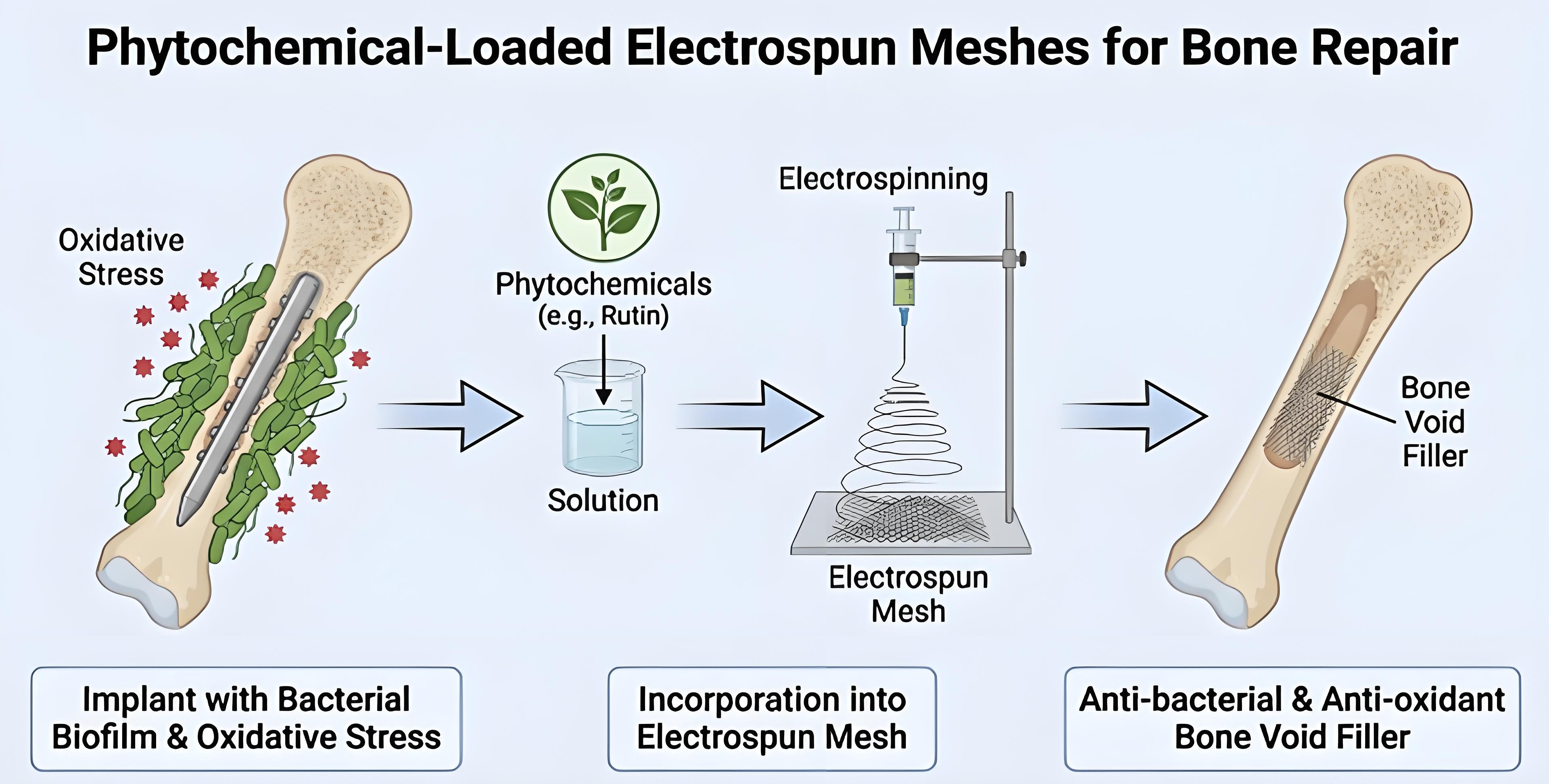
The application of electrospun scaffolds for musculoskeletal tissue repair, such as bone or tendon/ligament, is a growing field of research. They can mimic the fibrous architecture of native extracellular matrix, while simultaneously allowing to support cell attachment, proliferation, and ultimately tissue regeneration. However, implantation of electrospun scaffolds also faces the challenges of implant-associated infection or excessive oxidative stress. Both scenarios can result in serious inflammation, or fibrosis in the case of tendon injury repair, which might lead to failure of the implanted scaffold and strictly limited success of tissue healing. Modification of electrospun scaffolds via incorporation of compounds exerting antibacterial as well as antioxidant activity emerges as a promising approach to counteract these challenges. In this context, naturally derived compounds, such as those of phenolic or terpenic origin, are interesting candidates, because of their potential dual-action nature. In the first part of this review, the most promising candidate compounds to be incorporated into electrospun scaffolds for musculoskeletal tissue repair are examined. We discuss the mechanisms through which these compounds interact with Gram-positive and Gram-negative bacteria in regards to surface attachment, biofilm formation and virulence activity in general. We also highlight the most recent research on their antioxidant potential as well as their effect on cellular host inflammation pathways. In the second part of this review, particular emphasis is placed on strategies for incorporating these compounds into electrospun scaffolds. Finally, we propose future perspectives for research and development of functionalized electrospun scaffolds with bioactive character to advance translation into clinical application.
- Open Access
- Review
Bioactive Biomaterials: Phenolic and Terpenic Compounds as Prospective Antibacterial and Antioxidant Additives for Electrospun Scaffolds
Author Information
Received: 06 May 2026 | Revised: 20 Aug 2026 | Accepted: 25 Aug 2026 | Published: 11 Sep 2026
Abstract
Graphical Abstract
Keywords
electrospinning | antibacterial | oxidative stress | phytochemicals | musculoskeletal | tissue healing
References
- 1.
Halim, N.; Nallusamy, N.; Lakshminarayanan, R.; et al. Electrospinning in Drug Delivery: Progress and Future Outlook. Macromol. Rapid Commun. 2025, 46, e2400903. https://doi.org/10.1002/marc.202400903.
- 2.
Cho, Y.; Beak, J.W.; Sagong, M.; et al. Electrospinning and Nanofiber Technology: Fundamentals, Innovations, and Applications. Adv. Mater. 2025, 37, e2500162. https://doi.org/10.1002/adma.202500162.
- 3.
Pedram Rad, Z.; Mokhtari, J.; Abbasi, M. Calendula officinalis extract/PCL/Zein/Gum arabic nanofibrous bio-composite scaffolds via suspension, two-nozzle and multilayer electrospinning for skin tissue engineering. Int. J. Biol. Macromol. 2019, 135, 530–543. https://doi.org/10.1016/j.ijbiomac.2019.05.204.
- 4.
Cui, J.; Yu, X.; Yu, B.; et al. Coaxially Fabricated Dual-Drug Loading Electrospinning Fibrous Mat with Programmed Releasing Behavior to Boost Vascularized Bone Regeneration. Adv. Healthc. Mater. 2022, 11, e2200571. https://doi.org/10.1002/adhm.202200571.
- 5.
Zahran, S.M.E.; Abdel-Halim, A.H.; Nassar, K.; et al. Fabrication of nanofiltration membrane based on non-biofouling PVP/lecithin nanofibers reinforced with microcrystalline cellulose via needle and needle-less electrospinning techniques. Int. J. Biol. Macromol. 2020, 157, 530–543. https://doi.org/10.1016/j.ijbiomac.2020.04.152.
- 6.
Nutt, K.; Dombros-Ryan, Z.; Birea, R.; et al. Electrospun Polycaprolactone (PCL) Nanofibers Induce Elongation and Alignment of Co-Cultured Primary Cortical Astrocytes and Neurons. Micromachines 2025, 16, 256. https://doi.org/10.3390/mi16030256.
- 7.
Safari, P.; Rahimabadi, E.Z.; Vaezi, M.R.; et al. Development of ZnO-NPs reinforced chitosan nanofiber mats with improved antibacterial and biocompatibility properties. Sci. Rep. 2025, 15, 16567. https://doi.org/10.1038/s41598-025-01669-w.
- 8.
Näf, L.; Miescher, I.; Pfuderer, L.; et al. Pro-angiogenic and antibacterial copper containing nanoparticles in PLGA/amorphous calcium phosphate bone nanocomposites. Heliyon 2024, 10, e27267. https://doi.org/10.1016/j.heliyon.2024.e27267.
- 9.
Rieber, J.; Wolint, P.; Meier-Bürgisser, G.; et al. Synergistic Effects of Insulin-like Growth Factor-1 and Platelet-Derived Growth Factor-BB in Tendon Healing. Int. J. Mol. Sci. 2025, 26, 4039.
- 10.
Araújo, J.C.; Silva, P.M.; Cerqueira, M.A.; et al. Protective fibrous structures based on cellulose fibers functionalized with metal oxide nanoparticles by electrospinning and electrospray deposition. Int. J. Biol. Macromol. 2024, 282, 137218. https://doi.org/10.1016/j.ijbiomac.2024.137218.
- 11.
Kim, D.; Hwang, K.S.; Koh, W.G.; et al. Volatile Organic Compound Sensing Array and Optoelectronic Filter System using Ion-Pairing Dyes with a Wide Visible Spectrum. Adv. Mater. 2022, 34, e2203671. https://doi.org/10.1002/adma.202203671.
- 12.
Zhu, Y.; Dai, B.; Zhang, S.; et al. Tissue Mimetic Membranes for Healing Augmentation of Tendon-Bone Interface in Rotator Cuff Repair. Adv. Mater. 2025, 37, e2407358. https://doi.org/10.1002/adma.202407358.
- 13.
Chen, M.; Gao, S.; Wang, P.; et al. The application of electrospinning used in meniscus tissue engineering. J. Biomater. Sci. Polym. Ed. 2018, 29, 461–475. https://doi.org/10.1080/09205063.2018.1425180.
- 14.
Ivanisova, D.; Bevizova, K.; Vach Agocsova, S.; et al. Implications of Tissue Engineering for Tendon Repair and Regeneration. J. Funct. Biomater. 2025, 16, 403. https://doi.org/10.3390/jfb16110403.
- 15.
Soni, M.K.; Soni, V.K.; Barua, E. State of the Art Technology of Electroactive and Conductive Scaffolds for Bone Tissue Engineering. J. Biomed. Mater. Res. B Appl. Biomater. 2025, 113, e35663. https://doi.org/10.1002/jbm.b.35663.
- 16.
Altundag, Ö.; Öteyaka, M.; Çelebi-Saltik, B. Co- and Triaxial Electrospinning for Stem Cell-based Bone Regeneration. Curr. Stem Cell Res. Ther. 2024, 19, 865–878. https://doi.org/10.2174/1574888x18666230818094216.
- 17.
Liu, J.; Nie, H.; Xu, Z.; et al. Construction of PRP-containing nanofibrous scaffolds for controlled release and their application to cartilage regeneration. J. Mater. Chem. B 2015, 3, 581–591. https://doi.org/10.1039/c4tb00515e.
- 18.
Rieber, J.; Miescher, I.; Wolint, P.; et al. Electrospun DegraPol Tube Delivering Stem Cell/Tenocyte Co-Culture-Derived Secretome to Transected Rabbit Achilles Tendon—In Vitro and In Vivo Evaluation. Int. J. Mol. Sci. 2025, 26, 5457.
- 19.
Rieber, J.; Schweizer, T.A.; Kadler, G.; et al. Electrospun DegraPol® Meshes with Incorporated Bakuchiol: Characterization and Tissue Integration In Ovo—A Pilot Study. Micro 2026, 6, 18. https://doi.org/10.3390/micro6010018.
- 20.
Mele, E. Electrospinning of Essential Oils. Polymers 2020, 12, 908. https://doi.org/10.3390/polym12040908.
- 21.
Croitoru, A.M.; Ficai, D.; Ficai, A.; et al. Nanostructured Fibers Containing Natural or Synthetic Bioactive Compounds in Wound Dressing Applications. Materials 2020, 13, 2407. https://doi.org/10.3390/ma13102407.
- 22.
Anderson, J.M.; Rodriguez, A.; Chang, D.T. Foreign body reaction to biomaterials. Semin. Immunol. 2008, 20, 86–100. https://doi.org/10.1016/j.smim.2007.11.004.
- 23.
Kochar, S.P.; Reche, A.; Paul, P. The Etiology and Management of Dental Implant Failure: A Review. Cureus 2022, 14, e30455. https://doi.org/10.7759/cureus.30455.
- 24.
Pietropaoli, D.; Ortu, E.; Severino, M.; et al. Glycation and oxidative stress in the failure of dental implants: A case series. BMC Res. Notes 2013, 6, 296. https://doi.org/10.1186/1756-0500-6-296.
- 25.
Mierzejewska, Ż.A.; Woźniak, Ł.; Lechien, J.R.; et al. Beyond Biocompatibility: Immune Dysregulation, Oxidative Stress, and Tissue Intolerance Associated with Ti-6Al-4V Dental Implants—A Critical Review and Perspective. Antioxidants 2026, 15, 365.
- 26.
Kračun, D.; Görlach, A.; Snedeker, J.G.; et al. Reactive oxygen species in tendon injury and repair. Redox Biol. 2025, 81, 103568. https://doi.org/10.1016/j.redox.2025.103568.
- 27.
Liu, W.F.; Ma, M.; Bratlie, K.M.; et al. Real-time in vivo detection of biomaterial-induced reactive oxygen species. Biomaterials 2011, 32, 1796–1801. https://doi.org/10.1016/j.biomaterials.2010.11.029.
- 28.
Dunnill, C.; Patton, T.; Brennan, J.; et al. Reactive oxygen species (ROS) and wound healing: The functional role of ROS and emerging ROS-modulating technologies for augmentation of the healing process. Int. Wound J. 2017, 14, 89–96. https://doi.org/10.1111/iwj.12557.
- 29.
Latella, G. Redox Imbalance in Intestinal Fibrosis: Beware of the TGFβ-1, ROS, and Nrf2 Connection. Dig. Dis. Sci. 2018, 63, 312–320. https://doi.org/10.1007/s10620-017-4887-1.
- 30.
Tao, R.; Han, M.; Yuan, W.; et al. Fibrinogen-like protein 2 promotes proinflammatory macrophage polarization and mitochondrial dysfunction in liver fibrosis. Int. Immunopharmacol. 2023, 117, 109631. https://doi.org/10.1016/j.intimp.2022.109631.
- 31.
Ng, M.T.H.; Borst, R.; Gacaferi, H.; et al. A single cell atlas of frozen shoulder capsule identifies features associated with inflammatory fibrosis resolution. Nat. Commun. 2024, 15, 1394. https://doi.org/10.1038/s41467-024-45341-9.
- 32.
Hameister, R.; Kaur, C.; Dheen, S.T.; et al. Reactive oxygen/nitrogen species (ROS/RNS) and oxidative stress in arthroplasty. J. Biomed. Mater. Res. B Appl. Biomater. 2020, 108, 2073–2087. https://doi.org/10.1002/jbm.b.34546.
- 33.
Guabello, G.; Zuffetti, F.; Ravidà, A.; et al. Avoiding implant-related complications in medically compromised patients with or without unhealthy lifestyle/Elevated oxidative stress. Periodontol. 2000 2023, 92, 329–349. https://doi.org/10.1111/prd.12503.
- 34.
Cerqueni, G.; Scalzone, A.; Licini, C.; et al. Insights into oxidative stress in bone tissue and novel challenges for biomaterials. Mater. Sci. Eng. C Mater. Biol. Appl. 2021, 130, 112433. https://doi.org/10.1016/j.msec.2021.112433.
- 35.
Abdulhameed, E.A.; Al-Rawi, N.H.; Omar, M.; et al. Titanium dioxide dental implants surfaces related oxidative stress in bone remodeling: A systematic review. PeerJ 2022, 10, e12951. https://doi.org/10.7717/peerj.12951.
- 36.
Xie, D.M.; Zhong, Q.; Xu, X.; et al. Alpha lipoic acid-loaded electrospun fibrous patch films protect heart in acute myocardial infarction mice by inhibiting oxidative stress. Int. J. Pharm. 2023, 632, 122581. https://doi.org/10.1016/j.ijpharm.2023.122581.
- 37.
Zhang, J.; Xiao, C.; Zhang, X.; et al. An oxidative stress-responsive electrospun polyester membrane capable of releasing anti-bacterial and anti-inflammatory agents for postoperative anti-adhesion. J. Control. Release 2021, 335, 359–368. https://doi.org/10.1016/j.jconrel.2021.04.017.
- 38.
Oliva, A.; Miele, M.C.; Al Ismail, D.; et al. Challenges in the Microbiological Diagnosis of Implant-Associated Infections: A Summary of the Current Knowledge. Front. Microbiol. 2021, 12, 750460. https://doi.org/10.3389/fmicb.2021.750460.
- 39.
Ul Haq, I.; Khan, T.A.; Krukiewicz, K. Etiology, pathology, and host-impaired immunity in medical implant-associated infections. J. Infect. Public Health 2024, 17, 189–203. https://doi.org/10.1016/j.jiph.2023.11.024.
- 40.
Arciola, C.R.; Campoccia, D.; Montanaro, L. Implant infections: Adhesion, biofilm formation and immune evasion. Nat. Rev. Microbiol. 2018, 16, 397–409. https://doi.org/10.1038/s41579-018-0019-y.
- 41.
VanEpps, J.S.; Younger, J.G. Implantable Device-Related Infection. Shock 2016, 46, 597–608. https://doi.org/10.1097/shk.0000000000000692.
- 42.
Saleh, M.H.A.; Dias, D.R.; Kumar, P. The economic and societal impact of periodontal and peri-implant diseases. Periodontol. 2000 2025, 98, 100–118. https://doi.org/10.1111/prd.12568.
- 43.
Aftab, M.H.S.; Joseph, T.; Almeida, R.; et al. Periprosthetic Joint Infection: A Multifaceted Burden Undermining Arthroplasty Success. Orthop. Rev. 2025, 17, 138205. https://doi.org/10.52965/001c.138205.
- 44.
Moriarty, T.F.; Metsemakers, W.J.; Morgenstern, M.; et al. Fracture-related infection. Nat. Rev. Dis. Primers 2022, 8, 67. https://doi.org/10.1038/s41572-022-00396-0.
- 45.
Patel, R. Periprosthetic Joint Infection. N. Engl. J. Med. 2023, 388, 251–262. https://doi.org/10.1056/NEJMra2203477.
- 46.
Chavada, J.; Muneshwar, K.N.; Ghulaxe, Y.; et al. Antibiotic Resistance: Challenges and Strategies in Combating Infections. Cureus 2023, 15, e46013. https://doi.org/10.7759/cureus.46013.
- 47.
Costerton, J.W.; Montanaro, L.; Arciola, C.R. Biofilm in implant infections: Its production and regulation. Int. J. Artif. Organs 2005, 28, 1062–1068. https://doi.org/10.1177/039139880502801103.
- 48.
Zimmerli, W.; Lew, P.D.; Waldvogel, F.A. Pathogenesis of foreign body infection. Evidence for a local granulocyte defect. J. Clin. Invest. 1984, 73, 1191–1200. https://doi.org/10.1172/jci111305.
- 49.
Guarch-Pérez, C.; Riool, M.; de Boer, L.; et al. Bacterial reservoir in deeper skin is a potential source for surgical site and biomaterial-associated infections. J. Hosp. Infect. 2023, 140, 62–71. https://doi.org/10.1016/j.jhin.2023.07.014.
- 50.
Maurer, S.M.; Kursawe, L.; Rahm, S.; et al. Cutibacterium avidum resists surgical skin antisepsis in the groin-a potential risk factor for periprosthetic joint infection: A quality control study. Antimicrob. Resist. Infect. Control 2021, 10, 27. https://doi.org/10.1186/s13756-021-00883-1.
- 51.
Waldmann, I.; Schmid, T.; Prinz, J.; et al. Photodynamic therapy improves skin antisepsis as a prevention strategy in arthroplasty procedures: A pilot study. Photodiagnosis Photodyn. Ther. 2020, 31, 101941. https://doi.org/10.1016/j.pdpdt.2020.101941.
- 52.
Schweizer, T.A.; Würmli, J.S.; Prinz, J.; et al. Photodynamic Therapy with Protoporphyrin IX Precursors Using Artificial Daylight Improves Skin Antisepsis for Orthopedic Surgeries. Microorganisms 2025, 13, 204.
- 53.
Săndulescu, M.; Sîrbu, V.D.; Popovici, I.A. Bacterial species associated with peri-implant disease—A literature review. Germs 2023, 13, 352–361. https://doi.org/10.18683/germs.2023.1405.
- 54.
Hallmer, F.; Bjørnland, T.; Andersson, G.; et al. Bacterial diversity in medication-related osteonecrosis of the jaw. Oral Surg. Oral Med. Oral Pathol. Oral Radiol. 2017, 123, 436–444. https://doi.org/10.1016/j.oooo.2016.11.011.
- 55.
Tai, D.B.G.; Patel, R.; Abdel, M.P.; et al. Microbiology of hip and knee periprosthetic joint infections: A database study. Clin. Microbiol. Infect. 2022, 28, 255–259. https://doi.org/10.1016/j.cmi.2021.06.006.
- 56.
Depypere, M.; Sliepen, J.; Onsea, J.; et al. The Microbiological Etiology of Fracture-Related Infection. Front. Cell. Infect. Microbiol. 2022, 12, 934485. https://doi.org/10.3389/fcimb.2022.934485.
- 57.
Bowers, M.W.; Turner, N.S.; Ryssman, D.B.; et al. Outcomes Following Treatment of the Infected Achilles Tendon. Foot Ankle Orthop. 2019, 4, 2473011419846943. https://doi.org/10.1177/2473011419846943.
- 58.
Yeo, I.S.; Kim, H.Y.; Lim, K.S.; et al. Implant surface factors and bacterial adhesion: A review of the literature. Int. J. Artif. Organs 2012, 35, 762–772. https://doi.org/10.5301/ijao.5000154.
- 59.
Mohamed Zuki, F.; Edyvean, R.G.J.; Pourzolfaghar, H.; et al. Modeling of the Van Der Waals Forces during the Adhesion of Capsule-Shaped Bacteria to Flat Surfaces. Biomimetics 2021, 6, 5.
- 60.
Kadirvelu, L.; Sivaramalingam, S.S.; Jothivel, D.; et al. A review on antimicrobial strategies in mitigating biofilm-associated infections on medical implants. Curr. Res. Microb. Sci. 2024, 6, 100231. https://doi.org/10.1016/j.crmicr.2024.100231.
- 61.
Lorite, G.S.; Rodrigues, C.M.; de Souza, A.A.; et al. The role of conditioning film formation and surface chemical changes on Xylella fastidiosa adhesion and biofilm evolution. J. Colloid Interface Sci. 2011, 359, 289–295. https://doi.org/10.1016/j.jcis.2011.03.066.
- 62.
Joh, D.; Wann, E.R.; Kreikemeyer, B.; et al. Role of fibronectin-binding MSCRAMMs in bacterial adherence and entry into mammalian cells. Matrix Biol. 1999, 18, 211–223. https://doi.org/10.1016/s0945-053x(99)00025-6.
- 63.
Foster, T.J. The MSCRAMM Family of Cell-Wall-Anchored Surface Proteins of Gram-Positive Cocci. Trends Microbiol. 2019, 27, 927–941. https://doi.org/10.1016/j.tim.2019.06.007.
- 64.
Ghasemian, A.; Najar Peerayeh, S.; Bakhshi, B.; et al. The Microbial Surface Components Recognizing Adhesive Matrix Molecules (MSCRAMMs) Genes among Clinical Isolates of Staphylococcus aureus from Hospitalized Children. Iran. J. Pathol. 2015, 10, 258–264.
- 65.
Berne, C.; Ducret, A.; Hardy, G.G.; et al. Adhesins Involved in Attachment to Abiotic Surfaces by Gram-Negative Bacteria. Microbiol. Spectr. 2015, 3, 163–199. https://doi.org/10.1128/microbiolspec.MB-0018-2015.
- 66.
Preda, V.G.; Săndulescu, O. Communication is the key: Biofilms, quorum sensing, formation and prevention. Discoveries 2019, 7, e100. https://doi.org/10.15190/d.2019.13.
- 67.
Miescher, I.; Rieber, J.; Schweizer, T.A.; et al. In Vitro Assessment of Bacterial Adhesion and Biofilm Formation on Novel Bioactive, Biodegradable Electrospun Fiber Meshes Intended to Support Tendon Rupture Repair. ACS Appl. Mater. Interfaces 2024, 16, 6348–6355. https://doi.org/10.1021/acsami.3c15710.
- 68.
Biagini, F.; Calvigioni, M.; De Maria, C.; et al. Study of the Adhesion of the Human Gut Microbiota on Electrospun Structures. Bioengineering 2022, 9, 96.
- 69.
Clavijo-Grimaldo, D.; Ponce-Zapata, N.; Casadiego-Torrado, C. Control of Bacterial Proliferation and Formation of Biofilm in Membranes for Food Packaging Manufactured by Electrospinning. Chem. Eng. Trans. 2019, 75, 241–246. https://doi.org/10.3303/CET1975041.
- 70.
Sun, T.; Xue, B.; Shao, Z.; et al. Effect on the bacterial adhesion of PVA electrospinning membrane deposited on silicone catheter surface. Biomed. Phys. Eng. Express 2026, 12, 025003. https://doi.org/10.1088/2057-1976/ae3f35.
- 71.
Krasowska, A.; Sigler, K. How microorganisms use hydrophobicity and what does this mean for human needs? Front. Cell. Infect. Microbiol. 2014, 4, 112. https://doi.org/10.3389/fcimb.2014.00112.
- 72.
Abrigo, M.; Kingshott, P.; McArthur, S.L. Electrospun polystyrene fiber diameter influencing bacterial attachment, proliferation, and growth. ACS Appl. Mater. Interfaces 2015, 7, 7644–7652. https://doi.org/10.1021/acsami.5b00453.
- 73.
de Mesy Bentley, K.L.; MacDonald, A.; Schwarz, E.M.; et al. Chronic Osteomyelitis with Staphylococcus aureus Deformation in Submicron Canaliculi of Osteocytes: A Case Report. JBJS Case Connect. 2018, 8, e8. https://doi.org/10.2106/JBJS.CC.17.00154.
- 74.
de Mesy Bentley, K.L.; Trombetta, R.; Nishitani, K.; et al. Evidence of Staphylococcus Aureus Deformation, Proliferation, and Migration in Canaliculi of Live Cortical Bone in Murine Models of Osteomyelitis. J. Bone Miner. Res. 2017, 32, 985–990. https://doi.org/10.1002/jbmr.3055.
- 75.
Mufty, H.; Fourneau, I. Is silver still the Holy Grail for vascular grafts? J. Cardiovasc. Surg. 2023, 64, 304–309. https://doi.org/10.23736/S0021-9509.22.12538-3.
- 76.
Molacek, J.; Treska, V.; Houdek, K.; et al. Use of a Silver-Impregnated Vascular Graft: Single-Center Experience. Antibiotics 2022, 11, 386.
- 77.
Romanò, C.L.; Tsuchiya, H.; Morelli, I.; et al. Antibacterial coating of implants: Are we missing something? Bone Jt. Res. 2019, 8, 199–206. https://doi.org/10.1302/2046-3758.85.BJR-2018-0316.
- 78.
Ielo, I.; Bauso, L.V.; Laezza, A.; et al. In Vitro Assessment of Electrospun PVP + AgNPs Scaffolds for Bioactive Medical Use. Int. J. Mol. Sci. 2025, 26, 9114. https://doi.org/10.3390/ijms26189114.
- 79.
Elsheikh, R.; Makram, A.; Toth, L.; et al. Comparative effectiveness of antimicrobial implant surface coatings in preventing orthopaedic implant-associated infections: A network meta-analysis. Arch. Orthop. Trauma Surg. 2026, 146, 60. https://doi.org/10.1007/s00402-026-06225-3.
- 80.
Xi, W.; Hegde, V.; Zoller, S.D.; et al. Point-of-care antimicrobial coating protects orthopaedic implants from bacterial challenge. Nat. Commun. 2021, 12, 5473. https://doi.org/10.1038/s41467-021-25383-z.
- 81.
Clement, J.L.; Jarrett, P.S. Antibacterial silver. Met. Based Drugs 1994, 1, 467–482. https://doi.org/10.1155/MBD.1994.467.
- 82.
Villarreal-Gómez, L.J.; Pérez-González, G.L.; Bogdanchikova, N.; et al. Antimicrobial Effect of Electrospun Nanofibers Loaded with Silver Nanoparticles: Influence of Ag Incorporation Method. J. Nanomater. 2021, 2021, 9920755. https://doi.org/10.1155/2021/9920755.
- 83.
Ren, X.; Hu, Y.; Chang, L.; et al. Electrospinning of antibacterial and anti-inflammatory Ag@hesperidin core-shell nanoparticles into nanofibers used for promoting infected wound healing. Regen. Biomater. 2022, 9, rbac012. https://doi.org/10.1093/rb/rbac012.
- 84.
Yin, I.X.; Zhang, J.; Zhao, I.S.; et al. The Antibacterial Mechanism of Silver Nanoparticles and Its Application in Dentistry. Int. J. Nanomed. 2020, 15, 2555–2562. https://doi.org/10.2147/IJN.S246764.
- 85.
Mufty, H.; Van den Eynde, J.; Steenackers, H.P.; et al. A systematic review of preclinical data regarding commercial silver-coated vascular grafts. J. Vasc. Surg. 2021, 74, 1386–1393.e1. https://doi.org/10.1016/j.jvs.2021.04.055.
- 86.
Zhang, J.; Wang, F.; Yalamarty, S.S.K.; et al. Nano Silver-Induced Toxicity and Associated Mechanisms. Int. J. Nanomed. 2022, 17, 1851–1864. https://doi.org/10.2147/IJN.S355131.
- 87.
Paluch, E.; Rewak-Soroczyńska, J.; Jędrusik, I.; et al. Prevention of biofilm formation by quorum quenching. Appl. Microbiol. Biotechnol. 2020, 104, 1871–1881. https://doi.org/10.1007/s00253-020-10349-w.
- 88.
Sikdar, R.; Elias, M. Quorum quenching enzymes and their effects on virulence, biofilm, and microbiomes: A review of recent advances. Expert Rev. Anti Infect. Ther. 2020, 18, 1221–1233. https://doi.org/10.1080/14787210.2020.1794815.
- 89.
D’Aquila, P.; De Rose, E.; Sena, G.; et al. Quorum Quenching Approaches against Bacterial-Biofilm-Induced Antibiotic Resistance. Antibiotics 2024, 13, 619.
- 90.
Martelli, G.; Giacomini, D. Antibacterial and antioxidant activities for natural and synthetic dual-active compounds. Eur. J. Med. Chem. 2018, 158, 91–105. https://doi.org/10.1016/j.ejmech.2018.09.009.
- 91.
Ecevit, K.; Barros, A.A.; Silva, J.M.; et al. Preventing Microbial Infections with Natural Phenolic Compounds. Future Pharmacol. 2022, 2, 460–498.
- 92.
Kauffmann, A.C.; Castro, V.S. Phenolic Compounds in Bacterial Inactivation: A Perspective from Brazil. Antibiotics 2023, 12, 645.
- 93.
Foti, M.C. Antioxidant properties of phenols. J. Pharm. Pharmacol. 2007, 59, 1673–1685. https://doi.org/10.1211/jpp.59.12.0010.
- 94.
Marchese, A.; Orhan, I.E.; Daglia, M.; et al. Antibacterial and antifungal activities of thymol: A brief review of the literature. Food Chem. 2016, 210, 402–414. https://doi.org/10.1016/j.foodchem.2016.04.111.
- 95.
Anand, R.; Mohan, L.; Bharadvaja, N. Disease Prevention and Treatment Using β-Carotene: The Ultimate Provitamin A. Rev. Bras. Farmacogn. 2022, 32, 491–501. https://doi.org/10.1007/s43450-022-00262-w.
- 96.
Singh, R.V.; Sambyal, K. An overview of β-carotene production: Current status and future prospects. Food Biosci. 2022, 47, 101717. https://doi.org/10.1016/j.fbio.2022.101717.
- 97.
Dai, C.; Lin, J.; Li, H.; et al. The Natural Product Curcumin as an Antibacterial Agent: Current Achievements and Problems. Antioxidants 2022, 11, 459.
- 98.
Mitra, S.; Mateti, T.; Ramakrishna, S.; et al. A Review on Curcumin-Loaded Electrospun Nanofibers and their Application in Modern Medicine. JOM 2022, 74, 3392–3407. https://doi.org/10.1007/s11837-022-05180-9.
- 99.
Tamilarasi, G.P.; Krishnan, M.; Sabarees, G.; et al. Emerging Trends in Curcumin Embedded Electrospun Nanofibers for Impaired Diabetic Wound Healing. Appl. Nano 2022, 3, 202–232.
- 100.
Rostami, M.; Kolahi Azar, H.; Salehi, M.; et al. The food and biomedical applications of curcumin-loaded electrospun nanofibers: A comprehensive review. Crit. Rev. Food Sci. Nutr. 2024, 64, 12383–12410. https://doi.org/10.1080/10408398.2023.2251584.
- 101.
Fereydouni, N.; Darroudi, M.; Movaffagh, J.; et al. Curcumin nanofibers for the purpose of wound healing. J. Cell. Physiol. 2019, 234, 5537–5554. https://doi.org/10.1002/jcp.27362.
- 102.
Ubeyitogullari, A.; Ahmadzadeh, S.; Kandhola, G.; et al. Polysaccharide-based porous biopolymers for enhanced bioaccessibility and bioavailability of bioactive food compounds: Challenges, advances, and opportunities. Compr. Rev. Food Sci. Food Saf. 2022, 21, 4610–4639. https://doi.org/10.1111/1541-4337.13049.
- 103.
Sharifi, M.; Bahrami, S.H. Review on application of herbal extracts in biomacromolecules-based nanofibers as wound dressings and skin tissue engineering. Int. J. Biol. Macromol. 2024, 277, 133666. https://doi.org/10.1016/j.ijbiomac.2024.133666.
- 104.
Sinha, S.; Das, S.; Saha, B.; et al. Anti-microbial, anti-oxidant, and anti-breast cancer properties unraveled in yeast carotenoids produced via cost-effective fermentation technique utilizing waste hydrolysate. Front. Microbiol. 2023, 13, 1088477. https://doi.org/10.3389/fmicb.2022.1088477.
- 105.
Fernandez, A.; Torres-Giner, S.; Lagaron, J.M. Novel route to stabilization of bioactive antioxidants by encapsulation in electrospun fibers of zein prolamine. Food Hydrocoll. 2009, 23, 1427–1432. https://doi.org/10.1016/j.foodhyd.2008.10.011.
- 106.
Fabra, M.J.; López-Rubio, A.; Sentandreu, E.; et al. Development of multilayer corn starch-based food packaging structures containing β-carotene by means of the electro-hydrodynamic processing. Starch Stärke 2016, 68, 603–610. https://doi.org/10.1002/star.201500154.
- 107.
Dabouian, A.; Bakhshi, H.; Irani, S.; et al. β-Carotene: A natural osteogen to fabricate osteoinductive electrospun scaffolds. RSC Adv. 2018, 8, 9941–9945. https://doi.org/10.1039/C7RA13237A.
- 108.
Yildiz, Z.I.; Topuz, F.; Kilic, M.E.; et al. Encapsulation of antioxidant beta-carotene by cyclodextrin complex electrospun nanofibers: Solubilization and stabilization of beta-carotene by cyclodextrins. Food Chem. 2023, 423, 136284. https://doi.org/10.1016/j.foodchem.2023.136284.
- 109.
Yoshikawa, O.; Basoli, V.; Boschetto, F.; et al. Simple Electrospinning Method for Biocompatible Polycaprolactone β-Carotene Scaffolds: Advantages and Limitations. Polymers 2024, 16, 1371.
- 110.
Soni, N.; Roy, D.; Patel, D.M.; et al. Study on electrospinning of polyvinyl alcohol nanofibers loaded with carotenoids for enhanced stability and its multifarious applications. Discov. Chem. 2025, 2, 57. https://doi.org/10.1007/s44371-025-00132-z.
- 111.
Shafe, M.O.; Gumede, N.M.; Nyakudya, T.T.; et al. Lycopene: A Potent Antioxidant with Multiple Health Benefits. J. Nutr. Metab. 2024, 2024, 6252426. https://doi.org/10.1155/2024/6252426.
- 112.
Anlar, H.G.; Bacanli, M. Chapter 24—Lycopene as an antioxidant in human health and diseases. In Pathology, Preedy, V.R., Ed.; Academic Press: San Diego, CA, USA, 2020; pp. 247–254.
- 113.
Imran, M.; Ghorat, F.; Ul-Haq, I.; et al. Lycopene as a Natural Antioxidant Used to Prevent Human Health Disorders. Antioxidants 2020, 9, 706. https://doi.org/10.3390/antiox9080706.
- 114.
Bin-Jumah, M.N.; Nadeem, M.S.; Gilani, S.J.; et al. Lycopene: A Natural Arsenal in the War against Oxidative Stress and Cardiovascular Diseases. Antioxidants 2022, 11, 232.
- 115.
Pan, X.; Zhu, R.; Pei, J.; et al. Lycopene: A potent antioxidant to alleviate kidney disease. Int. Immunopharmacol. 2025, 151, 114363. https://doi.org/10.1016/j.intimp.2025.114363.
- 116.
Hraishawi, R.M.O.; Abdul-Razak, A.S.; Al-Hayder, M.N.; et al. Investigation the antimicrobial and antioxidant activity of lycopene extraction from Solanum Lycopersicum. Eurasian J. Biosci. 2020, 14, 5305–5310.
- 117.
Rasheed, A.A.H.A.A.; Al–Anbari, I.H. Study the Effect of Lycopene Extract and Nano-Lycopene Extracted from Tomato Waste in Inhibiting Microbial Growth and as an Intioxidant. Iraqi J. Agric. Sci. 2025, 56, 2180–2189.
- 118.
Tang, C.; Li, Q.; Lin, T. Lycopene attenuates Staphylococcus aureus-induced inflammation via inhibiting α-hemolysin expression. Microbes Infect. 2021, 23, 104853. https://doi.org/10.1016/j.micinf.2021.104853.
- 119.
Keddari, S.; Hamed, D.; Bouhend, A.; et al. Therapeutic Effects of Lycopene Alone or in Combination with Cephalexin on Chronic Prostatitis Caused by Staphylococcus aureus in a Rat Model. Microbiol. Res. 2025, 16, 114.
- 120.
Pawar, S.; Garge, S.; Shetty, S.; et al. Recent Advances in Lycopene Delivery Systems: Pharmaceutical, Nutraceutical, and Clinical Perspectives. Recent Adv. Drug Deliv. Formul. 2026. https://doi.org/10.2174/0126673878388760251126123939.
- 121.
Sensini, A.; Cristofolini, L.; Focarete, M.L.; et al. High-resolution X-ray tomographic morphological characterisation of electrospun nanofibrous bundles for tendon and ligament regeneration and replacement. J. Microsc. 2018, 272, 196–206. https://doi.org/10.1111/jmi.12720.
- 122.
Hajikhani, M.; Emam-Djomeh, Z.; Askari, G. Fabrication and Characterization of Gluten Film Reinforced by Lycopene-Loaded Electrospun Polylactic Acid Nano-fibers. Food Bioprocess Technol. 2020, 13, 2217–2227. https://doi.org/10.1007/s11947-020-02561-3.
- 123.
İnan-Çınkır, N.; Ağçam, E.; Altay, F.; et al. Emulsion electrospinning of zein nanofibers with carotenoid microemulsion: Optimization, characterization and fortification. Food Chem. 2024, 430, 137005. https://doi.org/10.1016/j.foodchem.2023.137005.
- 124.
de Farias, B.S.; Rizzi, F.Z.; Silva, P.P.; et al. Tri-layer nanostructured lycopene delivery system based on chitosan nanospheres and gelatin nanofibers. Food Res. Int. 2025, 220, 117099. https://doi.org/10.1016/j.foodres.2025.117099.
- 125.
Qi, W.; Qi, W.; Xiong, D.; et al. Quercetin: Its Antioxidant Mechanism, Antibacterial Properties and Potential Application in Prevention and Control of Toxipathy. Molecules 2022, 27, 6545.
- 126.
Nguyen, T.L.A.; Bhattacharya, D. Antimicrobial Activity of Quercetin: An Approach to Its Mechanistic Principle. Molecules 2022, 27, 2494.
- 127.
Veiko, A.G.; Olchowik-Grabarek, E.; Sekowski, S.; et al. Antimicrobial Activity of Quercetin, Naringenin and Catechin: Flavonoids Inhibit Staphylococcus aureus-Induced Hemolysis and Modify Membranes of Bacteria and Erythrocytes. Molecules 2023, 28, 1252.
- 128.
Musini, A.; Singh, H.N.; Vulise, J.; et al. Quercetin’s antibiofilm effectiveness against drug resistant Staphylococcus aureus and its validation by in silico modeling. Res. Microbiol. 2024, 175, 104091. https://doi.org/10.1016/j.resmic.2023.104091.
- 129.
Majumdar, G.; Mandal, S. Evaluation of broad-spectrum antibacterial efficacy of quercetin by molecular docking, molecular dynamics simulation and in vitro studies. Chem. Phys. Impact 2024, 8, 100501. https://doi.org/10.1016/j.chphi.2024.100501.
- 130.
Vijayakumar, K.; Ganesan, V.; Kannan, S. Antibacterial and antibiofilm efficacy of quercetin against Pseudomonas aeruginosa and methicillin resistant Staphylococcus aureus associated with ICU infections. Biofouling 2025, 41, 211–224. https://doi.org/10.1080/08927014.2025.2460491.
- 131.
Shastri, T.; Binsuwaidan, R.; Siddiqui, A.J.; et al. Quercetin Exhibits Broad-Spectrum Antibiofilm and Antiquorum Sensing Activities Against Gram-Negative Bacteria: In Vitro and In Silico Investigation Targeting Antimicrobial Therapy. Can. J. Infect. Dis. Med. Microbiol. 2025, 2025, 2333207. https://doi.org/10.1155/cjid/2333207.
- 132.
Zhang, Y.; Guo, L.; Wang, S.; et al. Quercetin Enhances the Antibacterial Activity of Polymyxin E Against MCR-1-Positive Bacteria by Inhibiting the Biological Functions of the Cell Membrane. Animals 2025, 15, 3491.
- 133.
Abdelnabi, H.; Mohsin, S. Phytochemicals in Bone Therapy: Exploring Natural Alternatives for Bone Health. Int. J. Nanomed. 2025, 20, 10831–10855. https://doi.org/10.2147/IJN.S524695.
- 134.
Zhou, P.; Yan, B.; Wei, B.; et al. Quercetin-solid lipid nanoparticle-embedded hyaluronic acid functionalized hydrogel for immunomodulation to promote bone reconstruction. Regen. Biomater. 2023, 10, rbad025. https://doi.org/10.1093/rb/rbad025.
- 135.
Stoyanova, N.; Spasova, M.; Manolova, N.; et al. Quercetin- and Rutin-Containing Electrospun Cellulose Acetate and Polyethylene Glycol Fibers with Antioxidant and Anticancer Properties. Polymers 2022, 14, 5380. https://doi.org/10.3390/polym14245380.
- 136.
Lv, Y.; Wu, W.; Liu, Z.; et al. Bilayer nanofibrous wound dressing prepared by electrospinning containing gallic acid and quercetin with improved biocompatibility, antibacterial, and antioxidant effects. Pharm. Dev. Technol. 2023, 28, 18–29. https://doi.org/10.1080/10837450.2022.2160734.
- 137.
Viscusi, G.; Paolella, G.; Lamberti, E.; et al. Quercetin-Loaded Polycaprolactone-Polyvinylpyrrolidone Electrospun Membranes for Health Application: Design, Characterization, Modeling and Cytotoxicity Studies. Membranes 2023, 13, 242. https://doi.org/10.3390/membranes13020242.
- 138.
Ferrari, P.F.; Aliakbarian, B.; Barisione, C.; et al. Engineering of poly(caprolactone) and poly(glycerol sebacate) small-diameter vascular prosthesis with quercetin. J. Biomed. Mater. Res. A 2023, 111, 1500–1512. https://doi.org/10.1002/jbm.a.37535.
- 139.
Karamian, M.; Alemzadeh, E.; Abedi, A.; et al. Glucantime and quercetin electrospun nanofiber membranes: Fabrication and their evaluation as dressing for cutaneous leishmaniasis. Mol. Biochem. Parasitol. 2025, 261, 111663. https://doi.org/10.1016/j.molbiopara.2024.111663.
- 140.
Li, S.F.; Hu, T.G.; Jin, Y.B.; et al. Fabrication and characterization of shellac nanofibers with colon-targeted delivery of quercetin and its anticancer activity. Int. J. Biol. Macromol. 2024, 265, 130789. https://doi.org/10.1016/j.ijbiomac.2024.130789.
- 141.
Lin, S.-H.; Ou, S.-L.; Hsu, H.-M.; et al. Preparation and Characteristics of Polyethylene Oxide/Curdlan Nanofiber Films by Electrospinning for Biomedical Applications. Materials 2023, 16, 3863. https://doi.org/10.3390/ma16103863.
- 142.
Amer, S.S.; Mamdouh, W.; Nasr, M.; et al. Quercetin loaded cosm-nutraceutical electrospun composite nanofibers for acne alleviation: Preparation, characterization and experimental clinical appraisal. Int. J. Pharm. 2022, 612, 121309. https://doi.org/10.1016/j.ijpharm.2021.121309.
- 143.
Li, S.F.; Wu, J.H.; Hu, T.G.; et al. Encapsulation of quercetin into zein-ethyl cellulose coaxial nanofibers: Preparation, characterization and its anticancer activity. Int. J. Biol. Macromol. 2023, 248, 125797. https://doi.org/10.1016/j.ijbiomac.2023.125797.
- 144.
Aleemardani, M.; Solouk, A.; Akbari, S.; et al. A hydrogel-fiber-hydrogel composite scaffold based on silk fibroin with the dual-delivery of oxygen and quercetin. Biotechnol. Bioeng. 2023, 120, 297–311. https://doi.org/10.1002/bit.28259.
- 145.
Hudecki, A.; Rzeszutek, I.; Lewińska, A.; et al. Electrospun fiber-based micro- and nano-system for delivery of high concentrated quercetin to cancer cells. Biomater. Adv. 2023, 153, 213582. https://doi.org/10.1016/j.bioadv.2023.213582.
- 146.
Al-Dhabi, N.A.; Arasu, M.V.; Park, C.H.; et al. An up-to-date review of rutin and its biological and pharmacological activities. EXCLI J. 2015, 14, 59–63. https://doi.org/10.17179/excli2014-663.
- 147.
Mane, M.D.; Patole, N.S.; Kodalkar, V.N.; et al. An Overview of Antimicrobial Properties of Rutin. Int. J. Res. Pharm. Allied Sci. 2024, 3, 17–23.
- 148.
Wang, Z.; Ding, Z.; Li, Z.; et al. Antioxidant and antibacterial study of 10 flavonoids revealed rutin as a potential antibiofilm agent in Klebsiella pneumoniae strains isolated from hospitalized patients. Microb. Pathog. 2021, 159, 105121. https://doi.org/10.1016/j.micpath.2021.105121.
- 149.
Miklasińska-Majdanik, M.; Kępa, M.; Wąsik, T.J.; et al. The Direction of the Antibacterial Effect of Rutin Hydrate and Amikacin. Antibiotics 2023, 12, 1469.
- 150.
Yi, L.; Bai, Y.; Chen, X.; et al. Synergistic Effects and Mechanisms of Action of Rutin with Conventional Antibiotics Against Escherichia coli. Int. J. Mol. Sci. 2024, 25, 13684.
- 151.
Chekuri, S.; Sirigiripeta, S.R.; Thupakula, S.; et al. Rutin isolated from Acalypha indica L.: A comprehensive analysis of its antibacterial and anticancer activities. Biochem. Biophys. Res. Commun. 2025, 765, 151833. https://doi.org/10.1016/j.bbrc.2025.151833.
- 152.
Ramezani, H.; Sazegar, H.; Rouhi, L. The antimicrobial efficacy of rutin encapsulated chitosan versus multidrug-resistant Pseudomonas aeruginosa. Sci. Rep. 2025, 15, 22047. https://doi.org/10.1038/s41598-025-06873-2.
- 153.
Li, N.; Qian, Y.; Zhang, Z.; et al. New Patent on Electrospinning for Increasing Rutin Loading in Nanofibers. Recent Pat. Nanotechnol. 2020, 14, 35–41. https://doi.org/10.2174/1872210513666191107101326.
- 154.
Li, B.; Yang, X. Rutin-loaded cellulose acetate/poly(ethylene oxide) fiber membrane fabricated by electrospinning: A bioactive material. Mater. Sci. Eng. C Mater. Biol. Appl. 2020, 109, 110601. https://doi.org/10.1016/j.msec.2019.110601.
- 155.
Özer, S.; Akyıl, E.; Arslan, R.; et al. Development of Nanofiber Patch Formulation Containing Rutin Hydrate, In Vitro and In Vivo Evaluation. ACS Omega 2025, 10, 29037–29058. https://doi.org/10.1021/acsomega.5c01101.
- 156.
Zhou, L.; Cai, L.; Ruan, H.; et al. Electrospun chitosan oligosaccharide/polycaprolactone nanofibers loaded with wound-healing compounds of Rutin and Quercetin as antibacterial dressings. Int. J. Biol. Macromol. 2021, 183, 1145–1154. https://doi.org/10.1016/j.ijbiomac.2021.05.031.
- 157.
Safdari, F.; Gholipour, M.D.; Ghadami, A.; et al. Multi-antibacterial agent-based electrospun polycaprolactone for active wound dressing. Prog. Biomater. 2022, 11, 27–41. https://doi.org/10.1007/s40204-021-00176-1.
- 158.
Dadgar, A.; Maleki Dizaj, S.; Alizadeh Oskoee, P.; et al. Gelatin nanofibrous scaffolds containing rutin nanoparticles: Physicochemical properties, antibacterial action, and anti-inflammatory effect on dental pulp stem cells. BMC Oral Health 2025, 25, 1129. https://doi.org/10.1186/s12903-025-06385-5.
- 159.
Alotaibi, B.S.; Khan, A.K.; Kharaba, Z.; et al. Development of Poly(vinyl alcohol)-Chitosan Composite Nanofibers for Dual Drug Therapy of Wounds. ACS Omega 2024, 9, 12825–12834. https://doi.org/10.1021/acsomega.3c08856.
- 160.
Tarabishi, A.A.; Mashhoud, J.; Tahan, Z.S. Quercetin and rutin as a dual approach to antibacterial and anti-biofilm activity via iron chelation mechanism. Discov. Food 2024, 4, 189. https://doi.org/10.1007/s44187-024-00265-7.
- 161.
Almuhanna, Y.; Alshalani, A.; AlSudais, H.; et al. Antibacterial, Antibiofilm, and Wound Healing Activities of Rutin and Quercetin and Their Interaction with Gentamicin on Excision Wounds in Diabetic Mice. Biology 2024, 13, 676.
- 162.
Gumisiriza, H.; Olet, E.A.; Mwikali, L.; et al. Antibacterial and Antioxidant Activities of Flavonoids, Phenolic and Flavonoid Glycosides from Gouania longispicata Leaves. Microbiol. Res. 2024, 15, 2085–2101.
- 163.
Borges, A.; Ferreira, C.; Saavedra, M.J.; et al. Antibacterial activity and mode of action of ferulic and gallic acids against pathogenic bacteria. Microb. Drug Resist. 2013, 19, 256–265. https://doi.org/10.1089/mdr.2012.0244.
- 164.
Vaikkathillam, P.; Mini, M.; Mohan, A.; et al. Anti-biofilm effect of ferulic acid against Enterobacter hormaechei and Klebsiella pneumoniae: In Vitro and in silico investigation. Biofouling 2025, 41, 157–170. https://doi.org/10.1080/08927014.2024.2446927.
- 165.
Zhang, Z.; Xu, J.; Wei, X.; et al. Antibiofilm Inhibitor Ferulic Acid as an Antibacterial Synergist against Escherichia coli. Biomolecules 2025, 15, 1253.
- 166.
Ibitoye, O.B.; Ajiboye, T.O. Ferulic acid potentiates the antibacterial activity of quinolone-based antibiotics against Acinetobacter baumannii. Microb. Pathog. 2019, 126, 393–398. https://doi.org/10.1016/j.micpath.2018.11.033.
- 167.
Ando, D.; Lwin, H.Y.; Aoki-Nonaka, Y.; et al. Ferulic acid suppresses Porphyromonas gingivalis biofilm formation via the inhibition of autoinducer-2 production and receptor activity. Arch. Oral Biol. 2025, 178, 106365. https://doi.org/10.1016/j.archoralbio.2025.106365.
- 168.
Tokam, C.R.K.; Ndezo Bisso, B.; Jahan, H.; et al. Evaluation of the Efficacy of Some Natural Products Against Methicillin-Resistant Staphylococcus aureus Biofilms by Scanning Electron Microscopy. APMIS 2025, 133, e70116. https://doi.org/10.1111/apm.70116.
- 169.
Sharma, P.; Haldiya, A.; Dubey, S.; et al. Computational Studies and Functional Validation of Eugenol and Ferulic Acid as Inhibitors of against Enterococcus faecalis Sortase A. Chembiochem 2025, 26, e202400554. https://doi.org/10.1002/cbic.202400554.
- 170.
Pinheiro, P.G.; Santiago, G.M.P.; da Silva, F.E.F.; et al. Antibacterial activity and inhibition against Staphylococcus aureus NorA efflux pump by ferulic acid and its esterified derivatives. Asian Pac. J. Trop. Biomed. 2021, 11, 405–413. https://doi.org/10.4103/2221-1691.321130.
- 171.
Pinheiro, P.G.; Santiago, G.M.P.; da Silva, F.E.F.; et al. Ferulic acid derivatives inhibiting Staphylococcus aureus tetK and MsrA efflux pumps. Biotechnol. Rep. 2022, 34, e00717. https://doi.org/10.1016/j.btre.2022.e00717.
- 172.
Yakub, G.; Ignatova, M.; Manolova, N.; et al. Chitosan/ferulic acid-coated poly(ε-caprolactone) electrospun materials with antioxidant, antibacterial and antitumor properties. Int. J. Biol. Macromol. 2018, 107, 689–702. https://doi.org/10.1016/j.ijbiomac.2017.08.183.
- 173.
Teymouri, S.; Pourhajibagher, M.; Bahador, A. Therapeutic Potential of Emodin: A Mini-Review of Its Anti-Biofilm and Antimicrobial Effects. Infect. Disord. Drug Targets 2025, 25, e18715265329198. https://doi.org/10.2174/0118715265329198241105030008.
- 174.
Lee, Y.S.; Kang, O.H.; Choi, J.G.; et al. Synergistic effect of emodin in combination with ampicillin or oxacillin against methicillin-resistant Staphylococcus aureus. Pharm. Biol. 2010, 48, 1285–1290. https://doi.org/10.3109/13880201003770150.
- 175.
Liu, M.; Peng, W.; Qin, R.; et al. The direct anti-MRSA effect of emodin via damaging cell membrane. Appl. Microbiol. Biotechnol. 2015, 99, 7699–7709. https://doi.org/10.1007/s00253-015-6657-3.
- 176.
Yang, Y.B.; Wang, S.; Wang, C.; et al. Emodin affects biofilm formation and expression of virulence factors in Streptococcus suis ATCC700794. Arch. Microbiol. 2015, 197, 1173–1180. https://doi.org/10.1007/s00203-015-1158-4.
- 177.
Wang, J.; Liu, Y.; Sun, C.; et al. Effect of emodin on Streptococcus suis by targeting β-ketoacyl-acyl carrier protein synthase Ⅱ. Phytomedicine 2025, 143, 156821. https://doi.org/10.1016/j.phymed.2025.156821.
- 178.
Li, L.; Song, X.; Yin, Z.; et al. The antibacterial activity and action mechanism of emodin from Polygonum cuspidatum against Haemophilus parasuis in vitro. Microbiol. Res. 2016, 186, 139–145. https://doi.org/10.1016/j.micres.2016.03.008.
- 179.
Xiang, H.; Cao, F.; Ming, D.; et al. Aloe-emodin inhibits Staphylococcus aureus biofilms and extracellular protein production at the initial adhesion stage of biofilm development. Appl. Microbiol. Biotechnol. 2017, 101, 6671–6681. https://doi.org/10.1007/s00253-017-8403-5.
- 180.
Yan, X.; Gu, S.; Shi, Y.; et al. The effect of emodin on Staphylococcus aureus strains in planktonic form and biofilm formation in vitro. Arch. Microbiol. 2017, 199, 1267–1275. https://doi.org/10.1007/s00203-017-1396-8.
- 181.
Zhao, M.; Chen, F.; Yang, W.; et al. Antibiofilm efficacy of emodin alone or combined with ampicillin against methicillin-resistant Staphylococcus aureus. Sci. Rep. 2025, 15, 21904. https://doi.org/10.1038/s41598-025-06800-5.
- 182.
Kan, Y.C.; Guo, R.; Xu, Y.; et al. Investigating the in vitro antibacterial efficacy of composite bone cement incorporating natural product-based monomers and gentamicin. J. Orthop. Surg. Res. 2024, 19, 169. https://doi.org/10.1186/s13018-024-04646-7.
- 183.
Tian, B.; Peng, D.; Wang, W.; et al. Particle size-dependent antibacterial activity of emodin nanocrystals against Staphylococcus aureus. Int. J. Pharm. 2025, 684, 126157. https://doi.org/10.1016/j.ijpharm.2025.126157.
- 184.
Ribeiro, T.A.N.; Dos Santos, G.A.; Dos Santos, C.T.; et al. Eugenol as a promising antibiofilm and anti-quorum sensing agent: A systematic review. Microb. Pathog. 2024, 196, 106937. https://doi.org/10.1016/j.micpath.2024.106937.
- 185.
Macêdo, N.S.; de Sousa Silveira, Z.; Cordeiro, P.P.M.; et al. Inhibition of Staphylococcus aureus Efflux Pump by O-Eugenol and Its Toxicity in Drosophila melanogaster Animal Model. BioMed Res. Int. 2022, 2022, 1440996. https://doi.org/10.1155/2022/1440996.
- 186.
Lady, J.; Nurcahyanti, A.D.R.; Tjoa, E. Synergistic Effect and Time-Kill Evaluation of Eugenol Combined with Cefotaxime Against Staphylococcus aureus. Curr. Microbiol. 2023, 80, 244. https://doi.org/10.1007/s00284-023-03364-3.
- 187.
Yadav, M.K.; Chae, S.W.; Im, G.J.; et al. Eugenol: A phyto-compound effective against methicillin-resistant and methicillin-sensitive Staphylococcus aureus clinical strain biofilms. PLoS ONE 2015, 10, e0119564. https://doi.org/10.1371/journal.pone.0119564.
- 188.
Chang, J.; Chen, B.; Du, Z.; et al. Eugenol targeting CrtM inhibits the biosynthesis of staphyloxanthin in Staphylococcus aureus. Food Sci. Hum. Wellness 2024, 13, 1368–1377. https://doi.org/10.26599/FSHW.2022.9250115.
- 189.
Kashi, M.; Farahani, A.; Ahmadi, A.; et al. Antibacterial and antibiofilm efficacy of eugenol, carvacrol, and cinnamaldehyde against colistin-resistant Klebsiella pneumoniae. Mol. Biol. Rep. 2025, 52, 480. https://doi.org/10.1007/s11033-025-10564-6.
- 190.
Hartman, K.; Świerczyńska, M.; Wieczorek, A.; et al. Preliminary Evaluation of the Synergistic Antibacterial Effects of Selected Commercial Essential Oil Compounds against Methicillin-Resistant Staphylococcus aureus ATCC 43300. Antibiotics 2025, 14, 733.
- 191.
Katsura, H.; Tsukiyama, R.I.; Suzuki, A.; et al. In Vitro antimicrobial activities of bakuchiol against oral microorganisms. Antimicrob. Agents Chemother. 2001, 45, 3009–3013. https://doi.org/10.1128/AAC.45.11.3009-3013.2001.
- 192.
Zeng, J.; Ma, X.; Zheng, Y.; et al. Traditional Chinese Medicine Monomer Bakuchiol Attenuates the Pathogenicity of Pseudomonas aeruginosa via Targeting PqsR. Int. J. Mol. Sci. 2025, 26, 243.
- 193.
Alasiri, A.; Soltane, R.; Taha, M.N.; et al. Bakuchiol inhibits Pseudomonas aeruginosa’s quorum sensing-dependent biofilm formation by selectively inhibiting its transcriptional activator protein LasR. Int. J. Biol. Macromol. 2024, 255, 128025. https://doi.org/10.1016/j.ijbiomac.2023.128025.
- 194.
Tripathi, N.; Roy, A.; Agnivesh, P.K.; et al. Pharmacological Evaluation of Bakuchiol from Psoralea corylifolia L. as Potent Antimicrobial Agent against Staphylococcus aureus. Chem. Biodivers. 2025, 22, e202401272. https://doi.org/10.1002/cbdv.202401272.
- 195.
Huang, X.; Li, Q.; Yun, S.; et al. Psoralea corylifolia seed extract bakuchiol hijacks bacterial metabolism to induce oxygen-dependent membrane lipid peroxidation in methicillin-resistant Staphylococcus aureus. Bioorg. Chem. 2025, 166, 109154. https://doi.org/10.1016/j.bioorg.2025.109154.
- 196.
Suh, K.; Lee, Y.; Baek, S.; et al. Bakuchiol kills Staphylococcus aureus persisters and potentiates colistin activity against Acinetobacter baumannii persisters. Front. Pharmacol. 2025, 16, 1592183. https://doi.org/10.3389/fphar.2025.1592183.
- 197.
Li, Q.; Yun, S.; Ren, X.; et al. Bakuchiol, a Natural Antioxidant, Synergizes with Colistin Against Multidrug-Resistant Gram-Negative Bacteria by Disrupting Iron Homeostasis and Membrane Integrity. Antioxidants 2025, 14, 1178.
- 198.
Valdés, F.; Muñoz, E.; Martinez, M.; et al. Synthetic bakuchiol derivatives: Ester and ether analogs with activity against clinically important bacteria. Front. Pharmacol. 2025, 16, 1619997. https://doi.org/10.3389/fphar.2025.1619997.
- 199.
Karpiński, T.M.; Adamczak, A. Fucoxanthin—An Antibacterial Carotenoid. Antioxidants 2019, 8, 239. https://doi.org/10.3390/antiox8080239.
- 200.
Karpiński, T.M.; Ożarowski, M.; Alam, R.; et al. What Do We Know about Antimicrobial Activity of Astaxanthin and Fucoxanthin? Mar. Drugs 2022, 20, 36.
- 201.
Liu, Z.; Sun, X.; Sun, X.; et al. Fucoxanthin Isolated from Undaria pinnatifida Can Interact with Escherichia coli and lactobacilli in the Intestine and Inhibit the Growth of Pathogenic Bacteria. J. Ocean. Univ. China 2019, 18, 926–932. https://doi.org/10.1007/s11802-019-4019-y.
- 202.
Nurcahyanti, A.D.R.; Lady, J.; Wink, M. Fucoxanthin Potentiates the Bactericidal Activity of Cefotaxime against Staphylococcus aureus. Curr. Microbiol. 2023, 80, 260. https://doi.org/10.1007/s00284-023-03381-2.
- 203.
Liu, B.; Teng, Z.; Wang, J.; et al. Inhibition of listeriolysin O oligomerization by lutein prevents Listeria monocytogenes infection. Fitoterapia 2017, 116, 45–50. https://doi.org/10.1016/j.fitote.2016.11.012.
- 204.
Sampathkumar, S.J.; Srivastava, P.; Ramachandran, S.; et al. Lutein: A potential antibiofilm and antiquorum sensing molecule from green microalga Chlorella pyrenoidosa. Microb. Pathog. 2019, 135, 103658. https://doi.org/10.1016/j.micpath.2019.103658.
- 205.
Mahavy, C.E.; Mol, A.; Andrianarisoa, B.; et al. The Xanthophyll Carotenoid Lutein Reduces the Invasive Potential of Pseudomonas aeruginosa and Increases Its Susceptibility to Tobramycin. Int. J. Mol. Sci. 2022, 23, 7199.
- 206.
Purushothaman, J.R.; Rizwanullah, M. Ferulic Acid: A Comprehensive Review. Cureus 2024, 16, e68063. https://doi.org/10.7759/cureus.68063.
- 207.
Zhai, Y.; Wang, T.; Fu, Y.; et al. Ferulic Acid: A Review of Pharmacology, Toxicology, and Therapeutic Effects on Pulmonary Diseases. Int. J. Mol. Sci. 2023, 24, 8011.
- 208.
Liu, J.; Guan, Y.; Yang, L.; et al. Ferulic Acid as an Anti-Inflammatory Agent: Insights into Molecular Mechanisms, Pharmacokinetics and Applications. Pharmaceuticals 2025, 18, 912.
- 209.
Nazaré, A.C.; De Faria, C.M.Q.G.; Chiari, B.G.; et al. Ethyl Ferulate, a Component with Anti-Inflammatory Properties for Emulsion-Based Creams. Molecules 2014, 19, 8124–8139.
- 210.
Picone, P.; Bondi, M.L.; Montana, G.; et al. Ferulic acid inhibits oxidative stress and cell death induced by Ab oligomers: Improved delivery by solid lipid nanoparticles. Free Radic. Res. 2009, 43, 1133–1145. https://doi.org/10.1080/10715760903214454.
- 211.
Hong, S.-K.; Yoon, M.-Y. A study on the antioxidant and anti-inflammatory activities of ferulic acid as a cosmetic material. J. Cosmet. Med. 2022, 6, 89–94. https://doi.org/10.25056/JCM.2022.6.2.89.
- 212.
Wang, B.; Liu, J.; Guo, C.; et al. Biomimetic dually cross-linked injectable poly(l-glutamic acid) based nanofiber composite hydrogels with self-healing, osteogenic and angiogenic properties for bone regeneration. Compos. Part B Eng. 2024, 280, 111492. https://doi.org/10.1016/j.compositesb.2024.111492.
- 213.
Talebi, A.; Nasab, P.M.; Labbaf, S.; et al. Polycaprolactone/Gelatin/Polypyrrole/Graphene Conductive Aligned Fibrous Scaffold with Ferulic Acid Encapsulation for Tissue Engineering Applications. Fibers Polym. 2023, 24, 2995–3006. https://doi.org/10.1007/s12221-023-00285-7.
- 214.
Jin, C.; Chen, D.; Zhu, T.; et al. Poly(ferulic acid)-hybrid nanofibers for reducing thrombosis and restraining intimal hyperplasia in vascular tissue engineering. Biomater. Adv. 2023, 146, 213278. https://doi.org/10.1016/j.bioadv.2023.213278.
- 215.
Jin, C.; Zhang, H.; Ren, F.; et al. Preparation and Characterization of Ferulic Acid Wheat Gluten Nanofiber Films with Excellent Antimicrobial Properties. Foods 2023, 12, 2778. https://doi.org/10.3390/foods12142778.
- 216.
Kumar, C.S.; Soloman, A.M.; Thangam, R.; et al. Ferulic acid-loaded collagen hydrolysate and polycaprolactone nanofibres for tissue engineering applications. IET Nanobiotechnol. 2020, 14, 202–209. https://doi.org/10.1049/iet-nbt.2019.0281.
- 217.
Anand, S.; Pandey, P.; Begum, M.Y.; et al. Electrospun Biomimetic Multifunctional Nanofibers Loaded with Ferulic Acid for Enhanced Antimicrobial and Wound-Healing Activities in STZ-Induced Diabetic Rats. Pharmaceuticals 2022, 15, 302. https://doi.org/10.3390/ph15030302.
- 218.
Dong, R.; Gong, W.; Guo, Q.; et al. Synergistic Effects of Radical Distributions of Soluble and Insoluble Polymers within Electrospun Nanofibers for an Extending Release of Ferulic Acid. Polymers 2024, 16, 2614. https://doi.org/10.3390/polym16182614.
- 219.
Yang, J.M.; Zha, L.S.; Yu, D.G.; et al. Coaxial electrospinning with acetic acid for preparing ferulic acid/zein composite fibers with improved drug release profiles. Colloids Surf. B Biointerfaces 2013, 102, 737–743. https://doi.org/10.1016/j.colsurfb.2012.09.039.
- 220.
Dong, X.; Fu, J.; Yin, X.; et al. Emodin: A Review of its Pharmacology, Toxicity and Pharmacokinetics. Phytother. Res. 2016, 30, 1207–1218. https://doi.org/10.1002/ptr.5631.
- 221.
Li, Q.; Gao, J.; Pang, X.; et al. Molecular Mechanisms of Action of Emodin: As an Anti-Cardiovascular Disease Drug. Front. Pharmacol. 2020, 11, 559607. https://doi.org/10.3389/fphar.2020.559607.
- 222.
Sharifi-Rad, J.; Herrera-Bravo, J.; Kamiloglu, S.; et al. Recent advances in the therapeutic potential of emodin for human health. Biomed. Pharmacother. 2022, 154, 113555. https://doi.org/10.1016/j.biopha.2022.113555.
- 223.
Gao, H.; Ren, Y.; Liu, C. Aloe-Emodin Suppresses Oxidative Stress and Inflammation via a PI3K-Dependent Mechanism in a Murine Model of Sepsis. Evid. Based Complement. Altern. Med. 2022, 2022, 9697887. https://doi.org/10.1155/2022/9697887.
- 224.
Zhu, X.; Guo, S.; Zhang, M.; et al. Emodin protects against apoptosis and inflammation by regulating reactive oxygen species-mediated NF-κB signaling in interleukin-1β-stimulated human nucleus pulposus cells. Hum. Exp. Toxicol. 2023, 42, 09603271221138552. https://doi.org/10.1177/09603271221138552.
- 225.
Liu, Y.; Ji, T.; Jiang, H.; et al. Emodin alleviates intestinal ischemia–reperfusion injury through antioxidant stress, anti-inflammatory responses and anti-apoptosis effects via Akt-mediated HO-1 upregulation. J. Inflamm. 2024, 21, 25. https://doi.org/10.1186/s12950-024-00392-z.
- 226.
Saha, P.; Ahmad, F. Emodin, a Phytotherapeutic Anthraquinone, Potently Attenuates Synaptic Redox Pathology in Aged Rats. Degener. Neurol. Neuromuscul. Dis. 2026, 16, 570320. https://doi.org/10.2147/DNND.S570320.
- 227.
Dong, X.; Wang, Y.; Li, Y.; et al. Therapeutic effect of sustained release of aloe emodin via novel grafted polymer in electrospun nanofiber membranes on peritendinous adhesions. Front. Bioeng. Biotechnol. 2026, 14, 1780630. https://doi.org/10.3389/fbioe.2026.1780630.
- 228.
Pan, Y.; He, M.; Chen, S.; et al. Preparation and Osteogenic Efficacy of Emodin-loaded Hydroxyapatite Electrospun Fibers. J. Bionic Eng. 2023, 20, 1060–1071. https://doi.org/10.1007/s42235-022-00319-5.
- 229.
Dai, X.-Y.; Nie, W.; Wang, Y.-C.; et al. Electrospun emodin polyvinylpyrrolidone blended nanofibrous membrane: A novel medicated biomaterial for drug delivery and accelerated wound healing. J. Mater. Sci. Mater. Med. 2012, 23, 2709–2716. https://doi.org/10.1007/s10856-012-4728-x.
- 230.
Ye, P.; Yusufu, R.; Guan, Z.; et al. Multifunctional Bioactivity Electrospinning Nanofibers Encapsulating Emodin Provide a Potential Postoperative Management Strategy for Skin Cancer. Pharmaceutics 2024, 16, 1131. https://doi.org/10.3390/pharmaceutics16091131.
- 231.
Ye, P.; Wei, S.; Luo, C.; et al. Long-Term Effect against Methicillin-Resistant Staphylococcus aureus of Emodin Released from Coaxial Electrospinning Nanofiber Membranes with a Biphasic Profile. Biomolecules 2020, 10, 362. https://doi.org/10.3390/biom10030362.
- 232.
Chen, M.; Nie, Y.K.; Liu, X.Y.; et al. Antiviral properties of the natural product eugenol: A review. Fitoterapia 2025, 185, 106674. https://doi.org/10.1016/j.fitote.2025.106674.
- 233.
Bezerra, D.P.; Militão, G.C.G.; De Morais, M.C.; et al. The Dual Antioxidant/Prooxidant Effect of Eugenol and Its Action in Cancer Development and Treatment. Nutrients 2017, 9, 1367.
- 234.
Barboza, J.N.; da Silva Maia Bezerra Filho, C.; Silva, R.O.; et al. An Overview on the Anti-inflammatory Potential and Antioxidant Profile of Eugenol. Oxid. Med. Cell. Longev. 2018, 2018, 3957262. https://doi.org/10.1155/2018/3957262.
- 235.
Port-Lougarre, Y.; Gourlaouen, C.; Vileno, B.; et al. Antioxidant Activity and Skin Sensitization of Eugenol and Isoeugenol: Two Sides of the Same Coin? Chem. Res. Toxicol. 2023, 36, 1804–1813. https://doi.org/10.1021/acs.chemrestox.3c00263.
- 236.
Damasceno, R.O.S.; Pinheiro, J.L.S.; Rodrigues, L.H.M.; et al. Anti-Inflammatory and Antioxidant Activities of Eugenol: An Update. Pharmaceuticals 2024, 17, 1505.
- 237.
Oroojan, A.A.; Chenani, N.; An’aam, M. Antioxidant Effects of Eugenol on Oxidative Stress Induced by Hydrogen Peroxide in Islets of Langerhans Isolated from Male Mouse. Int. J. Hepatol. 2020, 2020, 5890378. https://doi.org/10.1155/2020/5890378.
- 238.
Pires Costa, E.; Maciel Dos Santos, M.; de Paula, R.A.; et al. Antioxidant and Anti-inflammatory Activity of Eugenol, Bis-eugenol, and Clove Essential Oil: An In Vitro Study. ACS Omega 2025, 10, 31033–31045. https://doi.org/10.1021/acsomega.5c04146.
- 239.
de Oliveira Santos, M.V.; Gutierrez, K.; Glanzner, W.G.; et al. Eugenol as a natural antioxidant for porcine in vitro embryo production. Theriogenology 2026, 250, 117697. https://doi.org/10.1016/j.theriogenology.2025.117697.
- 240.
Figueroa-Lopez, K.J.; Cabedo, L.; Lagaron, J.M.; et al. Development of Electrospun Poly(3-hydroxybutyrate-co-3-hydroxyvalerate) Monolayers Containing Eugenol and Their Application in Multilayer Antimicrobial Food Packaging. Front. Nutr. 2020, 7, 140. https://doi.org/10.3389/fnut.2020.00140.
- 241.
Hu, G.; Yang, C.; Yi, H.; et al. Preparation of polylactic acid/chitosan oligosaccharide films loaded metal-organic framework composite L-theanine and eugenol and its antibacterial and antioxidant properties. Food Chem. 2025, 486, 144580. https://doi.org/10.1016/j.foodchem.2025.144580.
- 242.
Shirmohammadi, P.; Soltanizadeh, N.; Fathi, M.; et al. Nozzle-less electrospun eugenol-loaded gelatin nanofibers: Effect on fish preservation and quality enhancement. NPJ Sci. Food 2026, 10, 51. https://doi.org/10.1038/s41538-025-00700-8.
- 243.
Gupta, P.; Mishra, P.; Mehra, L.; et al. Eugenol-acacia gum-based bifunctional nanofibers as a potent antifungal transdermal substitute. Nanomedicine 2021, 16, 2269–2289. https://doi.org/10.2217/nnm-2021-0274.
- 244.
Celebioglu, A.; Yildiz, Z.I.; Uyar, T. Fabrication of Electrospun Eugenol/Cyclodextrin Inclusion Complex Nanofibrous Webs for Enhanced Antioxidant Property, Water Solubility, and High Temperature Stability. J. Agric. Food Chem. 2018, 66, 457–466. https://doi.org/10.1021/acs.jafc.7b04312.
- 245.
Liu, Z.; Qin, L.; Liu, S.; et al. Superhydrophobic and highly moisture-resistant PVA@EC composite membrane for air purification. RSC Adv. 2022, 12, 34921–34930. https://doi.org/10.1039/d2ra05798k.
- 246.
Zhao, F.; Fu, X.; Zhou, W.; et al. Synergistic Antibacterial and Pro-Healing Effects of a Novel Eugenol/Nano-Haliotidis Concha Electrospun Membrane for Vibrio vulnificus-Infected Wound. Polymers 2026, 18, 704. https://doi.org/10.3390/polym18060704.
- 247.
Wang, S.; Li, J.; Wang, P.; et al. Improvement in the Sustained-Release Performance of Electrospun Zein Nanofibers via Crosslinking Using Glutaraldehyde Vapors. Foods 2024, 13, 1583. https://doi.org/10.3390/foods13101583.
- 248.
Shi, Y.; Cao, X.; Zhu, Z.; et al. Fabrication of cellulose acetate/gelatin-eugenol core-shell structured nanofiber films for active packaging materials. Colloids Surf. B Biointerfaces 2022, 218, 112743. https://doi.org/10.1016/j.colsurfb.2022.112743.
- 249.
Pleva, P.; Bartošová, L.; Janalíková, M.; et al. Biodegradable zein/PEG nanofibers incorporated with natural antimicrobial compounds for eco-friendly food packaging. N. Biotechnol. 2025, 88, 12–21. https://doi.org/10.1016/j.nbt.2025.03.005.
- 250.
Mehta, G.; Nayak, U.R.; Dev, S. Bakuchiol, a novel monoterpenoid. Tetrahedron Lett. 1966, 7, 4561–4567. https://doi.org/10.1016/S0040-4039(00)70078-5.
- 251.
Park, S.J. A comprehensive review of topical bakuchiol for the treatment of photoaging: Retinol use may be limited by cutaneous side effects and concerns of teratogenicity. Bakuchiol may be a promising natural alternative to topical retinol in terms of efficacy and safety. J. Integr. Dermatol. 2022, 1. https://doi.org/10.64550/joid.9jag0x17.
- 252.
Nizam, N.N.; Mahmud, S.; Ark, S.M.A.; et al. Bakuchiol, a natural constituent and its pharmacological benefits. F1000Res 2023, 12, 29. https://doi.org/10.12688/f1000research.129072.2.
- 253.
Liu, H.; Guo, W.; Guo, H.; et al. Bakuchiol Attenuates Oxidative Stress and Neuron Damage by Regulating Trx1/TXNIP and the Phosphorylation of AMPK After Subarachnoid Hemorrhage in Mice. Front. Pharmacol. 2020, 11, 712. https://doi.org/10.3389/fphar.2020.00712.
- 254.
Cariola, A.; El Chami, M.; Granatieri, J.; et al. Anti-tyrosinase and antioxidant activity of meroterpene bakuchiol from Psoralea corylifolia (L.). Food Chem. 2023, 405, 134953. https://doi.org/10.1016/j.foodchem.2022.134953.
- 255.
Liu, Z.; Xu, X.; Liang, Z.; et al. Bakuchiol ameliorates glycolipid homeostasis by reducing inflammation. Food Sci. Hum. Wellness 2024, 13, 3159–3170. https://doi.org/10.26599/FSHW.2023.9250052.
- 256.
Saini, K.; Chauhan, S.; Dar, M.O.; et al. In Silico and In Vivo Evaluation of Anti-Arthritic Effects of Bakuchiol from Psoralea corylifolia Seeds in Experimental Rat Model. Chem. Biodivers. 2025, 22, e202401606. https://doi.org/10.1002/cbdv.202401606.
- 257.
Jiang, X.; Wang, W.; Wei, Y.; et al. BAK and ARG Have Moisturizing, Anti-Inflammatory, Antioxidant, and Antioxidant Bioactivities, in the Zebrafish Model. J. Cosmet. Dermatol. 2025, 24, e70128. https://doi.org/10.1111/jocd.70128.
- 258.
Ranjan, S.; Khan, S. Natural anti-aging innovations: Bakuchiol role in longevity and stress resilience through DAF-16 pathway activation. Biochem. Biophys. Res. Commun. 2025, 770, 151932. https://doi.org/10.1016/j.bbrc.2025.151932.
- 259.
Li, Q.; Chen, L.; Yu, J.; et al. Inhibiting Cell Inspection Points Intervention Via Injectable Short Fibers for Reversing Neural Cell Senescence. Adv. Fiber Mater. 2025, 7, 1766–1787. https://doi.org/10.1007/s42765-025-00513-0.
- 260.
Kaur, K.; Kant, S.; Chaudary, T.K.; et al. Bakuchiol nanoemulsion loaded electrospun nanofibers for the treatment of burn wounds. Naunyn-Schmiedeberg’s Arch. Pharmacol. 2024, 397, 6075–6091. https://doi.org/10.1007/s00210-024-03011-z.
- 261.
Kumarasinghe, H.S.; Gunathilaka, M. A systematic review of fucoxanthin as a promising bioactive compound in drug development. Phytochem. Lett. 2024, 61, 52–65. https://doi.org/10.1016/j.phytol.2024.03.009.
- 262.
Mohibbullah, M.; Haque, M.N.; Sohag, A.A.M.; et al. A Systematic Review on Marine Algae-Derived Fucoxanthin: An Update of Pharmacological Insights. Mar. Drugs 2022, 20, 279.
- 263.
Mumu, M.; Das, A.; Emran, T.B.; et al. Fucoxanthin: A Promising Phytochemical on Diverse Pharmacological Targets. Front. Pharmacol. 2022, 13, 929442. https://doi.org/10.3389/fphar.2022.929442.
- 264.
Wang, X.; Cui, Y.-j.; Qi, J.; et al. Fucoxanthin Exerts Cytoprotective Effects against Hydrogen Peroxide-induced Oxidative Damage in L02 Cells. BioMed Res. Int. 2018, 2018, 1085073. https://doi.org/10.1155/2018/1085073.
- 265.
Molina, N.; Morandi, A.C.; Bolin, A.P.; et al. Comparative effect of fucoxanthin and vitamin C on oxidative and functional parameters of human lymphocytes. Int. Immunopharmacol. 2014, 22, 41–50. https://doi.org/10.1016/j.intimp.2014.06.026.
- 266.
Neumann, U.; Derwenskus, F.; Flaiz Flister, V.; et al. Fucoxanthin, A Carotenoid Derived from Phaeodactylum tricornutum Exerts Antiproliferative and Antioxidant Activities In Vitro. Antioxidants 2019, 8, 183.
- 267.
Kim, M.-B.; Bae, M.; Hu, S.; et al. Fucoxanthin exerts anti-fibrogenic effects in hepatic stellate cells. Biochem. Biophys. Res. Commun. 2019, 513, 657–662. https://doi.org/10.1016/j.bbrc.2019.04.052.
- 268.
Li, Y.; Kim, M.-B.; Park, Y.-K.; et al. Fucoxanthin metabolites exert anti-fibrogenic and antioxidant effects in hepatic stellate cells. J. Agric. Food Res. 2021, 6, 100245. https://doi.org/10.1016/j.jafr.2021.100245.
- 269.
Koshak, M.F.; El-Readi, M.Z.; Elzubier, M.E.; et al. Antioxidative and Anti-Inflammatory Protective Effects of Fucoxanthin against Paracetamol-Induced Hepatotoxicity in Rats. Mar. Drugs 2023, 21, 592.
- 270.
Lee, N.; Youn, K.; Yoon, J.-H.; et al. The Role of Fucoxanthin as a Potent Nrf2 Activator via Akt/GSK-3β/Fyn Axis against Amyloid-β Peptide-Induced Oxidative Damage. Antioxidants 2023, 12, 629.
- 271.
Pruccoli, L.; Balducci, M.; Pagliarani, B.; et al. Antioxidant and Neuroprotective Effects of Fucoxanthin and Its Metabolite Fucoxanthinol: A Comparative In Vitro Study. Curr. Issues Mol. Biol. 2024, 46, 5984–5998. https://doi.org/10.3390/cimb46060357.
- 272.
Azarshah, A.; Moosavi-Nasab, M.; Khorram, M.; et al. Characterization of the produced electrospun fish gelatin nanofiber containing fucoxanthin. Food Sci. Biotechnol. 2023, 32, 329–339. https://doi.org/10.1007/s10068-022-01197-7.
- 273.
Wu, J.; Qin, Z.; Jiang, X.; et al. ROS-responsive PPGF nanofiber membrane as a drug delivery system for long-term drug release in attenuation of osteoarthritis. NPJ Regen. Med. 2022, 7, 66. https://doi.org/10.1038/s41536-022-00254-3.
- 274.
Li, J.; Li, Y.; Su, W.; et al. In Vivo anti-obesity efficacy of fucoxanthin/HP-β-CD nanofibers in high-fat diet induced obese mice. Food Chem. 2023, 429, 136790. https://doi.org/10.1016/j.foodchem.2023.136790.
- 275.
Prathyusha, P.; Viswanathan, G.; Tomcy, A.T.; et al. Lutein and inflammation: A comprehensive review of its mechanisms of action. Explor. Drug Sci. 2025, 3, 100885. https://doi.org/10.37349/eds.2025.100885.
- 276.
Gökalsın, B.; Aksoydan, B.; Erman, B.; et al. Reducing Virulence and Biofilm of Pseudomonas aeruginosa by Potential Quorum Sensing Inhibitor Carotenoid: Zeaxanthin. Microb. Ecol. 2017, 74, 466–473. https://doi.org/10.1007/s00248-017-0949-3.
- 277.
Ahn, Y.J.; Kim, H. Lutein as a Modulator of Oxidative Stress-Mediated Inflammatory Diseases. Antioxidants 2021, 10, 1448.
- 278.
Kusmiati; Ningsih, E.B.; Ramadhani, I.; et al. Antibacterial and antioxidant activity test of crude lutein extracted from sunflower (Helianthus annuus L.). AIP Conf. Proc. 2021, 2331, 050001. https://doi.org/10.1063/5.0041594.
- 279.
Pap, R.; Pandur, E.; Jánosa, G.; et al. Lutein Exerts Antioxidant and Anti-Inflammatory Effects and Influences Iron Utilization of BV-2 Microglia. Antioxidants 2021, 10, 363.
- 280.
Pap, R.; Pandur, E.; Jánosa, G.; et al. Lutein Decreases Inflammation and Oxidative Stress and Prevents Iron Accumulation and Lipid Peroxidation at Glutamate-Induced Neurotoxicity. Antioxidants 2022, 11, 2269.
- 281.
Cui, N.; Yang, C.; Chen, X.; et al. Anti-oxidative and anti-inflammatory effects of different lutein isomers in Caco-2 cell model. Food Biosci. 2023, 53, 102646. https://doi.org/10.1016/j.fbio.2023.102646.
- 282.
Mubarak, M.; Al-Zharani, M.; Rudayni, H.; et al. The Antioxidant Properties of Lutein Efficiently Mitigate the Cadmium-Induced Oxidative Stress through Restoring the Oxidant: Antioxidant Balance and Enhancing the Total Antioxidative Capacity in Wistar Rats. J. Food Nutr. Res. 2025, 13, 163–171.
- 283.
Han, X.; Huo, P.; Ding, Z.; et al. Preparation of Lutein-Loaded PVA/Sodium Alginate Nanofibers and Investigation of Its Release Behavior. Pharmaceutics 2019, 11, 449. https://doi.org/10.3390/pharmaceutics11090449.
- 284.
Carvalho, J.R.B.; Meireles, A.N.; Marques, S.S.; et al. Exploiting Kinetic Features of ORAC Assay for Evaluation of Radical Scavenging Capacity. Antioxidants 2023, 12, 505. https://doi.org/10.3390/antiox12020505.
- 285.
Csepregi, K.; Neugart, S.; Schreiner, M.; et al. Comparative Evaluation of Total Antioxidant Capacities of Plant Polyphenols. Molecules 2016, 21, 208. https://doi.org/10.3390/molecules21020208.
- 286.
Crowe, C.S.; Chiou, G.; McGoldrick, R.; et al. In Vitro Characteristics of Porcine Tendon Hydrogel for Tendon Regeneration. Ann. Plast. Surg. 2016, 77, 47–53. https://doi.org/10.1097/sap.0000000000000361.
- 287.
Lidya, A.A.; Fitriawati, A.; Ardiyantoro, B. Comparison of Antioxidant Activity Tests Using the DPPH, ABTS, and FRAP Methods on Ethanol Extracts of Cherry Leaves (Muntingia calabura L.) and Total Flavonoids. Int. J. Health Eng. Technol. 2025, 4, 385. https://doi.org/10.55227/ijhet.v4i3.385.
- 288.
Feng, C.; Luo, T.; Zhang, S.; et al. Lycopene protects human SH-SY5Y neuroblastoma cells against hydrogen peroxide-induced death via inhibition of oxidative stress and mitochondria-associated apoptotic pathways. Mol. Med. Rep. 2016, 13, 4205–4214. https://doi.org/10.3892/mmr.2016.5056.
- 289.
Ge, B.; Wang, W.; Gao, Y.; et al. Optimization of extraction of lycopene from carrot and determination of its antioxidant activity. J. Food Meas. Charact. 2023, 17, 5497–5505. https://doi.org/10.1007/s11694-023-02046-9.
- 290.
Kang, H.G.; Lee, S.; Jeong, P.S.; et al. Lycopene Improves In Vitro Development of Porcine Embryos by Reducing Oxidative Stress and Apoptosis. Antioxidants 2021, 10, 230. https://doi.org/10.3390/antiox10020230.
- 291.
Dos Santos, M.G.; Demonceaux, M.; Schimith, L.E.; et al. Pharmacokinetic Prediction and Cytotoxicity of New Quercetin Derivatives. Chem. Biodivers. 2025, 22, e202500119. https://doi.org/10.1002/cbdv.202500119.
- 292.
Rusmana, D.; Wahyudianingsih, R.; Elisabeth, M.; et al. Antioxidant Activity of Phyllanthus niruri Extract, Rutin and Quercetin. Indones. Biomed. J. 2017, 9, 84–90. https://doi.org/10.18585/inabj.v9i2.281.
- 293.
Gęgotek, A.; Ambrożewicz, E.; Jastrząb, A.; et al. Rutin and ascorbic acid cooperation in antioxidant and antiapoptotic effect on human skin keratinocytes and fibroblasts exposed to UVA and UVB radiation. Arch. Dermatol. Res. 2019, 311, 203–219. https://doi.org/10.1007/s00403-019-01898-w.
- 294.
Miljković, V.M.; Nikolić, L.; Mrmošanin, J.; et al. Chemical Profile and Antioxidant and Antimicrobial Activity of Rosa canina L. Dried Fruit Commercially Available in Serbia. Int. J. Mol. Sci. 2024, 25, 2518. https://doi.org/10.3390/ijms25052518.
- 295.
Shin, S.J.; Shim, G.Y.; Moon, S.H.; et al. Injectable Thermosensitive Hydrogel Containing Bakuchiol Reduces Periodontal Inflammation and Alveolar Bone Loss in a Rat Model. J. Funct. Biomater. 2025, 16, 292. https://doi.org/10.3390/jfb16080292.
- 296.
Li, G.; Lei, M. Exosome-Infused Scaffolding Systems in Chronic Wound Healing: Advances, Challenges, and Future Perspectives. J. Biomed. Mater. Res. B Appl. Biomater. 2026, 114, e70045. https://doi.org/10.1002/jbm.b.70045.
- 297.
Ullah, S.; Ali, H.G.; Hashmi, M.; et al. Electrospun composite nanofibers of deoxyribonucleic acid and polylactic acid for skincare applications. J. Biomed. Mater. Res. A 2023, 111, 1798–1807. https://doi.org/10.1002/jbm.a.37592.
- 298.
Yadav, S.K.; Saneja, A. Electrospun proliposomes and the quest to overcome liposome instability. Drug Discov. Today 2025, 30, 104548. https://doi.org/10.1016/j.drudis.2025.104548.
- 299.
Nayak, A.; Bhatt, A.; Bhat, K.; et al. Assessment of antibacterial effect of hydrogen water on plaque from patients with chronic periodontitis. J. Indian Soc. Periodontol. 2021, 25, 193–196. https://doi.org/10.4103/jisp.jisp_317_20.
- 300.
Shetty, K.; Bhandari, A.; Yadav, K.S. Nanoparticles incorporated in nanofibers using electrospinning: A novel nano-in-nano delivery system. J. Control. Release 2022, 350, 421–434. https://doi.org/10.1016/j.jconrel.2022.08.035.
- 301.
Streich, S.; Higuchi, J.; Opalińska, A.; et al. Ultrasonic Coating of Poly(D,L-lactic acid)/Poly(lactic-co-glycolic acid) Electrospun Fibers with ZnO Nanoparticles to Increase Angiogenesis in the CAM Assay. Biomedicines 2024, 12, 1155.
- 302.
Saiding, Q.; Cui, W. Functional nanoparticles in electrospun fibers for biomedical applications. Nano Sel. 2022, 3, 999–1011. https://doi.org/10.1002/nano.202100335.
- 303.
Marsi, T.C.O.; Ricci, R.; Toniato, T.V.; et al. Electrospun Nanofibrous Poly (Lactic Acid)/Titanium Dioxide Nanocomposite Membranes for Cutaneous Scar Minimization. Front. Bioeng. Biotechnol. 2019, 7, 421. https://doi.org/10.3389/fbioe.2019.00421.
- 304.
Gao, H.; Wang, L.; Lin, Z.; et al. Bi-lineage inducible and immunoregulatory electrospun fibers scaffolds for synchronous regeneration of tendon-to-bone interface. Mater. Today Bio 2023, 22, 100749. https://doi.org/10.1016/j.mtbio.2023.100749.
- 305.
Azhar, H.; Khadim, H.; Zeeshan, R.; et al. Bilayer electrospun membranes incorporated with chitosan-based nanoparticles, a novel approach targeting wound repair applications. Colloids Surf. A Physicochem. Eng. Asp. 2025, 719, 136989. https://doi.org/10.1016/j.colsurfa.2025.136989.
- 306.
Kotha, R.R.; Luthria, D.L. Curcumin: Biological, Pharmaceutical, Nutraceutical, and Analytical Aspects. Molecules 2019, 24, 2930.
- 307.
Laurindo, L.F.; de Carvalho, G.M.; de Oliveira Zanuso, B.; et al. Curcumin-Based Nanomedicines in the Treatment of Inflammatory and Immunomodulated Diseases: An Evidence-Based Comprehensive Review. Pharmaceutics 2023, 15, 229.
- 308.
Hu, P.; Su, L.; Wang, Y.; et al. Targeting Liver Fibrosis with Nanoparticle Technology: The Dual-Drug Strategy for Hepatic Stellate Cell Activation Inhibition. ACS Appl. Mater. Interfaces 2025, 17, 25071–25082. https://doi.org/10.1021/acsami.5c02796.
- 309.
Zhang, D.; Tian, W.; Chen, L.H.; et al. Synergistic effects of oleanolic acid and curcumin nanoparticles in gastric ulcer prevention. Int. J. Pharm. 2025, 674, 125465. https://doi.org/10.1016/j.ijpharm.2025.125465.
- 310.
Liu, H.; Cai, S.; Sun, Y.; et al. Macrophage membrane entrapped rapamycin-loaded TPGS/F127 micelles through intratracheal instillation for enhanced drug delivery and therapy to lung cancer with pulmonary fibrosis. Int. J. Pharm. 2025, 674, 125429. https://doi.org/10.1016/j.ijpharm.2025.125429.
- 311.
Liu, X.; Han, X.; Shang, Y.; et al. Hydrogen sulfide releasing poly(γ-glutamic acid) biocomposite hydrogel with monitoring, antioxidant, and antibacterial properties for diabetic wound healing. Int. J. Biol. Macromol. 2023, 253, 127053. https://doi.org/10.1016/j.ijbiomac.2023.127053.
- 312.
Tain, Y.-L.; Hsu, C.-N.; Hou, C.-Y.; et al. Antihypertensive Effects of a Sodium Thiosulfate-Loaded Nanoparticle in a Juvenile Chronic Kidney Disease Rat Model. Antioxidants 2024, 13, 1574.
- 313.
Liu, Y.F.; Zhang, Y.X.; Zhu, Y.W.; et al. Hydrogen Sulfide in Musculoskeletal Diseases: Molecular Mechanisms and Therapeutic Opportunities. Antioxid. Redox Signal 2025, 42, 321–340. https://doi.org/10.1089/ars.2024.0625.
- 314.
Yao, J.; Cheng, Y.; Zhou, M.; et al. ROS scavenging Mn3O4 nanozymes for in vivo anti-inflammation. Chem. Sci. 2018, 9, 2927–2933. https://doi.org/10.1039/C7SC05476A.
- 315.
Nikitchenko, Y.V.; Klochkov, V.K.; Kavok, N.S.; et al. CeO2 nanoparticles improve prooxidant/antioxidant balance, life quality and survival of old male rats. Biogerontology 2023, 24, 47–66. https://doi.org/10.1007/s10522-022-09987-6.
- 316.
Girigoswami, A.; Adhikesavan, H.; Mudenkattil, S.; et al. Role of Cerium Oxide Nanoparticles and Doxorubicin in Improving Cancer Management: A Mini Review. Curr. Pharm. Des. 2023, 29, 2640–2654. https://doi.org/10.2174/0113816128270290231029161741.
- 317.
Qi, M.; Li, W.; Zheng, X.; et al. Cerium and Its Oxidant-Based Nanomaterials for Antibacterial Applications: A State-of-the-Art Review. Front. Mater. 2020, 7, 213. https://doi.org/10.3389/fmats.2020.00213.
- 318.
Tomou, E.M.; Papakyriakopoulou, P.; Saitani, E.M.; et al. Recent Advances in Nanoformulations for Quercetin Delivery. Pharmaceutics 2023, 15, 1656. https://doi.org/10.3390/pharmaceutics15061656.
- 319.
Patel, J.J.; Bourne, L.E.; Davies, B.K.; et al. Differing calcification processes in cultured vascular smooth muscle cells and osteoblasts. Exp. Cell Res. 2019, 380, 100–113. https://doi.org/10.1016/j.yexcr.2019.04.020.
- 320.
Pathak, P.; Alam, K.; Beg, S.; et al. Chapter 18—Role of ferulic acid as nanomedicine in the management of various diseases. In Nanostructured Drug Delivery Systems in Infectious Disease Treatment; Beg, S., Shukla, R., Handa, M.; et al., Eds.; Academic Press: San Diego, CA, USA, 2024; pp. 387–394.
- 321.
Veneranda, M.; Hu, Q.; Wang, T.; et al. Formation and characterization of zein-caseinate-pectin complex nanoparticles for encapsulation of eugenol. LWT 2018, 89, 596–603. https://doi.org/10.1016/j.lwt.2017.11.040.
- 322.
Zhang, D.; Jiang, Y.; Xiang, M.; et al. Biocompatible Polyelectrolyte Complex Nanoparticles for Lycopene Encapsulation Attenuate Oxidative Stress-Induced Cell Damage. Front. Nutr. 2022, 9, 902208. https://doi.org/10.3389/fnut.2022.902208.
- 323.
Algan, A.H.; Gungor-Ak, A.; Karatas, A. Nanoscale Delivery Systems of Lutein: An Updated Review from a Pharmaceutical Perspective. Pharmaceutics 2022, 14, 1852. https://doi.org/10.3390/pharmaceutics14091852.
- 324.
Liu, Y.H.; Jin, L.; Wang, C.; et al. Thymol-functionalized hollow mesoporous silica spheres nanoparticles: Preparation, characterization and bactericidal activity. Bull. Mater. Sci. 2021, 44, 126. https://doi.org/10.1007/s12034-021-02425-2.
- 325.
Ataide, J.A.; Coco, J.C.; Dos Santos, É.M.; et al. Co-Encapsulation of Drugs for Topical Application—A Review. Molecules 2023, 28, 1449. https://doi.org/10.3390/molecules28031449.
- 326.
Zhao, C.; Chang, T.M.S. Biodegradable Polylactide Nanocapsules Containing Quercetin for In Vitro Suppression of Mouse B16F10 and Human Sk-Mel-28 Melanoma Cell Lines. Pharmaceuticals 2025, 18, 980. https://doi.org/10.3390/ph18070980.
- 327.
Rampelotto, C.R.; Pereira, V.G.; da Silva Silveira, L.; et al. Ferulic acid-loaded nanocapsules: Evaluation of mucosal interaction, safety and antioxidant activity in human mononucleated cells. Toxicol. In Vitro 2022, 78, 105259. https://doi.org/10.1016/j.tiv.2021.105259.
- 328.
Pramod, K.; Aji Alex, M.R.; Singh, M.; et al. Eugenol nanocapsule for enhanced therapeutic activity against periodontal infections. J. Drug Target. 2016, 24, 24–33. https://doi.org/10.3109/1061186x.2015.1052071.
- 329.
Johnston, N.R.; Strobel, S.A. Principles of fluoride toxicity and the cellular response: A review. Arch. Toxicol. 2020, 94, 1051–1069. https://doi.org/10.1007/s00204-020-02687-5.
- 330.
Samanta, A.; Chanda, S.; Bandyopadhyay, B.; et al. Establishment of drug delivery system nanocapsulated with an antioxidant (+)-catechin hydrate and sodium meta borate chelator against sodium fluoride induced oxidative stress in rats. J. Trace Elem. Med. Biol. 2016, 33, 54–67. https://doi.org/10.1016/j.jtemb.2015.09.003.
- 331.
Moreno-Vásquez, M.J.; Carretas-Valdez, M.I.; Luque-Alcaraz, A.G.; et al. Conjugation of Lysozyme and Epigallocatechin Gallate for Improving Antibacterial and Antioxidant Properties. Curr. Microbiol. 2024, 81, 264. https://doi.org/10.1007/s00284-024-03776-9.
- 332.
Kaur, S.; Goyal, A.; Rai, A.; et al. Quercetin nanoformulations: Recent advancements and therapeutic applications. Adv. Nat. Sci. Nanosci. Nanotechnol. 2023, 14, 033002. https://doi.org/10.1088/2043-6262/acedaa.
- 333.
Cota, D.; Patil, D. Antibacterial potential of ellagic acid and gallic acid against IBD bacterial isolates and cytotoxicity against colorectal cancer. Nat. Prod. Res. 2023, 37, 1998–2002. https://doi.org/10.1080/14786419.2022.2111560.
- 334.
Shamsi, S.; Abdul Ghafor, A.A.H.; Norjoshukrudin, N.H.; et al. Stability, Toxicity, and Antibacterial Potential of Gallic Acid-Loaded Graphene Oxide (GAGO) against Methicillin-Resistant Staphylococcus aureus (MRSA) strains. Int. J. Nanomed. 2022, 17, 5781–5807. https://doi.org/10.2147/IJN.S369373.
- 335.
Yan, M.; Huang, S.; Li, X.; et al. Gallic Acid Nanocrystal Hydrogel: A Novel Strategy for Promoting Wound Healing and Inhibiting Scar Formation. Int. J. Nanomed. 2025, 20, 4607–4626. https://doi.org/10.2147/ijn.S514961.
- 336.
Oliveira, F.M.; Sturala, J.; Wu, B.; et al. Electrospun PAN Membrane Loaded with AgInP2Se6 Nanosheets for Triboelectric Energy Harvesting. Small 2026, 22, e11763. https://doi.org/10.1002/smll.202511763.
- 337.
Pang, L.; Xiang, L.; Chen, G.; et al. In-situ hydrogen-generating injectable short fibers for osteoarthritis treatment by alleviating oxidative stress. Acta Biomater. 2024, 188, 406–419. https://doi.org/10.1016/j.actbio.2024.09.008.
- 338.
Liu, G.; Petrosko, S.H.; Zheng, Z.; et al. Evolution of Dip-Pen Nanolithography (DPN): From Molecular Patterning to Materials Discovery. Chem. Rev. 2020, 120, 6009–6047. https://doi.org/10.1021/acs.chemrev.9b00725.
- 339.
Engel, K.M.; Willi, P.O.; Grass, R.N.; et al. An automated platform for “on-demand” high-speed catalyst synthesis by flame spray pyrolysis. Digit. Discov. 2025, 4, 3478–3491. https://doi.org/10.1039/d5dd00042d.
- 340.
Wojnarowicz, J.; Chudoba, T.; Gierlotka, S.; et al. Effect of Microwave Radiation Power on the Size of Aggregates of ZnO NPs Prepared Using Microwave Solvothermal Synthesis. Nanomaterials 2018, 8, 343.
- 341.
Dallas, P.; Georgakilas, V. Interfacial polymerization of conductive polymers: Generation of polymeric nanostructures in a 2-D space. Adv. Colloid Interface Sci. 2015, 224, 46–61. https://doi.org/10.1016/j.cis.2015.07.008.
- 342.
Martínez Rivas, C.J.; Tarhini, M.; Badri, W.; et al. Nanoprecipitation process: From encapsulation to drug delivery. Int. J. Pharm. 2017, 532, 66–81. https://doi.org/10.1016/j.ijpharm.2017.08.064.
- 343.
Yu, B.; Liu, X.; Cong, H.; et al. Graphene-based multilayers constructed from layer-by-layer self-assembly techniques. J. Nanosci. Nanotechnol. 2014, 14, 1145–1153. https://doi.org/10.1166/jnn.2014.9110.
- 344.
Deepak, D.; Upadhyay, R.K.; Sankaran, K.J.; et al. Surfactant gel-based method: A universal soft method for the exfoliation of 2D materials. J. Colloid Interface Sci. 2023, 645, 906–917. https://doi.org/10.1016/j.jcis.2023.04.125.
- 345.
Zou, W.; Lu, J.; Zhang, L.; et al. Tetrahedral framework nucleic acids for improving wound healing. J. Nanobiotechnol. 2024, 22, 113. https://doi.org/10.1186/s12951-024-02365-z.
- 346.
Lin, S.; Zhang, Q.; Li, S.; et al. Tetrahedral framework nucleic acids-based delivery promotes intracellular transfer of healing peptides and accelerates diabetic would healing. Cell Prolif. 2022, 55, e13279. https://doi.org/10.1111/cpr.13279.
- 347.
Yao, L.; Li, J.J.; Qin, X.; et al. Antifibrotic and Antioxidant Effects of a Tetrahedral Framework Nucleic Acid-Based Chlorogenic Acid Delivery System. ACS Mater. Lett. 2023, 5, 1153–1163. https://doi.org/10.1021/acsmaterialslett.2c00839.
- 348.
Jin, Y.L.; Cai, T.; Huang, J.Y.; et al. Suppression of Retinal Neovascularisation by Tetrahedral Framework Nucleic Acids-Resveratrol Via Dual Anti-Angiogenesis and Anti-Inflammation. Cell Prolif. 2026, e70227. https://doi.org/10.1111/cpr.70227.
- 349.
Liu, Z.Q.; Chen, X.Y.; Ma, W.J.; et al. Suppression of Lipopolysaccharide-Induced Sepsis by Tetrahedral Framework Nucleic Acid Loaded with Quercetin. Adv. Funct. Mater. 2022, 32, 2204587. https://doi.org/10.1002/adfm.202204587.
- 350.
Liu, Y.; Sun, Y.; Li, S.; et al. Tetrahedral Framework Nucleic Acids Deliver Antimicrobial Peptides with Improved Effects and Less Susceptibility to Bacterial Degradation. Nano Lett. 2020, 20, 3602–3610. https://doi.org/10.1021/acs.nanolett.0c00529.
- 351.
Qi, C.; Sun, Q.; Xiao, D.; et al. Tetrahedral framework nucleic acids/hyaluronic acid-methacrylic anhydride hybrid hydrogel with antimicrobial and anti-inflammatory properties for infected wound healing. Int. J. Oral Sci. 2024, 16, 30. https://doi.org/10.1038/s41368-024-00290-3.
- 352.
Xie, Y.; Shi, S.; Lv, W.; et al. Tetrahedral Framework Nucleic Acids Delivery of Pirfenidone for Anti-Inflammatory and Antioxidative Effects to Treat Idiopathic Pulmonary Fibrosis. ACS Nano 2024, 18, 26704–26721. https://doi.org/10.1021/acsnano.4c06598.
- 353.
Ricci, A.; Stefanuto, L.; Gasperi, T.; et al. Lipid Nanovesicles for Antioxidant Delivery in Skin: Liposomes, Ufasomes, Ethosomes, and Niosomes. Antioxidants 2024, 13, 1516.
- 354.
Zhao, Y.; Gai, C.; Yu, S.; et al. Liposomes-Loaded miR-9-5p Alleviated Hypoxia-Ischemia-Induced Mitochondrial Oxidative Stress by Targeting ZBTB20 to Inhibiting Nrf2/Keap1 Interaction in Neonatal Mice. Antioxid. Redox Signal 2025, 42, 512–528. https://doi.org/10.1089/ars.2024.0640.
- 355.
Kono, Y. Apparent antibacterial activity of catalase: Role of lipid hydroperoxide contamination. J. Biochem. 1995, 117, 42–46. https://doi.org/10.1093/oxfordjournals.jbchem.a124718.
- 356.
Sun, H.; Xu, J.; Wang, Y.; et al. Bone microenvironment regulative hydrogels with ROS scavenging and prolonged oxygen-generating for enhancing bone repair. Bioact. Mater. 2023, 24, 477–496. https://doi.org/10.1016/j.bioactmat.2022.12.021.
- 357.
Li, Z.; Zhu, H.; Liu, H.; et al. Synergistic dual cell therapy for atherosclerosis regression: ROS-responsive Bio-liposomes co-loaded with Geniposide and Emodin. J. Nanobiotechnol. 2024, 22, 129. https://doi.org/10.1186/s12951-024-02389-5.
- 358.
Serati-Nouri, H.; Jafari, A.; Roshangar, L.; et al. Biomedical applications of zeolite-based materials: A review. Mater. Sci. Eng. C Mater. Biol. Appl. 2020, 116, 111225. https://doi.org/10.1016/j.msec.2020.111225.
- 359.
Servatan, M.; Zarrintaj, P.; Mahmodi, G.; et al. Zeolites in drug delivery: Progress, challenges and opportunities. Drug Discov. Today 2020, 25, 642–656. https://doi.org/10.1016/j.drudis.2020.02.005.
- 360.
Shamsudin, N.F.; Ahmed, Q.U.; Mahmood, S.; et al. Antibacterial Effects of Flavonoids and Their Structure-Activity Relationship Study: A Comparative Interpretation. Molecules 2022, 27, 1149.
- 361.
Nedić Vasiljević, B.; Takić, M.; Mijailović, N.R.; et al. Phenolics over Zeolites and Related Materials—Biomedical and Environmental Applications. Antioxidants 2024, 13, 1548.
- 362.
Cometa, S.; Bonifacio, M.A.; Bellissimo, A.; et al. A green approach to develop zeolite-thymol antimicrobial composites: Analytical characterization and antimicrobial activity evaluation. Heliyon 2022, 8, e09551. https://doi.org/10.1016/j.heliyon.2022.e09551.
- 363.
Jazi, R.D.; Rafienia, M.; Rozve, H.S.; et al. Fabrication and characterization of electrospun poly lactic-co-glycolic acid/zeolite nanocomposite scaffolds using bone tissue engineering. J. Bioact. Compat. Polym. 2018, 33, 63–78. https://doi.org/10.1177/0883911517707774.
- 364.
Wang, X.; Liu, X.; Yan, X.; et al. Enzyme-nanoporous gold biocomposite: Excellent biocatalyst with improved biocatalytic performance and stability. PLoS ONE 2011, 6, e24207. https://doi.org/10.1371/journal.pone.0024207.
- 365.
Jebahi, S.; Oudadesse, H.; Jardak, N.; et al. Biological therapy of strontium-substituted bioglass for soft tissue wound-healing: Responses to oxidative stress in ovariectomised rats. Ann. Pharm. Fr. 2013, 71, 234–242. https://doi.org/10.1016/j.pharma.2013.05.003.
- 366.
Alshammari, H.; Neilands, J.; Svensäter, G.; et al. Antimicrobial Potential of Strontium Hydroxide on Bacteria Associated with Peri-Implantitis. Antibiotics 2021, 10, 150.
- 367.
Wang, Y.; Wang, H.; Zhang, H.; et al. Metal-based micro/nanomaterials for hydrogen therapy and their biomedical applications. Nanoscale 2025, 17, 13594–13621. https://doi.org/10.1039/d5nr00271k.
- 368.
Gong, C.; Li, B.; Wang, Y.; et al. Novel Use of Magnesium Hydride to Prevent Peritoneal Adhesions by Regulating Inflammation and Oxidative Stress. Adv. Ther. 2025, 8, 2400319. https://doi.org/10.1002/adtp.202400319.
- 369.
Menachekanian, S.; Mora Perez, C.; Pennathur, A.K.; et al. Phenol as a Tethering Group to Gold Surfaces: Stark Response and Comparison to Benzenethiol. J. Phys. Chem. Lett. 2023, 14, 8353–8359. https://doi.org/10.1021/acs.jpclett.3c02058.
- 370.
Badmus, K.O.; Coetsee-Hugo, E.; Swart, H.; et al. Synthesis and characterisation of stable and efficient nano zero valent iron. Environ. Sci. Pollut. Res. Int. 2018, 25, 23667–23684. https://doi.org/10.1007/s11356-018-2119-7.
- 371.
Yu, D.G.; Branford-White, C.; White, K.; et al. Dissolution improvement of electrospun nanofiber-based solid dispersions for acetaminophen. AAPS PharmSciTech 2010, 11, 809–817. https://doi.org/10.1208/s12249-010-9438-4.
- 372.
Fröschen, F.S.; Randau, T.M.; Franz, A.; et al. Microbiological Profiles of Patients with Periprosthetic Joint Infection of the Hip or Knee. Diagnostics 2022, 12, 1654.
- 373.
Baltensperger, M.; Eyrich, G. Osteomyelitis of the Jaws: Definition and Classification. In Osteomyelitis of the Jaws; Baltensperger, M.M., Eyrich, G.K.H., Eds.; Springer Berlin Heidelberg: Berlin, Heidelberg, 2009; pp. 5–56.
- 374.
Fink, B.; Sevelda, F. Periprosthetic Joint Infection of Shoulder Arthroplasties: Diagnostic and Treatment Options. Biomed. Res. Int. 2017, 2017, 4582756. https://doi.org/10.1155/2017/4582756.
- 375.
de Almeida, A.J.P.O.; de Oliveira, J.C.P.L.; da Silva Pontes, L.V.; et al. ROS: Basic Concepts, Sources, Cellular Signaling, and its Implications in Aging Pathways. Oxid. Med. Cell. Longev. 2022, 2022, 1225578. https://doi.org/10.1155/2022/1225578.
- 376.
Zhou, Y.; Wei, Y.; Jiang, L.; et al. Anaerobic phloroglucinol degradation by Clostridium scatologenes. mBio 2023, 14, e01099-23. https://doi.org/10.1128/mbio.01099-23.
- 377.
Brackmann, R.; Fuchs, G. Enzymes of anaerobic metabolism of phenolic compounds. Eur. J. Biochem. 1993, 213, 563–571. https://doi.org/10.1111/j.1432-1033.1993.tb17795.x.
- 378.
Ellenrieder, M.; Lenz, R.; Haenle, M.; et al. Two-stage revision of implant-associated infections after total hip and knee arthroplasty. GMS Krankenhhyg. Interdiszip. 2011, 6, Doc17. https://doi.org/10.3205/dgkh000174.
- 379.
Grammatopoulos, G.; Bolduc, M.E.; Atkins, B.L.; et al. Functional outcome of debridement, antibiotics and implant retention in periprosthetic joint infection involving the hip: A case-control study. Bone Jt. J. 2017, 99-B, 614–622. https://doi.org/10.1302/0301-620X.99B5.BJJ-2016-0562.R2.
- 380.
Le Vavasseur, B.; Zeller, V. Antibiotic Therapy for Prosthetic Joint Infections: An Overview. Antibiotics 2022, 11, 486.
- 381.
Kračun, D.; Lopes, L.R.; Cifuentes-Pagano, E.; et al. NADPH oxidases: Redox regulation of cell homeostasis and disease. Physiol. Rev. 2025, 105, 1291–1428. https://doi.org/10.1152/physrev.00034.2023.
- 382.
Zhang, Q.; Chen, W.; Li, G.; et al. A Factor-Free Hydrogel with ROS Scavenging and Responsive Degradation for Enhanced Diabetic Bone Healing. Small 2024, 20, e2306389. https://doi.org/10.1002/smll.202306389.
- 383.
Marx, R.E. Bone and bone graft healing. Oral Maxillofac. Surg. Clin. North. Am. 2007, 19, 455–466. https://doi.org/10.1016/j.coms.2007.07.008.
- 384.
Voleti, P.B.; Buckley, M.R.; Soslowsky, L.J. Tendon healing: Repair and regeneration. Annu. Rev. Biomed. Eng. 2012, 14, 47–71. https://doi.org/10.1146/annurev-bioeng-071811-150122.
- 385.
Mrakic-Sposta, S.; Gussoni, M.; Montorsi, M.; et al. A quantitative method to monitor reactive oxygen species production by electron paramagnetic resonance in physiological and pathological conditions. Oxid. Med. Cell. Longev. 2014, 2014, 306179. https://doi.org/10.1155/2014/306179.
- 386.
Picou, F.; Vignon, C.; Debeissat, C.; et al. Bone marrow oxidative stress and specific antioxidant signatures in myelodysplastic syndromes. Blood Adv. 2019, 3, 4271–4279. https://doi.org/10.1182/bloodadvances.2019000677.
- 387.
Rinnerthaler, M.; Bischof, J.; Streubel, M.K.; et al. Oxidative stress in aging human skin. Biomolecules 2015, 5, 545–589. https://doi.org/10.3390/biom5020545.
- 388.
Wang, Y.; Cheng, H.; Wang, T.; et al. Oxidative stress in intervertebral disc degeneration: Molecular mechanisms, pathogenesis and treatment. Cell Prolif. 2023, 56, e13448. https://doi.org/10.1111/cpr.13448.
- 389.
Kim, R.J.; An, S.H.; Gwark, J.Y.; et al. Antioxidant effects on hypoxia-induced oxidative stress and apoptosis in rat rotator cuff fibroblasts. Eur. Cell Mater. 2021, 41, 680–693. https://doi.org/10.22203/eCM.v041a44.
- 390.
Luo, Q.; Yang, Y.; Ho, C.; et al. Dynamic hydrogel-metal-organic framework system promotes bone regeneration in periodontitis through controlled drug delivery. J. Nanobiotechnol. 2024, 22, 287. https://doi.org/10.1186/s12951-024-02555-9.
- 391.
Liu, Y.; Zang, J.; Abdullah, A.A.I.; et al. Design Strategies and Applications of ROS-Responsive Phenylborate Ester-Based Nanomedicine. ACS Biomater. Sci. Eng. 2020, 6, 6510–6527. https://doi.org/10.1021/acsbiomaterials.0c01190.
- 392.
Cremers, C.M.; Jakob, U. Oxidant sensing by reversible disulfide bond formation. J. Biol. Chem. 2013, 288, 26489–26496. https://doi.org/10.1074/jbc.R113.462929.

This work is licensed under a Creative Commons Attribution 4.0 International License.

 Suite 4002 Level 4, 447 Collins Street, Melbourne, Victoria 3000, Australia
 General Inquiries: info@sciltp.com