Hypokalaemia is an independent risk factor for sudden cardiac death, affecting 7–17% of cardiovascular patients. This review examines mechanisms of hypokalaemia-induced arrhythmias, focusing on the human ether-a-go-go-related gene (hERG) potassium channel. Low extracellular potassium inhibits the Na+/K+ -ATPase and suppresses repolarising potassium currents, converging on intracellular calcium overload and action potential prolongation. These changes trigger calcium/calmodulin-dependent protein kinase II (CaMKII) activation, which phosphorylates ion channels to amplify arrhythmia substrate through early and delayed afterdepolarisations and re-entry. Critically, hERG undergoes chronic internalisation and degradation in hypokalaemia, functioning as an acquired channelopathy. Current management is reactive and relies on potassium supplementation, which incompletely addresses these molecular remodelling processes. Emerging pharmacological strategies targeting RyR2, CaMKII, and the late sodium current show promise, but mechanistic insights suggest that multi-mechanistic strategies which also maintain hERG may offer superior protection, particularly in structurally remodelled hearts. Understanding these mechanisms identifies therapeutic targets beyond electrolyte replacement.
- Open Access
- Review
Hypokalaemia-Induced Arrhythmias: Mechanisms of hERG Channel Dysregulation and Current Therapeutic Strategies
- Jake Jacob and
- Katharine King *
Author Information
Received: 30 June 2026 | Revised: 09 September 2026 | Accepted: 09 September 2026 | Published: 13 November 2026
Abstract
Keywords
hypokalaemia | hERG | repolarisation reserve | CaMKII | afterdepolarisation
1. Introduction
Despite continuous advances in medicine, the long-term prognosis of patients with cardiovascular disease remains poor. Sudden cardiac death (SCD), driven by ventricular tachyarrhythmias (VT), is thought to affect three million people annually and is a major challenge within the scientific field [1]. Hypokalaemia, estimated to affect 7–17% of individuals with cardiovascular disease, has been well-established as a trigger for lethal arrhythmias, independently associated with increased all-cause mortality, cardiovascular mortality and heart failure mortality up to 10-fold [1].
The review examines current evidence for the mechanisms of hypokalaemia-induced arrhythmias, with a specific focus on the central role of the human ether-a-go-go-related gene (hERG) potassium channel. With this mechanistic understanding, potential pharmacological strategies as anti-arrhythmic targets will also be explored.
1.1. Hypokalaemia: Aetiology and Prevalence
Hypokalaemia is conventionally defined as serum potassium (K+) levels < 3.5 mmol/L and presents as a major burden in the health care setting, affecting an estimated 20% of hospitalised patients [2]. Critically, observational studies indicate that arrhythmic and mortality risk rise at even low-normal K+ levels (<4.0), suggesting the traditional threshold may be an underestimation [3,4].
The primary aetiology of hypokalaemia in cardiovascular patients is iatrogenic, driven mainly by non-potassium sparing diuretics [5]. Loop diuretics and thiazide diuretics inhibit sodium (Na+) and K+ reabsorption in the ascending limb and distal tubule respectively, increasing urinary potassium excretion. These populations demonstrate considerable variation in hypokalaemia prevalence, ranging from 7–56% based on pharmacological factors such as drug class, dose and duration of treatment [5]. The risk is further exacerbated in heart failure patients, where disruptions in the Renin-Angiotensin-Aldosterone System (RAAS) result in pathological increases in renal potassium excretion. This dual-pathway mechanism explains the elevated hypokalaemia burden in heart failure cohorts [6]. Neurohormonal activation in heart failure and intracellular potassium redistribution mediated by insulin or β-adrenergic stimulation can further reduce extracellular potassium, while inherited renal tubulopathies, such as Gitelman syndrome, represents a less common cause, often presenting in younger populations with atypical clinical patterns [1,7].
1.2. The Clinical Gap
There is a clear association between hypokalaemia and SCD [1], but current management is largely reactive, restricted to monitoring serum electrolytes and administering potassium supplements. This approach is fundamentally flawed as repletion is often low, poorly tolerated, and does not address the underlying molecular modification which is observed in cardiomyocytes during hypokalaemia [8]. Furthermore, studies document that 24% of hospitalised patients receive inadequate management by published guidelines, highlighting the need for novel therapies [8]. Importantly, as will be detailed in this review, low extracellular [K+] causes a physical reduction in the density of hERG (Kv11.1) channels on the cell membrane, functioning as an acquired channelopathy [9,10,11]. This vulnerability can persist even with fluctuating serum levels thus, there is an unmet clinical requirement for therapies to stabilise the membrane effects of hypokalaemia, independent of systemic electrolyte control [9].
2. Material/Methods
This mini-review is based on a targeted PubMed search in the English-language literature up to January 2026 using terms including ‘hypokalaemia’, ‘hERG’, ‘IKr’, ‘CaMKII’, ‘calcium handling’, ‘triggered activity’, ‘re-entry’, ‘afterdepolarisations’ and ‘torades de pointes’. Priority was given to mechanistic studies in mammalian cardiomyocytes and clinical cohorts with documented hypokalaemia and arrhythmia outcomes. Seminal older studies were included for historical context.
3. Hypokalaemia and Arrhythmogenesis
Potassium is a major intracellular cation in cardiomyocytes, and the precise regulation of extracellular [K+] concentration is critical for maintaining cardiac membrane potential and rhythm stability [1,12]. Epidemiological data suggests even mild-to-moderate hypokalaemia (2.5–3.5 mmol/L) is an independent risk factor for mortality and lethal arrhythmias, such as torsades de point and ventricular fibrillation (VF), exacerbated in patients with underlying structural heart disease or those prescribed QT-prolonging medications [8,13,14].
The arrhythmogenic potential of hypokalaemia arises from its multifaceted effects on cardiac electrophysiology, promoting the main mechanisms of arrhythmia: triggered activity and re-entry [15].
3.1. Triggered Activity
Triggered activity refers to the generation of ectopic impulses driven by pathological oscillations in membrane potential, commonly referred to as afterdepolarisations. Unlike automaticity, these events are not self-generating and require a preceding action potential to ‘trigger’ the instability [16]. The cause of triggered action potential is usually defined by two distinct types based on the timing of the oscillation relative to the repolarisation phase: early afterdepolarisation (EAD) and delayed afterdepolarisation (DAD) [16].
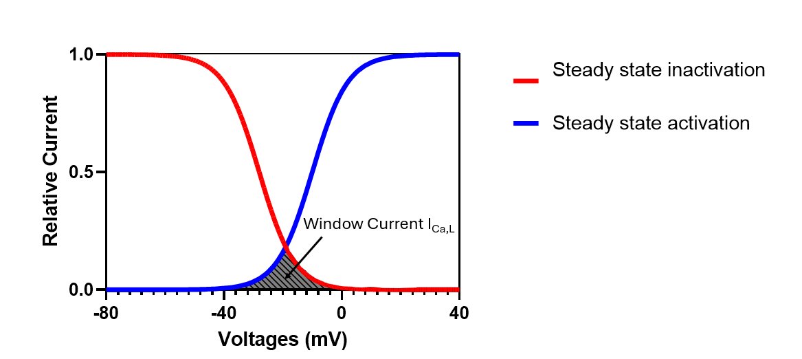
EADs typically occur during the plateau (phase 2) and rapid repolarisation phase (phase 3) of the action potential [16,17]. The pathophysiology involves disruption to the normal balance between inward (depolarising) and outward (repolarising) currents, resulting in the lengthening of the action potential. This delay in repolarisation maintains the membrane potential within a critical ‘window’ voltage range (approximately −30 to 0 mV) (Figure 1) [16,17]. In this zone, L-type calcium channels recover from inactivation yet remain in a voltage range sufficient for reactivation. This allows a subset of channels to reopen (re-activate), generating a secondary inward current that reverses repolarisation through calcium influx. While this EAD is localised within the myocyte, its arrhythmogenic consequence requires electronic transmission via gap junction, allowing neighbouring cells which have recovered from repolarisation to generate its own action potential, which can then propagate through the tissue [16,17].
DADs occur after full repolarisation of the action potential (phase 4) and are therefore observed in diastole [18]. These depolarisations are driven by a process called spontaneous calcium release (SCR) from the sarcoplasmic reticulum (SR), which, by definition occurs independently of an action potential during diastole (contrary to systolic calcium release that is triggered by an action potential) [19]. SCR is initiated when SR calcium ion (Ca2+) content reaches a critical concentration for release, also known as the threshold for SCR. SCR does not occur synchronously throughout the cell but rather begins in one area and propagates across the cell as a wave of calcium release. As this wave of calcium release propagates, it activates the electrogenic Na+/Ca2+ Exchanger (NCX), which removes one intracellular Ca2+ in exchange for three extracellular Na+, producing a net inward transient current (TI) [16]. This inward current underlies the membrane depolarisation that we record as a DAD. The onset of SCR depends on the interplay between SR Ca2+ content (concentration of Ca2+ in the SR) and the threshold for SCR [20]. Factors that increase ryanodine receptor type 2 (RyR2) open probability—such as CaMKII-mediated phosphorylation, certain naturally occurring mutations or caffeine—can lower the threshold for SCR and thereby increase the likelihood of DADs [21]. As we will see later, hypokalaemia can enhance both SR Ca2+ content and RyR2 open probability. The DADs ultimately can trigger a full ectopic action potential (AP) when the DAD amplitude reaches the threshold for activation of voltage-gated Na+ channels [16,18].
If spontaneous SR calcium release occurs during the repolarisation phase rather than in diastole, and the resulting inward NCX current is sufficiently large to overcome the outward potassium currents, the membrane voltage will reverse direction before complete repolarisation, manifesting as phase 3 EADs [16,18]. Late phase 3 EADs represent a less common variant occurring at very negative potentials wherein NCX-mediated depolarisation can trigger action potentials [16,18].
3.2. Re-Entry
Re-entry occurs when a cardiac impulse propagates in a circular pathway around an anatomical or functional obstacle, repeatedly re-exciting tissue that has recovered excitability, rather than extinguishing after depolarizing the heart [22,23]. For this to occur, three fundamental prerequisites must be met. First, a unidirectional conduction block, in which an impulse fails to conduct in one direction (typically due to tissue refractoriness) but can conduct in the opposite direction through an alternate pathway. Second, slow conduction through this alternate pathway, which provides sufficient delay to allow recovery of refractoriness at the site of initial block. Finally, an excitable gap, a temporal window during the re-entry cycle in which tissue downstream of the block site has recovered excitability and can be re-excited when the wavefront returns [22,23]. The excitation wavelength, defined as λ = conduction velocity (CV) × effective refractory period (ERP), is critical to re-entry mechanics because it defines the minimum path length required for a circuit. A reduction of wavelength, through either decreased CV or shortened refractoriness, permits re-entry around smaller anatomical obstacles or purely electrophysiological heterogeneity (functional re-entry) [22,23]. Re-entry can circulate around fixed anatomical barriers (scars, accessory pathways) or around functional cores consisting solely of heterogeneous tissue with disparate refractoriness and conduction properties [22,23]. Brugada syndrome is a clinically relevant example in which re-entry can arise from both functional and structural abnormalities in the right ventricular outflow tract [24].
4. Mechanistic Overview of Hypokalaemia-Induced Arrhythmia
Hypokalaemia employs a multifaceted and integrated approach to destabilise cardiac electrophysiology, promoting arrhythmias through mechanisms that operate across multiple temporal and spatial scales [25]. Rather than a single pathological process, contemporary evidence supports a multi-hit model in which reduced outward current, enhanced inward current, disturbed Ca2+ handling and tissue heterogeneity interact [22]. Within this framework, the relative contribution of each mechanism remains debated, likely to vary with hypokalaemia severity, structural disease and concomitant drug exposure.
4.1. Na+/K+-ATPase Inhibition in Hypokalaemia
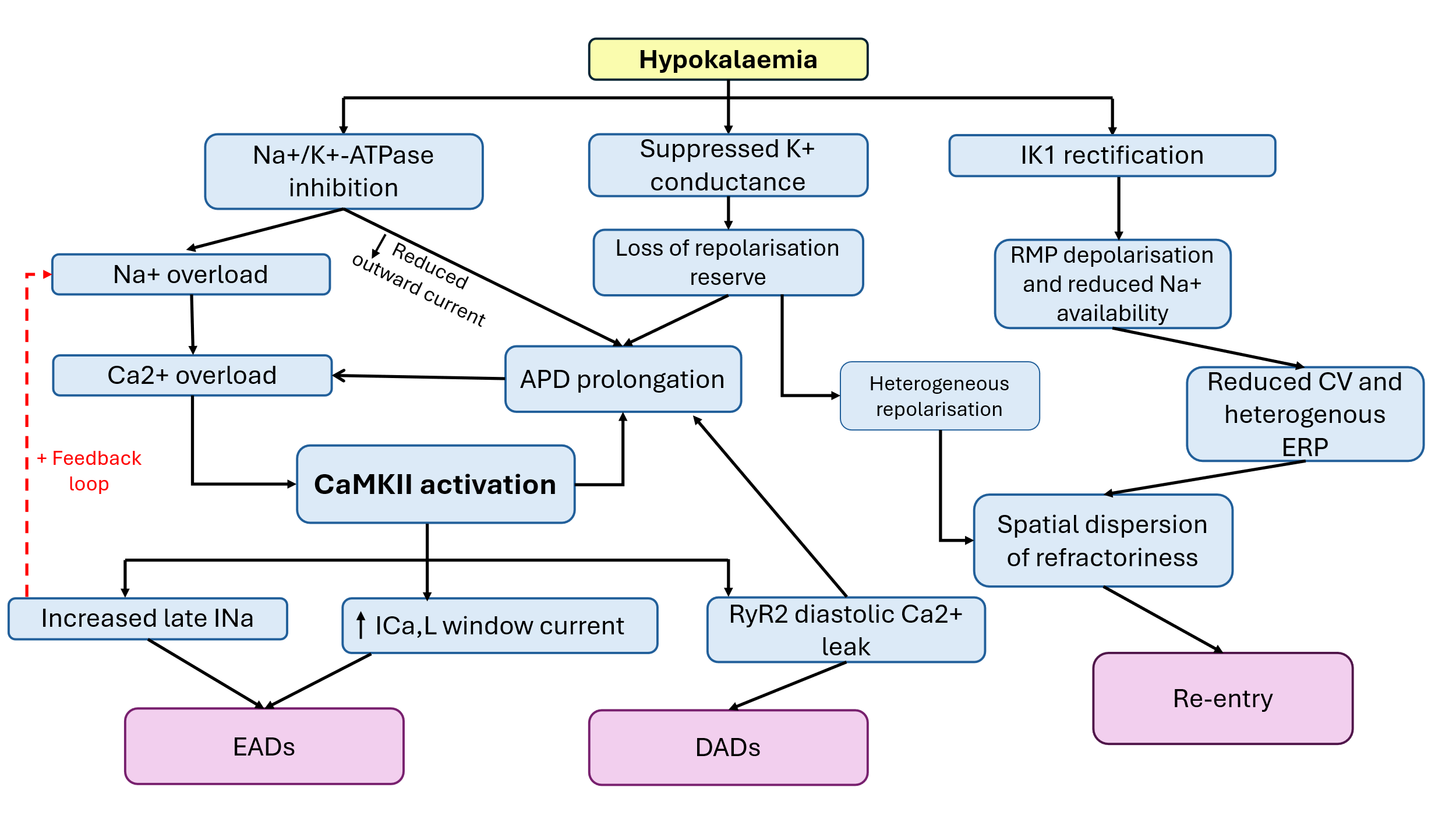
An important consequence of hypokalaemia is inhibition of the Na+/K+ -ATPase (NKA) (Figure 2), one of the suggested main contributors to calcium-mediated arrhythmogenesis [26]. The NKA is the cell’s principal regulator of intracellular Na+ and K+ concentration gradients essential for electrical excitability and calcium handling, functioning by hydrolysing ATP to extrude three Na+ in exchange for two K+ [27]. The action of this pump is essential in maintaining the resting membrane potential and provides the driving force for the action potential [27].
A seminal study by Aronsen and colleagues demonstrated that reducing extracellular [K+] in rat ventricular myocytes from 5 mM to 2.7 mM reduced NKA pump current by approximately 25% at constant membrane potential, further increasing to around 48% when the resting membrane potential (RMP) hyperpolarisation was accounted for [28]. This reduction can be explained through various physiological changes observed in hypokalaemia. First, the reduced extracellular [K+] and increased cytosolic [Na+] reduces the driving force for potassium uptake by the pump, reducing its forward activity [28]. Secondly, the hyperpolarisation of the resting membrane potential itself, which is expected according to the Nernst equation when extracellular [K+] is reduced, opposes the pump’s electrogenic activity, further inhibiting pump current [28].
The inhibition of NKA activity has two main consequences on electrolyte regulation and arrhythmia substrate formation. First, the intracellular Na+ accumulates due to the reduction in Na+ extrusion [26]. This rise in Na+ directly impairs the forward mode of the Na+/Ca2+ exchanger (NCX), which normally removes one intracellular Ca2+ ion in exchange for three Na+ ions, severely impairing the exchanger’s ability to extrude calcium (Figure 2) [26]. Under severe Na+ loading conditions, the NCX may even reverse direction, operating in the reverse mode, importing calcium into the cell and further exacerbating calcium overload [26]. Second, due to the electrogenic characteristic of the pump, its inhibition removes an important repolarising current from the action potential, directly contributing to APD prolongation (Figure 2) [26]. This maintains the membrane potential within the critical voltage window for, L-type Ca2+ channels, directly facilitating the generation of EADs (Figure 2) [26]. Thus, inhibition of NKA could establish the initiating events for the arrhythmogenic cascade of hypokalaemia through two parallel mechanisms: calcium overload and APD prolongation. These attributes prime the cell for triggered activity and activate downstream calcium mediated mechanisms, serving as a foundation.
4.2. Potassium Conductance
While NKA inhibition initiates the arrhythmogenic cascade, a second and complementary mechanism occurs—dysregulation of the repolarising K+ current which functions to terminate an action potential and return the resting membrane potential (Figure 2) [25]. This reduction in potassium current occurs paradoxically despite hyperpolarisation of the resting membrane potential and increased electrochemical driving force for potassium efflux [25]. This dysregulation is instead driven by the suppression of core repolarising potassium currents: the inward rectifier current (IK1), the delayed rectifier currents (IKr and IKs), and the transient outward current (Ito) [25]. Although cumulatively they create a loss of repolarisation reserve, each current has its unique mechanisms by which it is suppressed.
IK1, encoded primarily by Kir2.x genes, is the primary stabiliser of diastolic resting membrane potential and essential in myocyte repolarisation [29]. Mechanistically, low extracellular [K+] increases the stability of intracellular blocking cations (magnesium and polyamines) within the inward rectifier pore, preventing the normal electrostatic displacement of the blockers by extracellular [K+] [25]. Consequently, despite the increased electrochemical driving force for potassium efflux, the outward driving force is reduced [25]. This loss of repolarising current is also a critical contributor to the paradoxical depolarisation of the resting membrane potential which further exacerbates arrhythmogenesis [30].
The delayed inward rectifier currents consist of the IKr and IKs, comprised mainly by the pore forming alpha subunits hERG (encoded by KCNH2) and KvLQT1 (encoded by KCNQ1/KCNE1), and are essential for ventricular repolarisation and determining APD [31,32]. In hypokalaemia IKr is suppressed through accelerated inactivation kinetics and reduced recovery from inactivation, both resulting from low extracellular [K+] [31,32]. Additionally, critical for this review, the surface density of hERG channels is reduced through mechanisms involving conformational changes and endocytic degradation [31,32]. IKs is similarly affected, although the mechanism is more complex. Although IKs shows relative resistance to acute hypokalaemia when expressed alone, its suppression is accelerated parallel to IKr loss [31]. This can be explained by the macromolecular complex formed between hERG and KCNQ1 at the plasma membrane [31]. When endocytic degradation of hERG occurs, the co-assembled KCNQ1 (and KCNE1) are co-ordinately internalised and degraded [31]. Experimentally, it was demonstrated that HEK cells exposed to 0 mM extracellular [K+] for 6 h had reduced hERG-mediated IKr by approximately 55% and KCNQ1-mediated IKs by 76%, compared to only 20% reduction in IKs when KCNQ1 was expressed alone [31]. Importantly, the study also illustrated KCNQ1 delayed the acute reduction of hERG current, attributing a stabilising interaction between the channels, although this was lost following initial hERG degradation [31]. The suppression of both delayed rectifier components prolongs the action potential phase, delaying the return of the membrane potential to rest and maintaining the membrane within the voltage window for L-type calcium channel reactivation [25,31,32].
The transient outward current (Ito), mediated primarily by Kv4.2 and Kv4.3 channels, is responsible for the rapid phase 1 repolarisation (early outward current) following the action potential upstroke and contributes to the plateau voltage. Hypokalaemia has been suggested to slow reactivation of Ito in human atrial cells and further prolong APD [33]. However, compared with evidence for IKr and IK1, the contribution of Ito remains less clearly defined. Therefore, it would be beneficial to further investigate the channel’s role in ventricular hypokalaemia-based arrhythmias.
Overall, the combined suppression of potassium conductance creates a profound loss of repolarisation reserve. Critically, potassium current suppression alone is ineffective at causing EADs in isolated myocytes and rather synergistically acts with the calcium overload initiated by NKA inhibition [26]. As discussed, prolongation of APD maintains the membrane potential within the ‘calcium window’ voltage range, generating EADs. Furthermore, the reduced repolarisation removes the essential suppression of depolarising currents, allowing inward currents to dominate and exert great influence on the membrane potential. The prolongation of repolarisation also allows more time for calcium accumulation in the sarcoplasmic reticulum (SR), increasing the likelihood of spontaneous calcium release. Thus, suppression of repolarising potassium currents acts synergistically with other key mechanisms to establish a highly arrhythmogenic substrate [25].
4.3. Depolarisation of the Resting Membrane Potential
Although low extracellular [K+] is expected to hyperpolarise the membrane potential in accordance with the Nernst equation, at least initially, a paradoxical depolarisation of the resting membrane potential (RMP) is observed under sustained hypokalaemia conditions [30]. This counterintuitive phenomenon can be credited primarily to the non-linear nature of the inward rectifier current (IK1), mediated by Kir2.1 channels [30]. During hypokalaemia, the rectifying properties of the channel at depolarised potentials become pathological, shifting the balance between inward and outward currents at the resting potential, manifesting as depolarisation toward more positive voltages (Figure 2) [30]. An experimental study using human iPSC-derived cardiomyocytes outlined overexpressing Kir2.1 channels manifested as depolarisation of the RMP while the mutant channels, lacking the rectification component, displayed hyperpolarised RMP [30].
This depolarisation of the RMP could carry significant consequences for conduction. By shifting the membrane potential closer to threshold for Na+-channel inactivation, there could be a reduction in the number of Nav1.5 channels available to open during phase 0 of the action potential, thereby decreasing the upstroke velocity and local CV while prolonging the ERP. In spatially heterogeneous tissues, regions of slower conduction are more prone to unidirectional block and supporting re-entry [22,23,30]. As this progression has not been demonstrated fully within a tissue model, it should be interpreted as a mechanistically supported pathway requiring confirmation in multicellular preparations with more mature conduction properties.
4.4. CaMKII Activation and Downstream Phosphorylation
The progressive accumulation of calcium through NKA inhibition and APD prolongation increase the amount of calcium stored in the SR and, therefore, increases systolic calcium release. This sustained systolic elevation triggers activation of calcium/calmodulin-dependent protein kinase II (CaMKII), a protein that integrates calcium signalling with phosphorylation of multiple downstream targets and pathways [26,34,35]. Each of these phosphorylation events further contributes to the destabilisation of cardiac electrophysiology, forming the vicious cycle of calcium overload and arrhythmia formation [26,34,35].
4.4.1. Late Sodium Current Activation
Sodium channels represent the core depolarising current of the action potential and are a major contributor to arrhythmogenesis within the cell. CaMKII phosphorylates Nav1.5 channels at important serine residues which promotes opening of the late sodium current (INa,L) [26]. In addition, CaMKII-dependent phosphorylation has the effect of slowing the recovery of Nav1.5 channels from inactivation, reducing the availability of sodium channels and thereby slowing conduction velocity, a substrate to facilitate re-entry [22,23,36]. This aggravates the progressive loss of repolarisation reserve and potentiates the [Na+] loading of NKA [26] (Figure 2). In animal models, suppressing INa,L through selective inhibitors prevented VT/VF and reduced EADs in isolated myocytes, illustrating their key role in initiating triggered activity [26]. There is also evidence to suggest that within atrial cells, which inherently lack the tubules of the ventricular cells, the main mechanism of arrhythmias in hypokalaemia is through reactivation of non-equilibrium sodium channel rather than the traditional calcium overload, further highlighting their mechanistic importance [37]. These findings demonstrate that the Na+ channel dysfunction is an independent source of inward current and conduction disturbance. However, the dominant Na+ channel mechanism may differ from atrial and ventricular tissue, limiting direct extrapolation.
4.4.2. L-type Calcium Channel Enhancement
CaMKII phosphorylation of the L-type calcium channel (Cav1.2) widens the “calcium window”—the voltage range (−30 to 0 mV) at which the channel can reactivate from inactivation (Figure 2) [26,34,35]. This occurs because phosphorylation slows channel inactivation kinetics (prolonging channel availability) and increases open probability (lowering the threshold for opening). Consequently, Cav1.2 channels pathologically reactivate during the repolarisation phase at normally inactivating voltages, exacerbating inward calcium current and driving EAD formation (Figure 2) [26,34,35].
4.4.3. Ryanodine Receptor Type 2 Activation
The RyR2, the primary calcium release channel of the cardiac sarcoplasmic reticulum, represents a third critical substrate of CaMKII phosphorylation in hypokalaemia arrhythmogenesis (Figure 2) [26,34,35]. This increases the open probability of the channel during diastole and decreases the threshold for SCR, facilitating the onset of spontaneous release of SR-stored Ca2+ and DAD formation (Figure 2). The most consequential feature of the hypokalaemia-CaMKII pathway is the positive feedback loop it establishes as shown in Figure 2 [26,34,35]. The continuous intracellular Na+ and Ca2+ loading establish a vicious cycle in which each component perpetuates the activation of the next, fuelling further afterdepolarisation events.
4.5. Re-Entry and Hypokalaemia
The cascade of electrophysiological changes outlined do not merely trigger isolated premature beats but rather establish a tissue level substrate permissive for re-entrant arrhythmias. The core mechanisms which facilitate this likely involve simultaneously creating spatial heterogeneity in refractoriness and CV, thereby reducing the excitation wavelength, all of which are essential for re-entry initiation and maintenance [12,25].
The suppression of repolarising potassium currents in hypokalaemia is not uniformly distributed across cardiac tissue [12,15,25]. Heterogeneous expression of hERG, IK1, and Ito channels across the ventricular wall and between cardiac chambers creates regions with disparate repolarisation kinetics and action potential durations (Figure 2) [18,25]. Consequently, adjacent tissue regions may recover excitability at markedly different rates, establishing the necessary spatial gradient in refractoriness that permits unidirectional conduction block—a prerequisite for re-entry [12,25].
Although APD prolongation would be expected to increase the absolute refractory period, experimental evidence demonstrates that changes in the ERP are often heterogeneous and depend on the resting membrane potential [15,30,37]. More importantly, depolarisation-induced reductions in Na+ channel ability and consequent conduction slowing, together with spatial differences in ADP/ERP, could play a major role in creating the conditions necessary for unidirectional block and re-entry.
4.6. Age-Related Changes
Experimental work in Langendorff-perfused rat hearts has illustrated the profound effect of age-associated fibrosis in lowering the threshold for EAD-mediated ventricular arrhythmias in angiotensin II-induced oxidative stress and, critically, hypokalaemia [38]. In this model, the aged ventricles exhibited interstitial fibrosis and reduced connexin-43 density, with most developing EAD-mediated VF secondary to a reduction in extracellular potassium compared to young hearts which consistently observed sinus rhythm or required a longer stress exposure before VF [38]. However, patch-clamp recordings from isolated myocytes illustrated a broadly similar tendency for both young and old cells to promote EADs and triggered activity, implying that cellular electrophysiological remodelling alone does not explain the pronounced age-dependent differences in VF susceptibility [38]. Rather, the age-related changes between cells alter source-sink relationships such that afterdepolarisations that would normally be suppressed by neighbouring cells can escape, propagate as premature beats and degenerate into VF [38]. This is consistent with a “multi-hit” paradigm, where structural remodelling is the first hit and reduced repolarisation reserve the second to induce VF [38].
This finding integrates closely with the mechanisms previously described in this review, with intracellular calcium overload acting as the “second hit” necessary to initiate arrhythmias alongside reduced repolarisation reserve. This convergence of structural and ionic remodelling may outline how more modest hypokalaemia can be particularly malignant in elderly patients with fibrotic cardiomyopathy and highlights the need for therapies that address both components of the substrate.
4.7. Integrated Ionic and Signalling Mechanisms
From the available evidence, the arrhythmogenic consequence of hypokalaemia is likely an integrated network rather than a signal-channel disorder. Reduced NKA activity promotes intracellular Na+ accumulation and impaired Ca2+ extrusion through the NCX, suppression of IK1, IKr and IKs and potentially Ito reduce the repolarisation reserve and activation of CaMKII amplifies inward current and spontaneous release. These processes converge to promote triggered activity, conduction slowing and spatially heterogenous recovery of excitability.
Within this network, evidence strongly supports NKA inhibition as a critical pathway for EAD formation and arrhythmogenesis. However, these findings alone do not establish NKA inhibition as the universal initiating or dominant mechanism of hypokalaemia-induced arrhythmia, as the relative contributions of individual pathways are likely to vary by biological context. Loss of IKr may have greater importance when repolarisation reserve is already compromised, such as exposure to hERG-blocking drugs. Conversely, CaMKII signalling may contribute more prominently in settings characterised by pre-existing Na+ and Ca2+ loading, including heart failure [39]. Hypokalaemia should therefore be regarded as a context-dependent, multi-hit process in which altered repolarisation, intracellular Ca2+ handling and the tissue substrate interact to determine arrhythmic risk.
5. The Critical Role of hERG Channel Stability
The loss of hERG-mediated IKr in hypokalaemia represents a multifactorial disruption to cardiac repolarisation that extends beyond simple suppression of one among several outward currents. Fundamentally, hERG dysfunction differs from other repolarising current such as IK1 or Ito, involving both acute functional changes and chronic remodelling of hERG trafficking machinery [9,10]. This temporal multiplicity creates a distinctive pathophysiological scenario where acute serum potassium normalisation may restore electrochemical gradients and transiently reverse IKr suppression yet fails to address the underlying remodelling of hERG stability and trafficking [9,10,40]. This mechanistic disconnect may explain the clinical observation that conventional potassium replacement is incomplete in eliminating arrhythmia risk in hypokalaemia patients [41]. Therefore, a more profound understanding of the unique structural and functional biology of the hERG channel is required.
5.1. Historical Identification
The human ether-à-go-go-related gene (KCNH2), widely known as hERG (Kv11.1), was first identified in 1994 by Warmke and Ganetzky [42,43] and, over the decades since its discovery, has been identified to play a major role in the action potential by conducting the rapid component of the delayed rectifier K+ current [44]. With the concurrent identification of the complementary slow component, IKs [45], the distinctive properties of hERG, particularly its rapid inactivation followed by slow deactivation (commonly recognised as the characteristic hook-tail or inward rectification), highlighted is physiological importance [46]. This inward rectification enables hERG to conduct maximal repolarising current during late phases of the action potential when the membrane potential becomes negative, making hERG the primary repolarisation reserve current in cardiomyocytes [43].
5.2. Structural Characterisation
5.2.1. Tetrameric Architecture and Domain Organisation
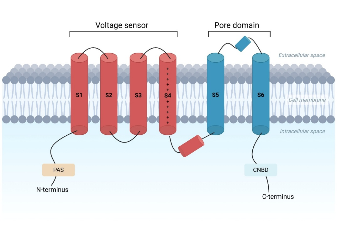
As a member of the voltage-gated K+ channel family (Kv), hERG is a tetrameric protein composed of four identical α-subunits [43,47]. Each subunit contains six transmembrane segments (S1–S6), where S1–S4 form the voltage-sensing domain (VSD), and S5–S6 constitute the pore domain (PD), consisting of the selectivity filter (SF) responsible for K+ specificity (Figure 3) [43,47]. Within the VSD, S4 plays a vital role, being the primary voltage sensor through its positively charged residues that respond to the transmembrane potential changes (Figure 3) [48]. A distinctive feature of KCNH family channels, including hERG, is the presence of two critical cytoplasmic domains: an N-terminal Per-Arnt-Sim (PAS), essential for channel stability when present, and a C-terminal cyclic nucleotide-binding domain (CNBD) that regulates gating kinetics and deactivation (Figure 3) [43,49].
Early electrophysiological and mutagenesis studies identified key functional residues such as Y652 and F656 within the pore as high-affinity drug-binding sites [51,52] and, critically, F627 in the selectivity filter as the primary mediator of rapid C-type inactivation [43,53].
5.2.2. Cryo-EM and Mechanistic Understanding
Our understanding of hERG’s architecture was fundamentally revolutionized in 2017 when Wang and MacKinnon published the first high-resolution cryo-electron microscopy (cryo-EM) structure, resolved at 3.8 Å with the channel in the open, conducting state [47]. This landmark study utilized a truncated, hERGT, construct maintaining near-native gating kinetics (minimal +5 mV shift in voltage-dependent inactivation) while improving sample preparation [47,54]. This revealed several architectural features with direct mechanistic implications.
One of the key findings was how, unlike domain-swapped Kv channels, hERG exhibits a non-domain-swapped arrangement where each voltage-sensing domain is packed directly against the pore domain of the same subunit [47]. This intimate coupling is enabled by the remarkably short S4–S5 linker and the close apposition of S4 to the C-terminal end of S6, together allowing voltage-sensor movement to be transmitted directly to the activation gate [47,55,56]. Unlike classical domain-swapped Kv channels, hERG relies on direct S4–S6 interactions within the same subunit rather than a long lever, potentially contributing to its atypical gating kinetics [47].
The cryo-EM structure also revealed four deep, hydrophobic pockets extending into the central cavity below the selectivity filter, with Y652 and F656 asymmetrically positioned on the inner surfaces of these pockets [47]. The central cavity possesses an unusually small volume and an extreme negative electrostatic potential, working in tandem to create the ideal drug binding environment [47,56]. This structural susceptibility becomes clinically relevant in hypokalaemia, where many potentially anti-arrhythmic drugs have an affinity for hERG blockade and QT prolongation [51,52].
5.3. Gating Kinetics
The physiological significance of hERG as a major contributor to repolarisation reserve current is fundamentally determined by its distinctive gating kinetics: slow activation, rapid inactivation, rapid recovery from inactivation and slow deactivation [44,48]. This specific kinetic profile enables hERG to preserve the depolarising currents of the action potential while simultaneously generating the inward tail currents essential for repolarisation.
5.3.1. Activation, Inactivation and Recovery
hERG activation is kinetically slow relative to other cardiac K+ channels [44,46,57]. This slow activation reflects conformational changes within the VSD that are coupled to the pore via interaction between the S4–S5 linker and the cytoplasmic end of S6 [47,54]. During the action potential plateau phase, hERG channels open gradually whilst simultaneously undergoing rapid C-type inactivation [43,44,48,57]. This dual kinetics ensures minimal outward current during the plateau, allowing the inward L-type calcium current (ICa,L) to dominate unopposed and preserve myocardial contractility [44,46]. The rapid inactivation is mediated by conformational changes in the selectivity filter, particularly the asymmetrical positioning of F627, which rapidly occludes the conduction pathway [53].
Recovery from inactivation is rapid and voltage dependent, occurring in a few milliseconds during the repolarisation phase [43,44,49]. This strong voltage dependence reflects the mechanism of C-type inactivation, with more negative potentials progressively destabilising the inactivated conformation and allowing rapid gate reversal. Rapid recovery from inactivation is essential for repolarisation initiation. When the membrane potential rapidly becomes negative, the large cohort of channels trapped in the inactivated state rapidly recover to the open state. This produces the characteristic “hook” of the hERG tail current—a large, brief K+ tail current that initiates rapid repolarisation [43,44,46,48]. The hook component is particularly relevant to ventricular phase 3 repolarisation and thus to T-wave morphology on the electrocardiogram and, importantly, preventing dangerous EADs that occur with repolarisation delay [44,45,47].
5.3.2. Slow Deactivation
hERG deactivation is characteristically slow and is thought to be regulated by cooperative interactions between the N-terminal, PAS domain and C-terminal CNBD, together forming the PAS/CNBD complex that contacts the VSD-pore interface [49,58]. Deletion of the eag region markedly accelerates deactivation (approximately 5-fold), and exogenous PAS fragments or mutations within the CNBD that disrupt this complex similarly alter deactivation kinetics, demonstrating their central role in gating kinetics [49,58]. Slow deactivation is critical for hERG’s role as a repolarisation reserve. During late repolarisation, hERG remains open and able to conduct sustained outward K+ current for prolonged periods during late repolarisation and early diastole. This prolonged tail current provides the continuous repolarising drive that returns the membrane potential to resting potential, helping to prevent dangerous arrhythmias [43,44,46,57].
5.4. Dysregulation of hERG in Low Extracellular [K+]
The dependence of hERG on extracellular [K+] was recognised shortly after its initial cloning and was initially attributed to the rectification properties intrinsic to the channel’s rapid inactivation kinetics, specifically, the voltage-dependent trapping of the channel in a non-conducting, inactivated state [46]. However, over the past two decades, this understanding has undergone a fundamental shift, driven by the discovery of a coordinated dysregulation of plasma membrane (PM) hERG expression through accelerated endocytosis and protein degradation [9]. This system presents a second, independent mechanism of hERG current loss, operating in parallel with the acute kinetic suppression [32].
5.4.1. Acute Conformational Destabilization of the Selectivity Filter
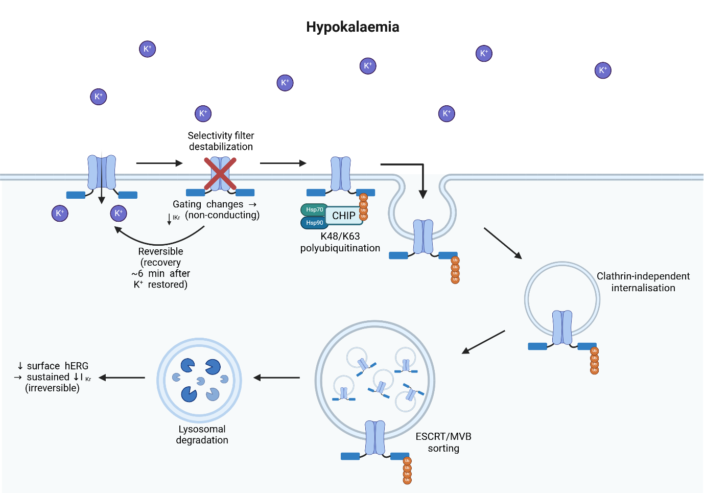
The initial mechanism by which low extracellular [K+] initiates hERG internalisation involves conformational destabilisation of the mature channel protein (Figure 4). Massaeli and colleagues demonstrated through site directed mutagenesis and functional analysis that specific residues within the hERG selectivity filter are critical for the K+-dependent stabilisation of the hERG protein [10]. At physiological extracellular [K+] (5 mM), these residues can maintain a stable, plasma membrane (PM) bound channel configuration. However, at low extracellular [K+], the selectivity filter loses K+ coordination geometry, resulting in conformation destabilisation and a non-conducting state of the channel (Figure 4) [10]. Critically, this acute suppression is reversible with recovered hERG current in ~6 min of restoring normal low extracellular [K+] [10].
Parallel to the conformational destabilisation, low extracellular [K+] disrupts the gating kinetics properties previously discussed [32]. The compromised selectivity filter geometry accelerates inactivation and slows recovery from inactivation, producing functional IKr loss before trafficking mechanisms engage [32]. This acute kinetic suppression compounds the electrochemical gradient loss from hypokalaemia itself [32].
5.4.2. Chronic Internalisation Cascade: Conformational Recognition and Polyubiquitination
Under chronic hypokalaemia, the acute conformational destabilization is recognised as abnormal by the cellular quality control machinery, initiating an irreversible endocytic cascade that removes hERG from the plasma membrane and targets it for degradation (Figure 4) [9,10]. While early work indicated monoubiquitylation in hypokalaemia-induced hERG down-regulation, more recent studies using direct immunodetection with polyubiquitin-specific antibodies have demonstrated the critical role of polyubiquitination [60,61]. Under low extracellular [K+], the K+ depletion-induced conformational change in mature hERG is detected by CHIP E3 ubiquitin ligase (carboxyl terminus of Hsc70-interacting protein) in complex with Hsp70/Hsp90 chaperone proteins, components of the peripheral control (QC) system [61]. This triggers K48- and K63-linked polyubiquitination, serving as the molecular signal for internalisation. This polyubiquitination occurs within ~40 min of acute low extracellular [K+] exposure, demonstrating the rapidity of the initial trigger mechanism [61].
5.4.3. Clathrin-Independent Internalisation and Degradation
Upon polyubiquitination, current evidence suggests hERG is internalised via a clathrin-independent pathway, likely involving direct ESCRT (Endosomal Sorting Complexes Required for Transport) recruitment or other non-classical endocytic routes (Figure 4) [62]. Internalised hERG is then sorted into the multivesicular Body (MVB) pathway and targeted for lysosomal degradation [62]. The ubiquitinated channel is recognized by the ESCRT machinery, sorted into intraluminal vesicles of the MVB, and subsequently degraded by lysosomal [62], delaying the restoration of surface IKr until replacement channels are synthesised and trafficked.
5.4.4. Recycling of hERG
Internalised hERG channels are not inevitably committed to lysosomal degradation. In hERG-expressing HEK293 cells, internalised channels were returned to plasma membrane through a Rab11-associated pathway, while disruption of Rab11 function delayed the recovery of hERG surface expression and current following restoration of extracellular [K+] [40]. Moreover, recovery persisted during inhibition of protein synthesis, supporting recycling of an intracellular pool rather than hERG synthesis [40].
These findings indicate that hERG surface density reflects a dynamic balance between internalisation, recycling and degradation. However, it remains unclear how low extracellular [K+] regulate the shift between Rab11-dpeendent recycling and ubiquitin-directed lysosomal sorting. Future studies should therefore define the concentration dependence, time course and functional consequences of hERG recycling during hypokalaemia and subsequent [K+] normalisation.
5.4.5. Translational Evidence Gaps
Acute suppression of hERG-mediated IKr by low extracellular [K+] is well supported by electrophysiological studies while sustained reductions in hERG surface expression have been demonstrated principally in heterologous expression systems, isolated cardiomyocytes and experimental rabbit models. Furthermore, many trafficking experiments employed extreme conditions, including 0 mmol/L extracellular [K+] for several hours, to maximise internalisation. These protocols exceed the severity of most clinical hypokalaemia and may overestimate the magnitude and irreversibility of hERG loss.
Finally, the contribution of hERG trafficking to human arrhythmogenesis remains uncertain because of hypokalaemia’s simultaneous alteration of several pathways as previously discussed. Studies in mature human cardiomyocytes are therefore required to establish the concentration dependence, time course and reversibility of hERG surface loss and functional recovery under clinically relevant hypokalaemia. Linking these changes to IKr, action potential duration and triggered activity will also be necessary before altered hERG trafficking can be regarded as an established clinical mechanism rather than a compelling experimental contributor.
6. Pharmacological Strategies and Potential Therapies
6.1. Potassium Supplementation
Despite these developments in the mechanistic understanding of hypokalaemia-induced arrhythmia, current management remains fundamentally limited to potassium supplementation. Recent evidence does confirm that potassium repletion is effective for arrhythmia prevention, particularly in high-risk populations, demonstrating that targeting high normal K+ (4.5–5.0 mmol/L) reduced arrhythmia burden by 24% in implantable cardioverter-defibrillator (ICD) patients [63]. However, this repletion alone addresses only the acute electrochemical phase of hypokalaemia-induced dysfunction and has no preventative effect on the chronic structural remodelling of hERG channels or the calcium-mediated mechanisms that operate in parallel.
The mechanistic complexity of hypokalaemia creates multiple therapeutic targets rather than a single solution. Unlike traditional thinking that frames hypokalaemia management as a K+ problem, modern understanding reveals that multimodal intervention targeting different mechanisms simultaneously likely provides superior arrhythmia prevention compared to a simpler approach. This section reviews the current pharmacological landscape for hypokalaemia-induced arrhythmia prevention, discussing emerging agents.
6.2. hERG-Blocking Drugs during Hypokalaemia
A critical paradox defines hypokalaemia pharmacotherapy as many antiarrhythmics block hERG channels, the same channel already suppressed by hypokalaemia. The most striking evidence for this comes from Pezhouman and colleagues, demonstrating that dofetilide (a class III antiarrhythmic drug) worsens hypokalaemia-induced arrhythmia in isolated rabbit hearts [26]. At K+ 2.7mmol/L, around 50% of cardiomyocytes developed VT/VF compared to 80% in a population treated with 1 μM dofetilide, with additional prolongation of APD observed [26]. Dofetilide achieves this by adding additional hERG blockade through preferential binding to inactivated state of depolarisation. Furthermore, other studies reported that dofetilide, and other similar class III antiarrhythmics, could also have a synergistic effect of activating INa,L through phosphoinositide 3-kinase inhibition [64]. These effects compound with the additional properties of hypokalaemia to promote EAD formation and arrhythmia.
6.3. Preclinical Emerging Agents
The recognition that arrhythmia in hypokalaemia involves intracellular Na+ and Ca2+ loading in addition to reduced repolarisation reserve has identified several potential therapeutic targets including CaMKII, INa and RyR2. However, most of these approaches remain preclinical, deriving principally from isolated cardiomyocytes and Langendorff-perfused animal hearts [26,65].
6.3.1. CaMKII and late Na+ Current Inhibition
In isolated rabbit and rat hearts, the CaMKII inhibitor KN-93 prevented hypokalaemia induced EADs and VT/VF, while its inactive analogue KN-92 was ineffective. The more selective CaMKII-inhibitory peptide AIP also suppressed EADs in isolated rabbit ventricular myocytes, supporting CaMKII-dependent signalling as a mechanistic contributor rather than attributing the effects solely to KN-93 [26]. In the same study, the INa,L inhibitor GS-967 suppressed EADs in isolated ventricular myocytes and prevented VT/VF in rabbit hearts exposed to severe hypokalaemia, supporting INa,L as a further potential target [26].
Nevertheless, this evidence should be interpreted cautiously considering the small number of isolated hearts and cardiomyocytes tested with each inhibitor, and the experiments assessing short term prevention in animal preparations rather than established arrhythmia in vivo. Furthermore, the therapeutics involved are largely research tools, with KN-93 having pharmacological actions beyond selective CaMKII inhibition [66]. Consequently, these findings establish experimental proof of concept but do not demonstrate that CaMKII or INa,L inhibition is safe or effective in hypokalaemia.
6.3.2. RyR2-Directed Therapy
RyR2 modulation represents a complementary approach directed at spontaneous SR Ca2+ release. In Langendorff-perfused mouse hearts, pretreatment with dantrolene reduced the overall burden of hypokalaemia-induced ventricular arrhythmias by 94%, although afterdepolarisations persisted and VT remained detectable in some hearts [65]. Dantrolene also failed to prevent action-potential prolongation and afterdepolarisations consistently in isolated murine and canine cardiomyocytes, suggesting that RyR2 modulation may reduce arrhythmia burden without correcting the wider loss of repolarisation reserve [65].
6.3.3. The Mechanistic Gap
Collectively, these findings indicate that CaMKII, INa,L and RyR2 are potential therapeutic targets for managing hypokalaemia-induced arrhythmia. However, there is currently no evidence that any medications reverse hypokalaemia-induced hERG loss which can be considered a ‘second hit’ alongside other pathological processes and an important contributor to loss of repolarisation reserve. A multi-mechanistic strategy targeting the cellular processes discussed, while also maintaining hERG, is therefore likely to provide superior protection, especially in the context of structural remodelling.
6.4. Translational Gap
A substantial gap separates mechanistic evidence from clinical application. Current potassium replacement is supported by clinical practice and human outcome data while the therapies discussed are mostly experimental strategies. Progression towards clinical testing will require efficacy under clinically relevant degrees and durations of hypokalaemia, confirmation in mature human cardiomyocytes, multicellular cardiac preparations and simultaneous assessment of the ion channels involved in hypokalaemia.
7. Conclusions: Deficiencies in Current Understanding
Current management of hypokalaemia-induced arrhythmia remains inadequate despite detailed mechanistic understating of the underlying electrophysiological cascade. At present, there is no antiarrhythmic therapy that has proven safely to counteract the pro-arrhythmic effects of hypokalaemia, and clinical practice largely relies on recognising this imbalance once it has occurred and correcting serum K+ or treating arrhythmias when they manifest. Moreover, potassium supplementation addresses only the acute electrochemical disturbance and, although sustained normalisation of K+ can gradually reverse many electrophysiological abnormalities, it does not target the chronic structural remodelling of hERG trafficking, calcium handling and the fibrotic substrate that amplifies EAD-mediated arrhythmogenesis in aged hearts. Given the high prevalence of hypokalaemia in cardiovascular disease patients and its strong association with malignant arrhythmias, there is a clear need to develop targeted therapies that can stabilise hypokalaemia-prone, structurally remodelled hearts, rather than relying solely on episodic potassium supplementation. Bearing this in mind, three major gaps in our current knowledge and therapeutic strategies can be identified.
First, the molecular basis by which hypokalaemia triggers hERG internalisation and degradation remains incompletely characterized. Whilst the conformational destabilisation of the selectivity filter and subsequent polyubiquitination have been identified as key steps, the precise molecular events are not fully understood. Understanding how these components integrate with acute suppression of IKr and with broader cellular pathways will be essential for designing intervention that can stably preserve repolarisation reserve during recurrent hypokalaemia episodes.
Second, the methods of pharmacological hERG trafficking rescue remain poorly understood. A structure-activity relationship linking specific therapeutics to a measurable restoration of surface hERG without off-target ion channel block has not yet been established and compounds being tested in the context of hypokalaemia are largely confined to preclinical models. Discovering potential agents, particularly those that influence calcium handling, will be critical for the rational design of multi-target therapies that avoid pro-arrhythmic liabilities.
Third, and finally, the applicability of in vitro cellular models to chronic, patient-relevant hypokalaemia is limited. Most mechanistic studies employ acute hypokalaemia (minutes to hours) in isolated myocytes or heterologous expression systems, whereas clinical hypokalaemia develops over days and occurs in structurally remodelled myocardium with heterogeneous ion channel expression and impaired intercellular coupling. Human iPSC-derived cardiomyocytes and aged, fibrotic whole heart preparations offer complementary platforms that more faithfully recapitulate human electrophysiology and the “multi-hit” interplay between ionic and structural stress, but they have not been systematically used to model chronic hypokalaemia. Future work should focus on integrating these models to define how long-term hypokalaemia reshapes hERG trafficking axis, CaMKII signalling and fibrotic modelling, and on developing multi-mechanistic strategies capable of safely stabilising cardiac electrophysiology during hypokalaemia beyond simple electrolyte replacement.
Author Contributions
J.J.: writing—original draft preparation; K.K.: supervision, conceptualisation. All authors have read and agreed to the published version of the manuscript.
Funding
This research received no external funding.
Institutional Review Board Statement
Not applicable.
Informed Consent Statement
Not applicable.
Data Availability Statement
Not applicable.
Conflicts of Interest
The authors declare no conflicts of interest.
Use of AI and AI-Assisted Technologies
No AI tools were utilized for this paper.
References
- 1.
Kjeldsen, K. Hypokalemia and Sudden Cardiac Death. Exp. Clin. Cardiol. 2010, 15, e96–e99.
- 2.
Bhargava, J.; Viswanathan, S. Incidence, Risk Factors and Prognosis of Hypokalaemia in Patients with Normokalaemia at Hospital Admission. Indian J. Endocrinol. Metab. 2023, 27, 537–543. https://doi.org/10.4103/ijem.ijem_159_23.
- 3.
Abensur Vuillaume, L.; Ferreira, J.P.; Asseray, N.; et al. Hypokalemia Is Frequent and Has Prognostic Implications in Stable Patients Attending the Emergency Department. PLoS ONE 2020, 15, e0236934. https://doi.org/10.1371/journal.pone.0236934.
- 4.
Matsushita, K.; Sang, Y.; Yang, C.; et al. Dyskalemia, Its Patterns, and Prognosis among Patients with Incident Heart Failure: A Nationwide Study of US Veterans. PLoS ONE 2019, 14, e0219899. https://doi.org/10.1371/journal.pone.0219899.
- 5.
Lin, Z.; Wong, L.; Cheung, B. Diuretic-Induced Hypokalaemia: An Updated Review. Postgrad. Med. J. 2022, 98, 477–482.
- 6.
Ferreira, J.P.; Butler, J.; Rossignol, P.; et al. Abnormalities of Potassium in Heart Failure: JACC State-of-the-Art Review. J. Am. Coll. Cardiol. 2020, 75, 2836–2850. https://doi.org/10.1016/j.jacc.2020.04.024.
- 7.
Correia, A.L.; Marques, M.G.; Alves, R. Gitelman Syndrome—A New Mutation in the SLC12A3 Gene. Nefrología 2022, 42, 490–492. https://doi.org/10.1016/j.nefro.2021.01.005.
- 8.
Paltiel, O.; Salakhov, E.; Ronen, I.; et al. Management of Severe Hypokalemia in Hospitalized Patients. Arch. Intern. Med. 2001, 161, 1089–1095. https://doi.org/10.1001/archinte.161.8.1089.
- 9.
Guo, J.; Massaeli, H.; Xu, J.; et al. Extracellular K+ Concentration Controls Cell Surface Density of IKr in Rabbit Hearts and of the HERG Channel in Human Cell Lines. J. Clin. Investig. 2009, 119, 2745–2757. https://doi.org/10.1172/jci39027.
- 10.
Massaeli, H.; Guo, J.; Xu, J.; et al. Extracellular K+ Is a Prerequisite for the Function and Plasma Membrane Stability of HERG Channels. Circ. Res. 2010, 106, 1072–1082.
- 11.
Zhang, K.; Yang, B.; Li, B. Translational Toxicology and Rescue Strategies of the hERG Channel Dysfunction: Biochemical and Molecular Mechanistic Aspects. Acta Pharmacol. Sin. 2014, 35, 1473–1484.
- 12.
Tse, G.; Li, K.H.C.; Cheung, C.K.Y.; et al. Arrhythmogenic Mechanisms in Hypokalaemia: Insights from Pre-Clinical Models. Front. Cardiovasc. Med. 2021, 8, 620539. https://doi.org/10.3389/fcvm.2021.620539.
- 13.
Skogestad, J.; Aronsen, J.M.; Rourke, B.C.; et al. Hypokalemia-Induced Arrhythmias and Heart Failure: New Insights and Implications for Therapy. Front. Physiol. 2018, 9, 1500. https://doi.org/10.3389/fphys.2018.01500.
- 14.
Collins, A.J.; Pitt, B.; Reaven, N.; et al. Association of Serum Potassium with All-Cause Mortality in Patients with and without Heart Failure, Chronic Kidney Disease, and/or Diabetes. Am. J. Nephrol. 2018, 46, 213–221.
- 15.
Osadchii, O.E. Mechanisms of Hypokalemia-Induced Ventricular Arrhythmogenicity. Fundam. Clin. Pharmacol. 2010, 24, 547–559. https://doi.org/10.1111/j.1472-8206.2010.00835.x.
- 16.
Lerman, B.; Markowitz, S.; Cheung, J.; et al. Ventricular Tachycardia Due to Triggered Activity: Role of Early and Delayed Afterdepolarisations. JACC Clin. Electrophysiol. 2024, 10, 379–401. https://doi.org/10.1016/j.jacep.2023.10.033.
- 17.
Weiss, J.N.; Garfinkel, A.; Karagueuzian, H.S.; et al. Early Afterdepolarizations and Cardiac Arrhythmias. Heart Rhythm. 2010, 7, 1891–1899. https://doi.org/10.1016/j.hrthm.2010.09.017.
- 18.
Qu, Z.; Xie, L.H.; Olcese, R.; et al. Early Afterdepolarizations in Cardiac Myocytes: Beyond Reduced Repolarization Reserve. Cardiovasc. Res. 2013, 99, 6–15. https://doi.org/10.1093/cvr/cvt104.
- 19.
Venetucci, L.; Trafford, A.; O’Neill, S.; et al. The Sarcoplasmic Reticulum and Arrhythmogenic Calcium Release. Cardiovasc. Res. 2008, 77, 285–292.
- 20.
Venetucci, L.; Trafford, A.; Eisner, D. Increasing Ryanodine Receptor Open Probability Alone Does Not Produce Arrhythmogenic Calcium Waves: Threshold Sarcoplasmic Reticulum Calcium Content Is Required. Circ. Res. 2007, 100, 105–111.
- 21.
Kashimura, T.; Briston, S.J.; Trafford, A.W.; et al. In the RyR2 R4496C Mouse Model of CPVT, β-Adrenergic Stimulation Induces Ca Waves by Increasing SR Ca Content and Not by Decreasing the Threshold for Ca Waves. Circ. Res. 2010, 107, 1483–1489. https://doi.org/10.1161/circresaha.110.227744.
- 22.
Antzelevitch, C.; Burashnikov, A. Overview of Basic Mechanisms of Cardiac Arrhythmia. Card. Electrophysiol. Clin. 2011, 3, 23–45.
- 23.
Tse, G. Mechanisms of Cardiac Arrhythmias. J. Arrhythm. 2016, 32, 75–81.
- 24.
Bueno-Orovio, A.; Cherry, E.M.; Evans, S.J.; et al. Basis for the Induction of Tissue-Level Phase-2 Reentry as a Repolarization Disorder in the Brugada Syndrome. BioMed Res. Int. 2015, 2015, 197586. https://doi.org/10.1155/2015/197586.
- 25.
Weiss, J.N.; Qu, Z.; Shivkumar, K. Electrophysiology of Hypokalemia and Hyperkalemia. Circ. Arrhythm. Electrophysiol. 2017, 10, e004667. https://doi.org/10.1161/circep.116.004667.
- 26.
Pezhouman, A.; Singh, N.; Song, Z.; et al. Molecular Basis of Hypokalaemia-Induced Ventricular Fibrillation. Circulation 2015, 132, 1528–1537.
- 27.
Bueno-Orovio, A.; Sánchez, C.; Pueyo, E.; et al. Na/K Pump Regulation of Cardiac Repolarisation: Insights from a Systems Biology Approach. Pflugers Arch. Eur. J. Physiol. 2014, 466, 183–193. https://doi.org/10.1007/s00424-013-1330-0.
- 28.
Aronsen, J.M.; Skogestad, J.; Lewalle, A.; et al. Hypokalaemia Induces Ca2+ Overload and Ca2+ Waves in Ventricular Myocytes by Reducing Na+, K+-ATPase α2 Activity. J. Physiol. 2015, 593, 1509–1521. https://doi.org/10.1113/jphysiol.2014.279034.
- 29.
Sun, Y.; Timofeyev, V.; Dennis, A.; et al. A Singular Role of IK1 Promoting the Development of Cardiac Automaticity during Cardiomyocyte Differentiation by IK1–Induced Activation of Pacemaker Current. Stem Cell Rev. Rep. 2017, 13, 631–643. https://doi.org/10.1007/s12015-017-9745-1.
- 30.
Lv, J.; Jiang, Y.; Zeng, J.; et al. Kir2.1 Channel Contributes to Hypokalaemia-Induced Abnormal Pacemaker Activities of Cardiomyocytes with Inward Rectification. Front. Biosci. 2025, 30, 39879. https://doi.org/10.31083/fbl39879.
- 31.
Guo, J.; Wang, T.; Yang, T.; et al. Interaction between the Cardiac Rapidly (IKr) and Slowly (IKs) Activating Delayed Rectifier Potassium Channels Revealed by Low K+-Induced hERG Endocytic Degradation. J. Biol. Chem. 2011, 286, 34664–34674. https://doi.org/10.1074/jbc.m111.253351.
- 32.
Melgari, D.; Du, C.; El Harchi, A.; et al. Suppression of the hERG Potassium Channel Response to Premature Stimulation by Reduction in Extracellular Potassium Concentration. Physiol. Rep. 2014, 2, e12165. https://doi.org/10.14814/phy2.12165.
- 33.
Firek, L.; Giles, W.R. Outward Currents Underlying Repolarization in Human Atrial Myocytes. Cardiovasc. Res. 1995, 30, 31–38. https://doi.org/10.1016/0008-6363(95)00014-3.
- 34.
Li, L.; Satoh, H.; Ginsburg, K.; et al. The Effect of Ca2+-Calmodulin-Dependent Protein Kinase II on Cardiac Excitation-Contraction Coupling in Ferret Ventricular Myocytes. J. Physiol. 1997, 501, 17–31. https://doi.org/10.1111/j.1469-7793.1997.017bo.x.
- 35.
Swaminathan, P.D.; Purohit, A.; Hund, T.J.; et al. Calmodulin-Dependent Protein Kinase II: Linking Heart Failure and Arrhythmias. Circ. Res. 2012, 110, 1661–1677. https://doi.org/10.1161/circresaha.111.243956.
- 36.
Wagner, S.; Dybkova, N.; Rasenack, E.C.L.; et al. Ca2+/Calmodulin-Dependent Protein Kinase II Regulates Cardiac Na+ Channels. J. Clin. Investig. 2006, 116, 3127–3138. https://doi.org/10.1172/jci26620.
- 37.
Tazmini, K.; Frisk, M.; Lewalle, A.; et al. Hypokalemia Promotes Arrhythmia by Distinct Mechanisms in Atrial and Ventricular Myocytes. Circ. Res. 2020, 126, 889–906. https://doi.org/10.1161/circresaha.119.315641.
- 38.
Bapat, A.; Nguyen, T.P.; Lee, J.H.; et al. Enhanced Sensitivity of Aged Fibrotic Hearts to Angiotensin II- and Hypokalemia-Induced Early Afterdepolarization-Mediated Ventricular Arrhythmias. Am. J. Physiol. Heart Circ. Physiol. 2012, 302, H2331–H2340. https://doi.org/10.1152/ajpheart.00094.2012.
- 39.
Pogwizd, S.M.; Sipido, K.R.; Verdonck, F.; et al. Intracellular Na in Animal Models of Hypertrophy and Heart Failure: Contractile Function and Arrhythmogenesis. Cardiovasc. Res. 2003, 57, 887–896. https://doi.org/10.1016/s0008-6363(02)00735-6.
- 40.
Chen, J.; Guo, J.; Yang, T.; et al. Rab11-Dependent Recycling of the Human Ether-a-Go-Go-Related Gene (hERG) Channel. J. Biol. Chem. 2015, 290, 21101–21113.
- 41.
Kyaw, M.T.; Maung, Z.M. Hypokalemia-Induced Arrhythmia: A Case Series and Literature Review. Cureus 2022, 14, e22940. https://doi.org/10.7759/cureus.22940.
- 42.
Warmke, J.W.; Ganetzky, B. A Family of Potassium Channel Genes Related to Eag in Drosophila and Mammals. Proc. Natl. Acad. Sci. USA 1994, 91, 3438–3442. https://doi.org/10.1073/pnas.91.8.3438.
- 43.
Vandenberg, J.I.; Perry, M.D.; Perrin, M.J.; et al. hERG K+ Channels: Structure, Function, and Clinical Significance. Physiol. Rev. 2012, 92, 1393–1478. https://doi.org/10.1152/physrev.00036.2011.
- 44.
Lu, Y.; Mahaut-Smith, M.P.; Varghese, A.; et al. Effects of Premature Stimulation on HERG K+ Channels. J. Physiol. 2001, 537, 843–851. https://doi.org/10.1113/jphysiol.2001.012690.
- 45.
Barhanin, J.; Lesage, F.; Guillemare, E.; et al. KvLQT1 and IsK (minK) Proteins Associate to Form the IKS Cardiac Potassium Current. Nature 1996, 384, 78–80. https://doi.org/10.1038/384078a0.
- 46.
Yang, T.; Snyders, D.J.; Roden, D.M. Rapid Inactivation Determines the Rectification and [K+]O Dependence of the Rapid Component of the Delayed Rectifier K+ Current in Cardiac Cells. Circ. Res. 1997, 80, 782–789.
- 47.
Wang, W.; MacKinnon, R. Cryo-EM Structure of the Open Human Ether-à-Go-Go-Related K+ Channel hERG. Cell 2017, 169, 422–430.e10. https://doi.org/10.1016/j.cell.2017.03.048.
- 48.
Vandenberg, J.I.; Torres, A.M.; Campbell, T.J.; et al. The HERG K+ Channel: Progress in Understanding the Molecular Basis of Its Unusual Gating Kinetics. Eur. Biophys. J. 2004, 33, 89–97.
- 49.
Gustina, A.S.; Trudeau, M.C. hERG Potassium Channel Gating Is Mediated by N- and C-Terminal Region Interactions. J. Gen. Physiol. 2011, 137, 315–325. https://doi.org/10.1085/jgp.201010582.
- 50.
Jacob, J. Schematic Architecture of a Single hERG (Kv11.1) Channel Subunit. Available online: https://BioRender.com/79tr0sl (accessed on 1 July 2026).
- 51.
Mitcheson, J.S.; Chen, J.; Lin, M.; et al. A Structural Basis for Drug-Induced Long QT Syndrome. Proc. Natl. Acad. Sci. USA 2000, 97, 12329–12333. https://doi.org/10.1073/pnas.210244497.
- 52.
Kamiya, K.; Niwa, R.; Morishima, M.; et al. Molecular Determinants of hERG Channel Block by Terfenadine and Cisapride. J. Pharmacol. Sci. 2008, 108, 301–307. https://doi.org/10.1254/jphs.08102fp.
- 53.
Li, J.; Shen, R.; Reddy, B.; et al. Mechanism of C-Type Inactivation in the hERG Potassium Channel. Sci. Adv. 2021, 7, eabd6203. https://doi.org/10.1126/sciadv.abd6203.
- 54.
Butler, A.; Helliwell, M.; Zhang, Y.; et al. An Update on the Structure of hERG. Front. Pharmacol. 2019, 10, 1572.
- 55.
Ng, C.A.; Perry, M.D.; Tan, P.S.; et al. The S4–S5 Linker Acts as a Signal Integrator for hERG K+ Channel Activation and Deactivation Gating. PLoS ONE 2012, 7, e31640. https://doi.org/10.1371/journal.pone.0031640.
- 56.
Vandenberg, J.I.; Perozo, E.; Allen, T.W. Towards a Structural View of Drug Binding to hERG K+ Channels. Trends Pharmacol. Sci. 2017, 38, 899–907. https://doi.org/10.1016/j.tips.2017.06.004.
- 57.
Subbiah, R.N.; Clarke, C.E.; Smith, D.J.; et al. Molecular Basis of Slow Activation of the Human Ether-a-Go-Go Related Gene Potassium Channel. J. Physiol. 2004, 558, 417–431. https://doi.org/10.1113/jphysiol.2004.062588.
- 58.
Gustina, A.S.; Trudeau, M.C. A Recombinant N-Terminal Domain Fully Restores Deactivation Gating in N-Truncated and Long QT Syndrome Mutant hERG Potassium Channels. Proc. Natl. Acad. Sci. USA 2009, 106, 13082–13087. https://doi.org/10.1073/pnas.0900180106.
- 59.
Jacob, J. hERG Internalisation in Hypokalaemia. Available online: https://BioRender.com/dnykho6 (accessed on 1 July 2026).
- 60.
Sun, T.; Guo, J.; Shallow, H.; et al. The Role of Monoubiquitination in Endocytic Degradation of Human Ether-a-Go-Go-Related Gene (hERG) Channels under Low K+ Conditions. J. Biol. Chem. 2011, 286, 6751–6759.
- 61.
Apaja, P.M.; Foo, B.; Okiyoneda, T.; et al. Ubiquitination-Dependent Quality Control of hERG K+ Channel with Acquired and Inherited Conformational Defect at the Plasma Membrane. Mol. Biol. Cell 2013, 24, 3787–3804. https://doi.org/10.1091/mbc.e13-07-0417.
- 62.
Foo, B.; Williamson, B.; Young, J.; et al. hERG Quality Control and the Long QT Syndrome. J. Physiol. 2016, 594, 2469–2481.
- 63.
Jøns, C.; Zheng, C.; Winsløw, U.C.G.; et al. Increasing the Potassium Level in Patients at High Risk for Ventricular Arrhythmias. N. Engl. J. Med. 2025, 393,1979–1989.
- 64.
Yang, T.; Roden, D.M. Extracellular Potassium Modulation of Drug Block of IKr: Implications for Torsade de Pointes and Reverse Use-Dependence. Circulation 1996, 93, 407–411. https://doi.org/10.1161/01.cir.93.3.407.
- 65.
Robinson, V.; Alsalahat, I.; Freeman, S.; et al. A Carvedilol Analogue, VK-II-86, Prevents Hypokalaemia-Induced Ventricular Arrhythmia through Novel Multi-Channel Effects. Br. J. Pharmacol. 2022, 179, 2713–2732.
- 66.
Pellicena, P.; Schulman, H. CaMKII Inhibitors: From Research Tools to Therapeutic Agents. Front. Pharmacol. 2014, 5, 21. https://doi.org/10.3389/fphar.2014.00021.

This work is licensed under a Creative Commons Attribution 4.0 International License.