Background: G protein-coupled receptors (GPCRs) can recognize endogenous small-molecule metabolites and are important regulators of cardiovascular diseases associated with metabolic disorders; however, their comprehensive classification and translational value remain to be clarified. Methods: This narrative review synthesizes recent research findings on endogenous metabolite-activated GPCRs, summarizes ligand-receptor pairings for the various GPCR classes, analyzes downstream signaling pathways and the regulatory mechanisms underlying cardiovascular pathology, and evaluates the evidence supporting these receptors as potential drug targets. Only studies addressing endogenous metabolite-GPCR interactions were included; exogenous drugs and synthetic ligands were excluded unless used as pharmacological tools. Results: Metabolites such as short-chain fatty acids, ceramides, bile acids, and succinate interact with their respective GPCRs and are involved in cardiovascular pathophysiology, including blood pressure regulation, atherosclerosis, myocardial injury, and aortic dissection. Some of these metabolites can serve as diagnostic biomarkers, and intervention strategies targeting this axis hold therapeutic potential. Through structured appraisal of evidence strength, druggability, and tissue-specific expression, we identify SUCNR1 (GPR91), GPBAR1 (TGR5), and the ceramide receptor complex CYSLTR2/P2RY6 as high-priority candidates for drug development, with GPR35, LPAR subtypes, and hydroxycarboxylic acid receptor 2 (HCAR2, also known as HCA2/GPR109A) considered moderate-priority candidates. Conclusions: The metabolite-GPCR axis links metabolism and cardiovascular disease, providing new targets and approaches for its diagnosis and treatment.
- Open Access
- Review
Endogenous Metabolite-Activated G Protein-Coupled Receptors in Cardiovascular Disease: Mechanisms, Evidence Appraisal, and Potential Drug Target Identification
- Nan Li 1,
- Weijie Yan 2,†,
- Shihao Fu 1,†,
- Zequn Yin 2,
- Yanxiang Wang 3,
- Yajun Duan 2,
- Zhenxiao Ma 1,4,* and
- Zhiwei Zhao 1,*
Author Information
Received: 29 July 2026 | Revised: 11 September 2026 | Accepted: 11 September 2026 | Published: 17 November 2026
Abstract
Keywords
G protein-coupled receptors | cardiovascular disease | succinate | short-chain fatty acids | potential drug target identification | endogenous metabolites
1. Introduction
Cardiovascular diseases pose a serious threat to human health, and metabolic disorders are a major underlying cause of these diseases [1,2]. Recent studies have reported that various endogenous small-molecule metabolites can act as ligands to activate GPCRs, establishing a regulatory link between metabolic phenotypes and cardiovascular physiology and pathology; this area of research has attracted considerable attention [3,4]. This article focuses exclusively on endogenous metabolites (i.e., molecules produced by host metabolism or the gut microbiota) and their cognate GPCRs; exogenous pharmaceutical agents and synthetic compounds are discussed only as pharmacological probes or therapeutic candidates derived from these endogenous axes. This class of metabolite-sensing GPCRs is widely expressed in vascular endothelial cells, smooth muscle cells, cardiomyocytes, and immune cells. By coupling with different G protein subtypes, they mediate various biological effects, including regulation of inflammation, metabolic reprogramming, and cell proliferation and apoptosis [5]. This narrative review covers the classification, mechanisms of action, and progress in targeted interventions for metabolite-activated GPCRs, and explores their translational value.
2. Classification and Mechanisms of Action of Metabolite-Derived GPCR Ligands
2.1. Lipid Metabolites and Their Receptors
2.1.1. Short-Chain Fatty Acids (SCFAs)
Short-chain fatty acids can activate signaling pathways within host cells; this regulatory process depends mainly on two receptors, free fatty acid receptors, GPR41 (FFAR3) and GPR43 (FFAR2), which have overlapping functions [6,7]. Both receptors are highly expressed on the surface of various immune cells, including macrophages [8].
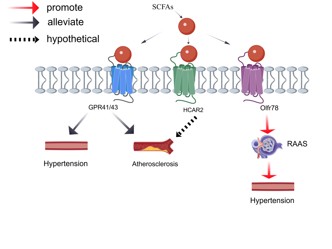
Emerging evidence indicates that short-chain fatty acids (SCFAs), primarily acetate, propionate, and butyrate, generally exert protective effects against hypertension through activation of GPR41 (FFAR3) and GPR43 (FFAR2) [9] (Figure 1). Pluznick and colleagues demonstrated a dual-receptor sensing mechanism in blood pressure regulation: Olfr78 (an olfactory GPCR localized in renal afferent arterioles and vascular smooth muscle cells) promotes renin secretion and elevates vascular tone, whereas GPR41 signaling predominantly promotes vasodilation and counterbalances excessive hypertensive responses [10] (Figure 1).
Oral butyrate slows atherosclerosis in Apoe−/− mice, reducing aortic lesion area by ~50%, accompanied by lower CD36, VCAM-1, CCL2 and NF-κB p65 nuclear translocation and increased plaque collagen [11]. Butyrate and niacin are endogenous and pharmacological ligands of HCAR2, and Gpr109a signaling promotes anti-inflammatory programming in colonic macrophages and dendritic cells [12]. Whether HCAR2-dependent M2 polarization occurs in vascular macrophages and stabilizes atherosclerotic plaques has not been directly tested.
Furthermore, SCFAs can inhibit the release of inflammatory cytokines from vascular endothelial cells via GPR43, thereby improving endothelium-dependent vasodilation and slowing the progression of early atherosclerotic lesions [13].
In a UK Biobank whole-exome sequencing study of 393,649 participants of white-European ancestry, rare pathogenic variants in SCFA-sensing GPCR genes were associated with a modestly higher prevalence of hypertension (OR 1.12) and MACE (OR 1.18); in single-gene analyses, FFAR2 variants were associated with hypertension (OR 1.23) whereas HCAR2 variants were associated with heart failure [14]. Of note, prospective evidence directly linking circulating or faecal SCFA concentrations to incident cardiovascular events is currently lacking [15], and higher faecal SCFA levels have paradoxically been associated with gut dysbiosis, obesity, hypertension and cardiometabolic risk factors in a cross-sectional study [16].
2.1.2. Ceramide
This lipid molecule can mediate the onset and progression of metabolic disorders; elevated circulating ceramide levels in humans are associated with increased cardiovascular risk [17,18].
In addition, a research team has identified two GPCRs—cysteinyl leukotriene receptor 2 (CYSLTR2) and pyrimidinergic receptor P2Y6 (P2RY6)—as endogenous cell membrane receptors for circulating long-chain ceramides. These dual ceramide-binding receptors activate the Gq signaling pathway, triggering NOD-, LRR- and pyrin domain-containing protein 3 (NLRP3) inflammasome activation and exacerbating atherosclerosis; inhibition of these dual receptors reduces vascular plaques independently of cholesterol levels, particularly ameliorating atherosclerotic lesions associated with kidney disease [19,20].
2.1.3. Bile Acids and Their Receptor, TGR5
Bile acids can act as endogenous ligands to activate the G protein-coupled bile acid receptor 1 (GPBAR1, also known as TGR5) [21,22].
GPBAR1/TGR5 is widely expressed in various cell types throughout the body, including vascular endothelial cells, vascular smooth muscle cells, cardiomyocytes, and macrophages; the core molecular mechanism by which this receptor mediates downstream signaling involves coupling to Gs-type G proteins, thereby activating the cyclic adenosine monophosphate-protein kinase A (cAMP-PKA) signaling cascade to exert biological effects [23]. Notably, bile acid excess per se can induce cardiomyopathy, underscoring the need for tissue-restricted agonists [24,25].
In macrophages, TGR5 activation suppresses oxidized LDL uptake by downregulating the scavenger receptors CD36 and SR-A, thereby inhibiting foam cell formation; this effect is mediated by TGR5-induced cAMP signaling and consequent NF-κB inhibition [26]. In Apoe−/− mice fed a Western diet, the selective GPBAR1 agonist BAR501 (30 mg/kg for 14 weeks) reduced aortic thickness, attenuated atherosclerotic lesion severity, and decreased plaque macrophage content, while reshaping the aortic transcriptome toward an anti-inflammatory profile (e.g., IL-10) [27]. Complementing these findings, the TGR5 agonist INT-777 attenuated atherosclerosis in Ldlr−/− mice, an effect associated with reduced intraplaque inflammation and plaque macrophage content and abolished in Ldlr−/−Tgr5−/− mice [26]. Beyond atherosclerosis, TGR5 shapes post-infarction immune responses: Tgr5−/− mice displayed persistently greater cardiac recruitment of CD4+ and CD8+ T cells up to day 7 after myocardial infarction, and CD4-specific TGR5 depletion aggravated ischemic injury, indicating that TGR5 modulates CD4+ T-cell function and subset distribution to protect against myocardial infarction [28]. Consistently, the TGR5 ligand deoxycholic acid improved cardiac function and reduced infarct size after MI in a TGR5-dependent manner [29].
The bile acid profile regulated by the gut microbiota can modulate platelet activation via TGR5, and an imbalance in bile acid composition increases the risk of arterial thrombosis, providing a new mechanism by which the gut microbiota-bile acid axis regulates cardiovascular events [30].
2.1.4. Lysophosphatidic Acid (LPA)
LPA exerts its effects through six GPCR subtypes (LPAR1–6), which couple to different G proteins and mediate effects such as cell proliferation, migration, inflammation, and fibrosis [31].
The signaling axis comprising LPA and its cognate lysophosphatidic acid receptors (LPARs) is involved in multiple pathological processes underlying the onset and progression of atherosclerosis in cardiovascular disease [32].
Individual LPAR subtypes mediate distinct pathological functions: LPAR1 and LPAR3 can induce directed migration of monocytes and promote foam cell formation [32]; and LPAR6 is primarily expressed in the vascular endothelium and regulates vascular wall permeability [31]. Based on these pathological mechanisms, targeting the autotaxin (ATX)-LPAR signaling pathway has emerged as a promising strategy for the prevention and treatment of atherosclerosis and calcific aortic valve disease [33,34,35].
LPA derived from oxidized low-density lipoprotein can induce osteogenic differentiation of aortic valve stromal cells via the LPAR1-Ras homolog family member A (RhoA)-nuclear factor κ-light-chain-enhancer of activated B cells (NF-κB) pathway, representing a pathogenic mechanism in calcific aortic stenosis; targeting LPAR1 can significantly delay the progression of valve calcification [36]. Tissue metabolomics studies have shown that LPA levels are significantly elevated in atherosclerotic plaques, and that plasma LPA subtypes may serve as biomarkers for cardiovascular risk assessment [37].
2.1.5. Prostaglandins (PGs)
After prostacyclin (PGI2) binds to the prostacyclin receptor (IP receptor), it initiates an intracellular cyclic adenosine monophosphate (cAMP) signaling cascade, which dilates blood vessels, inhibits abnormal platelet adhesion and aggregation, and blocks pathological damage associated with atherosclerosis [38]; PGE2 binds to four receptors—prostaglandin E receptors 1–4 (EP1–EP4)—to mediate diverse physiological and pathological responses. Among these, excessive activation of EP3 exacerbates ventricular remodeling following myocardial infarction and induces impaired cardiac systolic and diastolic function [39]; once TXA2 binds to the thromboxane A2 receptor (TP receptor), it induces vascular smooth muscle contraction and promotes platelet aggregation and thrombus formation [40,41].
Suppression of the PGI2-IP/type I interferon axis induces splenic extramedullary hematopoiesis, promotes M2 macrophage infiltration, and improves cardiac function after ischemia [42].
Prostaglandin-related signaling pathways are well-established targets for drug intervention in pulmonary arterial hypertension (PAH), and prostacyclin (PGI2) derivatives and IP receptor agonists are now in routine clinical use [43]. After PGI2 binds to the IP receptor, it activates the downstream cAMP signaling cascade, which dilates pulmonary blood vessels while inhibiting excessive vascular smooth muscle proliferation and abnormal platelet aggregation [43,44].
2.1.6. Leukotrienes and Their GPCRs
Leukotrienes (LTs) are bioactive lipid mediators derived from arachidonic acid via the 5-lipoxygenase (5-LO) pathway. They are classified into two major groups: leukotriene B4 (LTB4), a dihydroxy acid, and the cysteinyl leukotrienes (LTC4, LTD4, LTE4). Both groups signal through distinct GPCR subfamilies and have been implicated in cardiovascular inflammation, atherosclerosis, and ischemic injury [45,46].
LTB4 exerts its effects through two GPCR subtypes: the high-affinity receptor BLT1 (LTB4R) and the low-affinity receptor BLT2 (LTB4R2). BLT1 is primarily expressed on leukocytes, including neutrophils, monocytes, and macrophages, and mediates chemotaxis, degranulation, and production of reactive oxygen species. In atherosclerotic lesions, BLT1-expressing monocytes are recruited to the vascular intima, where they differentiate into macrophages and promote foam cell formation. BLT1 deficiency or pharmacological antagonism reduces atherosclerotic plaque burden in mouse models [45,47]. BLT2 is more ubiquitously expressed and has been linked to vascular smooth muscle cell proliferation and endothelial dysfunction, although its precise role in cardiovascular disease remains less well defined [46].
Cysteinyl leukotrienes (LTC4, LTD4, LTE4) signal through the GPCRs CysLT1 (CYSLTR1) and CysLT2 (CYSLTR2). In the cardiovascular system, CysLT receptors are expressed on endothelial cells, vascular smooth muscle cells, macrophages, and platelets. Cysteinyl LTs mediate vasoconstriction, increase vascular permeability, and enhance endothelial-leukocyte interactions. Human atherosclerotic lesions show increased expression of 5-LO pathway enzymes and CysLT receptors, and CysLT1 antagonism reduces macrophage foam cell formation and plaque inflammation in preclinical models [45,48]. CYSLTR2 has recently been identified as a dual receptor for both cysteinyl leukotrienes and long-chain ceramides in the context of atherosclerosis [19,20], highlighting receptor pleiotropy in metabolite sensing.
Despite strong preclinical evidence, clinical translation of leukotriene-targeted therapies for cardiovascular disease has been limited. The 5-LO inhibitor zileuton and CysLT1 antagonists (montelukast, zafirlukast) are approved for asthma but have not been adequately tested in cardiovascular outcomes trials. Emerging epidemiological data suggest that 5-LO pathway genetic variants are associated with increased cardiovascular risk, supporting further investigation of this axis as a potential therapeutic target [45,49].
2.1.7. Endocannabinoids and Cannabinoid-Sensitive GPCRs
Endocannabinoids (eCBs) are endogenous lipid signaling molecules derived from arachidonic acid, the most well-characterized being N-arachidonoylethanolamine (anandamide, AEA) and 2-arachidonoylglycerol (2-AG). They signal primarily through two GPCRs: cannabinoid receptor type 1 (CB1, CNR1) and cannabinoid receptor type 2 (CB2, CNR2). The endocannabinoid system (ECS) is increasingly recognized as a significant modulator of cardiovascular function, with divergent and often opposing effects mediated by CB1 versus CB2 receptor activation [50,51].
CB1 receptors are expressed in vascular endothelial cells, smooth muscle cells, cardiomyocytes, and macrophages, and their expression is upregulated in atherosclerotic plaques, hypertensive vessels, and failing hearts. CB1 activation is generally considered pro-atherogenic: it promotes endothelial dysfunction, oxidative stress, macrophage foam cell formation, and vascular inflammation. In human coronary artery disease, CB1 receptor expression is increased in unstable plaques, and CB1 blockade reduces macrophage inflammatory cytokine production. Peripherally restricted CB1 antagonists (e.g., JD5037, AM6545) have shown atheroprotective effects in preclinical models without the central nervous system side effects that led to the withdrawal of rimonabant [50,52].
In contrast, CB2 receptors are predominantly expressed on immune cells and are upregulated in atherosclerotic lesions and ischemic myocardium. CB2 activation is generally anti-inflammatory and atheroprotective: it inhibits macrophage chemotaxis, reduces pro-inflammatory cytokine release, and promotes plaque stability. CB2 agonists reduce infarct size and improve cardiac function in animal models of myocardial ischemia-reperfusion injury, in part by attenuating inflammatory cell infiltration and oxidative stress [50,51]. However, the role of CB2 in cardiovascular disease is context-dependent, and some studies have reported pro-fibrotic effects of CB2 activation in the heart, underscoring the need for tissue-specific and cell-type-specific approaches [53].
Endocannabinoids also exert hemodynamic effects: AEA produces a triphasic blood pressure response (brief hypotension, transient hypertension, and prolonged hypotension) mediated by CB1-dependent and CB1-independent mechanisms, including activation of transient receptor potential vanilloid 1 (TRPV1) channels. Under pathological conditions such as cardiogenic shock and advanced heart failure, eCBs produced by activated immune cells contribute to hypotension and negative inotropy. The balance between CB1-mediated detrimental effects and CB2-mediated protective effects makes the ECS a complex but potentially valuable target for cardiovascular drug development, with peripherally restricted CB1 antagonists and selective CB2 agonists representing the most promising therapeutic strategies [50,54].
2.2. Amino Acid–Derived Metabolites and Their Receptors
Amino acids and their metabolic derivatives mediate intracellular signal transduction through GPCR pathways. These receptors include the calcium-sensing receptor (CaSR), GPR35 (activated by the tryptophan metabolite kynurenic acid) and the GABAB receptor complex. The signaling pathways mediated by these receptors regulate normal physiological functions and modulate a range of disease-related pathological processes [55,56].
The CaSR, whose ligands include L-amino acids and polyamines, is widely expressed throughout the body [57]. Calcium-sensing receptors are highly expressed in the parathyroid glands and kidneys, where they exert a pro-calcium effect by regulating systemic calcium homeostasis. Abnormal function or expression of calcium-sensing receptors in non-calcifying organs has been associated with cardiovascular disease, gastrointestinal disorders, Alzheimer’s disease, impaired glucose tolerance, and cancer [58,59,60,61].
In the cardiovascular system, CaSR regulates macrophage polarization, ameliorates myocardial remodeling in hypertension, and participates in various vascular pathological processes, including vascular calcification and pulmonary arterial hypertension [62,63].
2.2.1. Kynurenic Acid and GPR35
G protein-coupled receptor 35 (GPR35) is widely expressed in human tissues, including digestive-tract organs and various immune cells [64,65].
Kynurenic acid (KynA), a tryptophan metabolite, was the first endogenous ligand reported to bind GPR35 [66]. A recent study then showed that the KynA–GPR35 axis protects the infarcted heart: KynA suppressed pro-inflammatory macrophage infiltration and downregulated the cGAS/STING/TBK1/IκBα/p65 pathway by inhibiting voltage-dependent anion channel 1 (VDAC1) oligomerization and preventing mitochondrial DNA (mtDNA) leakage into the cytoplasm, thereby reducing infarct scarring and fibrosis and improving cardiac function in a mouse model of myocardial infarction; these effects were abrogated by macrophage depletion and by GPR35-targeted siRNA [67]. In a separate myocardial ischaemia–reperfusion model, KynA preserved mitochondrial structure and function through GPR35 [68].
The atherosclerotic microenvironment induces indoleamine 2,3-dioxygenase (IDO) expression, thereby promoting kynurenine production and AhR activation, which has been reported to aggravate local vascular inflammation and foam cell formation [69]. Notably, the net direction of this pathway is context-dependent: IDO-mediated tryptophan catabolism also induces IL-10 production in B cells and constrains atherogenesis in Apoe−/− mice [70].
A functional imbalance in the KynA–GPR35 signaling axis has been associated with incident atrial fibrillation risk in cohort studies [71,72]; excess KynA accumulation in epicardial adipose tissue can bind GPR35, disrupting normal mitochondrial function in lymphatic endothelial cells, impairing lymphatic drainage, and ultimately inducing formation of a pathological substrate for atrial fibrillation [73,74].
2.2.2. γ-Aminobutyric Acid
γ-Aminobutyric acid (GABA), synthesized and released by vascular endothelial cells, is a small-molecule metabolite that maintains intravascular homeostasis [75]. GABA levels are significantly reduced in both endothelial tissue from patients with aortic dissection and peripheral plasma [76].
GABA signals through the γ-aminobutyric acid type B (GABAB) receptor complex, in which GABBR2 (GABAB receptor subunit 2) is the obligatory heterodimerization partner of GABBR1 (GABAB receptor subunit 1); the functional GABAB receptor is a GABBR1–GABBR2 heterodimer, and GABBR2 alone does not constitute a complete receptor. Through this heterodimeric complex, GABA regulates mitochondrial function and maintains endothelial and mitochondrial stability, thereby blocking the mitogen-activated protein kinase (MAPK)/c-Fos-mediated endothelial inflammatory response; the complex also activates the Notch receptor 3 (Notch3) pathway through paracrine mechanisms, protecting vascular smooth muscle cells. GABA levels in peripheral plasma can serve as a noninvasive diagnostic marker for early screening of acute aortic dissection. Targeting the GABA/GABAB receptor (GABBR1/GABBR2) signaling axis also offers new insights for the prevention and clinical management of aortic dissection [76].
2.3. Energy-Metabolism Intermediates and Other Small-Molecule Metabolites
2.3.1. Succinic Acid and SUCNR1 (GPR91)
Succinate signals through the succinate receptor 1 (SUCNR1, also known as GPR91), a G protein-coupled receptor for this metabolite. Under ischemic and hypoxic conditions, succinic acid accumulates both intracellularly and extracellularly, and is now recognized as a signaling metabolite that participates in the regulation of cardiac physiological and pathological functions [77,78,79].
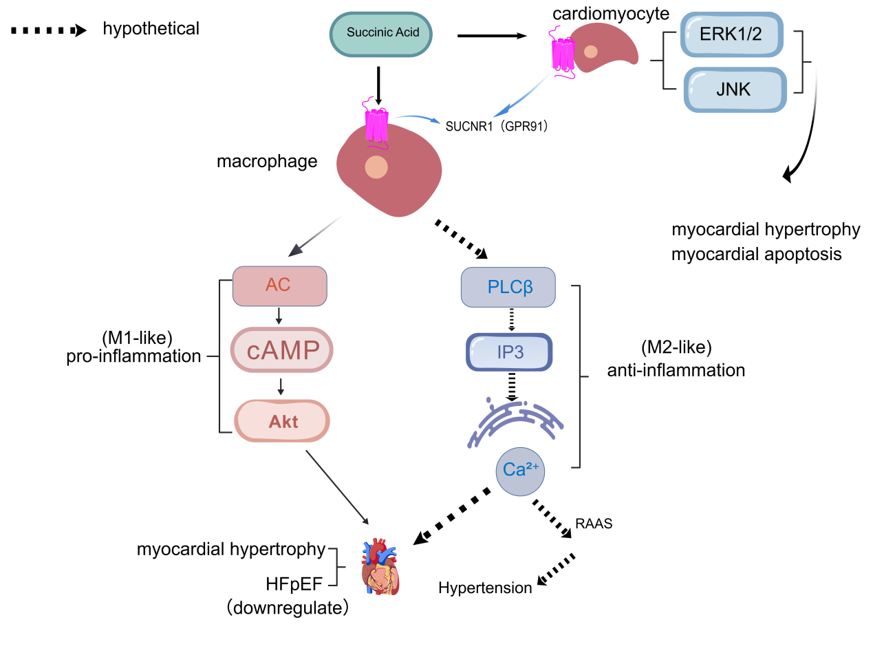
Both in vitro and in vivo studies have shown that succinic acid induces myocardial hypertrophy, and prolonged incubation of cardiomyocytes with high concentrations of succinic acid induces apoptosis [78,80]. The succinate–GPR91 signaling pathway is downregulated in heart failure with preserved ejection fraction (HFpEF); restoring this pathway exerts cardioprotective effects by promoting metabolic reprogramming of cardiomyocytes and increasing nicotinamide adenine dinucleotide (NAD+) production [81,82].
Exogenous succinate activates SUCNR1 and raises plasma renin activity and mean arterial pressure in rats, an effect abolished in Gpr91-deficient animals [83]. Disturbed systemic succinate handling caused by Slc26a6 deletion is likewise associated with elevated renin secretion and blood pressure [84]. However, the hypertensive phenotype in this model was unaffected by dietary salt intake, and a direct SUCNR1–Gαq–PLC–IP3–Ca2+ cascade linking salt loading to juxtaglomerular renin secretion has not been demonstrated (Figure 2).
Studies have reported that succinate levels in the extracellular microenvironment of ischaemic myocardial regions are significantly elevated and that this metabolite activates the ERK1/2 and JNK signaling pathways in cardiomyocytes via SUCNR1, accelerating cardiomyocyte apoptosis and exacerbating inflammatory cell infiltration [78,85,86,87,88] (Figure 2). Consistent with a causal role, genetic ablation of Sucnr1 attenuated myocardial ischemia-reperfusion injury in diabetic mice, reducing infarct size, serum lactate dehydrogenase and cardiac troponin T levels, and cardiomyocyte apoptosis while suppressing M1 macrophage polarization [86].
2.3.2. Ketone Bodies
β-Hydroxybutyrate (BHB) is an endogenous ligand of HCAR2, a Gi/o-coupled receptor through which BHB inhibits adipocyte lipolysis at concentrations reached during fasting [89]. In macrophages and in cardiovascular preclinical models, HCAR2 activation by BHB or butyrate has been reported to exert anti-inflammatory effects and to attenuate atherogenesis, vascular calcification and myocardial inflammation, although the contribution of HCAR2 varies by model [12,89,90,91]. BHB also competitively antagonizes free fatty acid receptor 3 (FFAR3, GPR41). By blocking FFAR3-mediated signaling in sympathetic ganglia, it suppresses propionate-induced sympathetic outflow, thereby regulating blood pressure and lowering peripheral vascular pressure [92,93].
Beyond its effects on sympathetic ganglia, BHB has recently been shown to modulate catecholamine secretion from adrenal chromaffin cells. A 2024 study using PC12 cells as a model of adrenal chromaffin cells demonstrated that propionate-activated FFAR3 promotes catecholamine secretion, whereas BHB—acting as an FFAR3 antagonist—suppresses this FFAR3-dependent catecholamine production. When combined with α2A-adrenergic receptor functional restoration (via GRK2 blockade), BHB-mediated FFAR3 antagonism profoundly suppresses catecholamine secretion. These findings suggest that BHB may regulate peripheral vascular resistance not only by reducing sympathetic neuronal norepinephrine release but also by inhibiting adrenal chromaffin cell catecholamine secretion, representing an additional mechanism by which the ketone body–FFAR3 axis modulates cardiovascular homeostasis [94].
Preclinical studies have shown that BHB suppresses NLRP3 inflammasome activation [95], thereby alleviating myocardial inflammatory infiltration and fibrosis [91,96]. Importantly, in the study that established this mechanism, BHB inhibited the NLRP3 inflammasome by preventing K+ efflux and reducing ASC oligomerisation and speck formation, and this effect was independent of chirality, AMPK, ROS, autophagy, UCP2, SIRT2 and—critically—of HCAR2 [95]. In a randomized crossover study of 12 patients with HFrEF, 3-OHB infusion acutely increased cardiac output (+2.3 L/min) and lowered free fatty acids by 58%; however, these hemodynamic effects were not blocked by aspirin and were not reproduced by the HCAR2 agonist niacin, indicating that they are not HCAR2-dependent [97].
Guo and colleagues reported in 2020 that a ketogenic diet produces cardioprotective effects in db/db mice with type 2 diabetes, alleviating oxidative stress-induced damage and inhibiting programmed cell death in cardiomyocytes [98]; nutritional ketosis has additionally been proposed to signal through the BHB–HCAR2 axis, although this link awaits further validation [99].
2.3.3. Lactate and Its Receptors (GPR132/G2A and HCAR1/GPR81)
G protein-coupled receptor 132 (GPR132, also known as G2A) is a lactate-sensing membrane receptor expressed primarily on the surface of various immune cells. Recent experimental data indicate that lactate levels are significantly elevated in the diabetic pathological environment. Excess lactate activates the GPR132–proto-oncogene tyrosine-protein kinase Src (Src) pathway in macrophages, which, on the one hand, induces macrophage senescence and the senescence-associated secretory phenotype (SASP), and, on the other, promotes extensive foam cell formation. These effects collectively accelerate the onset and progression of atherosclerotic lesions [100].
Lactate signaling through the hydroxycarboxylic acid receptor 1 (HCAR1, also known as GPR81) exerts anti-inflammatory effects in macrophages, as reviewed elsewhere [101,102]. Upon binding GPR81, lactate downregulates the transcription of multiple pro-inflammatory genes in macrophages while promoting adaptive remodeling of cellular metabolism by stabilizing the hypoxia-inducible factor 1-alpha (HIF-1α) protein [103,104]. These findings support the hypothesis that extensive lactate accumulation at ischemic sites may mediate endogenous anti-inflammatory protection via the GPR81 pathway [105,106,107].
Under myocardial ischemia, anaerobic glycolysis is markedly activated, producing large amounts of lactate as a metabolic byproduct [103]. GPR81 expression is upregulated in ischaemic myocardial tissue, and through this receptor lactate blocks lipolysis and reduces free fatty acid release, thereby limiting fatty-acid lipotoxic injury to cardiomyocytes and attenuating arrhythmogenesis in preclinical models [101,103,108,109]. These findings, however, rest largely on preclinical models and a proof-of-concept conference report [108], and confirmation in independent cohorts is required.
2.3.4. Adenosine
Adenosine exerts its effects through four GPCR subtypes (A1, A2A, A2B, and A3), of which A1 and A3 couple to Gi/o proteins, whereas A2A and A2B couple to Gs proteins [110]. Activation of the A1 receptor slows heart rate, reduces myocardial oxygen consumption, and exerts antiarrhythmic effects; the A2A receptor mediates coronary vasodilation and anti-inflammatory effects; and the A2B receptor is involved in vascular remodeling and fibrosis [111,112].
Adenosine receptor subtypes exert bidirectional regulation in cardiovascular disease, and the development of subtype-selective agonists and antagonists is an important direction for future drug development [112,113].
The ligand–receptor pairs discussed above, together with their potency, species and cell type, disease model, downstream signaling, evidence level, pharmacological tools and major limitations, are summarised in Table 1.
Comprehensive summary of endogenous metabolite-GPCR ligand-receptor pairs in cardiovascular disease.
| Metabolite | Receptor (Gene/Protein) | Potency/Affinity | Species & Cell Type | Disease Model | Downstream Signaling | Evidence Level | Pharmacological Tool | Major Limitation | Key Reference(s) |
| SCFAs (acetate, propionate, butyrate) | FFAR2/GPR43; FFAR3/GPR41 | Propionate EC50 ~6–290 μM (assay-dependent; FFAR2/3) | Mouse; immune cells, endothelium, VSMCs | Ang II–induced HTN, Apoe−/− atherosclerosis | FFAR2: Gq/Gi; FFAR3: Gi/o | Level I–II | 4-CMTB (FFAR2 agonist-positive allosteric modulator); FFAR3 agonist (AR420626) | Receptor redundancy; opposing BP effects of FFAR3 vs. Olfr78 | [6,7,9,10,11,12,13,14,15,16] |
| SCFAs (propionate) | Olfr78 (olfactory receptor) | Propionate EC50 ~0.6–0.9 mM | Mouse; juxtaglomerular cells, VSMCs | BP regulation, renin secretion | Gαolf → AC → cAMP | Level II (KO mice) | No selective pharmacological tool available | Low ligand affinity; species differences in expression | [10] |
| Butyrate | HCAR2 | Butyrate EC50 ~0.7–1.6 mM | Mouse; macrophages | Atherosclerosis | Gi/o (↓cAMP) | Level II–III | Niacin (non-selective agonist) | HCAR2 also activated by BHB; broad tissue expression | [11,12,90,91,92,93,94,96,97,98,99] |
| Long-chain ceramides | CYSLTR2 + P2RY6 (dual receptor) | C16:0 ceramide EC50 ~μM range | Mouse; macrophages, endothelium | Atherosclerosis (Apoe−/−) | Gq (PLC–IP3–Ca2+), NLRP3 | Level I–II | Dual receptor antagonists (experimental) | Dual receptor complexity; limited human data | [17,18,19,20] |
| Bile acids | GPBAR1/TGR5 | LCA EC50 ~0.5 μM; DCA ~1 μM | Mouse/human; endothelium, VSMCs, cardiomyocytes, macrophages | Atherosclerosis, MI, HF, HTN | Gs (cAMP–PKA) | Level II–III | BAR501 (selective agonist); INT-777 (agonist, Ldlr−/− data) | Systemic effects; gallstone risk with agonists; agonist- and model-dependent effects (BAR501/Apoe−/− vs. INT-777/Ldlr−/−) | [21,22,23,24,25,26,27,28,29,30] |
| LPA | LPAR1–6 | LPA(18:1) EC50 ~1–100 nM (subtype-dependent) | Mouse/human; monocytes, platelets, endothelium, valve cells | Atherosclerosis, CAVD, thrombosis | LPAR1/3: Gq/Gi | Level II–III | Ki16425 (LPAR1/3 antagonist); BMS-986020 (LPAR1) | Subtype redundancy; LPA species heterogeneity | [31,32,33,34,35,36,37] |
| PGI2 | PTGIR/IP receptor | PGI2 EC50 ~nM | Human/mouse; VSMCs, platelets, endothelium | PAH, atherosclerosis, MI | Gs (cAMP) | Level I (clinical drugs) | Epoprostenol, treprostinil, selexipag | Short half-life; systemic vasodilation | [38,41,42,43,44] |
| PGE2 | PTGER1–4/EP1–EP4 | PGE2 EC50 ~nM (subtype-dependent) | Mouse; cardiomyocytes, fibroblasts, immune cells | MI, ventricular remodeling | EP3: Gi; EP4: Gs | Level II | EP3 antagonist (DG-041); EP4 agonist (CAY10580) | Subtype-specific opposing effects | [38,39] |
| TXA2 | TBXA2R/TP receptor | TXA2 analog U46619 EC50 ~nM | Human/mouse; platelets, VSMCs | Thrombosis, vasospasm | Gq/G12/13 | Level I (aspirin, upstream COX inhibition) | SQ29548 (antagonist) | TXA2 instability; upstream COX inhibition used clinically | [40,41] |
| LTB4 | LTB4R/BLT1; LTB4R2/BLT2 | LTB4 EC50 ~0.1–1 nM (BLT1) | Mouse/human; neutrophils, monocytes, macrophages, VSMCs | Atherosclerosis, MI | BLT1: Gi; BLT2: Gq/Gi | Level II–III | CP-105,696 (BLT1 antagonist) | Limited clinical cardiovascular outcomes data | [45,46,47,49] |
| Cysteinyl LTs (LTC4/D4/E4) | CYSLTR1/CysLT1; CYSLTR2/CysLT2 | LTD4 EC50 ~0.4–1 nM (CysLT1) | Human/mouse; endothelium, VSMCs, macrophages, platelets | Atherosclerosis, ischemia | CysLT1: Gq; CysLT2: Gq/Gi | Level II–III | Montelukast, zafirlukast (CysLT1 antagonists) | Approved for asthma only; no cardiovascular outcomes trials | [45,46,48,49] |
| Anandamide (AEA), 2-AG | CNR1/CB1; CNR2/CB2 | AEA Ki ~60–540 nM (CB1), ~280–1940 nM (CB2); 2-AG Ki ~60–470 nM (CB1), ~150–1400 nM (CB2) | Human/mouse; endothelium, VSMCs, cardiomyocytes, macrophages | Atherosclerosis, MI, HF, shock | CB1: Gi/o; CB2: Gi/o | Level II–III | JD5037 (peripheral CB1 antagonist); HU-308 (CB2 agonist) | CB1 central side effects; CB2 context-dependent effects | [50,51,52,53,54] |
| Kynurenic acid (KynA) | GPR35 | KynA EC50 ~40 μM (human GPR35) | Human/mouse; macrophages, lymphatic endothelial cells | MI, atrial fibrillation | Gi/o (β-arrestin) | Level I–III | Zaprinast (agonist); pamoic acid (agonist) | Low potency; species differences (human GPR35 requires higher kynurenic acid concentrations than rodent orthologs; EC50 ~40 μM vs. ~7–11 μM) | [64,65,66,67,68,69,70,71,72,73,74,114] |
| GABA | GABBR1/GABBR2 heterodimer (GABAB receptor) | GABA EC50 ~μM | Human/mouse; endothelial cells, VSMCs | Aortic dissection | Gi/o (↓cAMP), MAPK inhibition | Level II–III | Baclofen (GABAB agonist); CGP55845 (antagonist) | GABBR2 is a subunit, not a standalone receptor; central nervous system effects | [75,76] |
| Succinate | SUCNR1/GPR91 | Succinate EC50 ~20–50 μM | Mouse/human; cardiomyocytes, juxtaglomerular cells, endothelium, immune cells | HTN, MI/I/R, HFpEF, aortic dissection | Gq (PLC–IP3–Ca2+); ERK1/2; JNK | Level II | cis-epoxysuccinate (agonist); NF-56-EJ40, compound 4c (antagonists) | Context-dependent (pathological vs repair phase); tissue-specific effects | [77,78,79,80,81,82,83,84,85,86,87,88,115,116,117,118] |
| β-hydroxybutyrate (BHB) | HCAR2; FFAR3/GPR41 (antagonist) | BHB EC50 ~100–700 μM (HCAR2); IC50 ~mM (FFAR3) | Mouse/human; macrophages, cardiomyocytes, sympathetic neurons, chromaffin cells | Atherosclerosis, HF, MI, vascular calcification | HCAR2: Gi/o; FFAR3: antagonism of Gi/o | Level II–III | Niacin (HCAR2 agonist); AR420626 (FFAR3 agonist) | High concentrations required; multiple receptor targets; NLRP3 inhibition is HCAR2-independent | [90,91,92,93,94,95,96,97,98,99] |
| Lactate | GPR132/G2A; HCAR1/GPR81 | Lactate EC50 ~1–10 mM (GPR81); ~mM (GPR132) | Mouse; macrophages, cardiomyocytes, endothelium | Diabetes-accelerated atherosclerosis, MI, ischemia | GPR132: Src; GPR81: Gi/o | Level III–IV | 3,5-DHBA (GPR81 agonist) | High physiological concentrations; GPR132 also senses oxidized phospholipids | [100,101,102,103,104,105,106,107,108,109] |
| Adenosine | ADORA1/A1; ADORA2A/A2A; ADORA2B/A2B; ADORA3/A3 | Adenosine EC50 ~nM–μM (subtype-dependent) | Human/mouse; cardiomyocytes, endothelium, VSMCs, immune cells | Arrhythmia, MI, HF, vascular fibrosis | A1/A3: Gi/o; A2A/A2B: Gs | Level I (adenosine for SVT) | Adenosine (non-selective); regadenoson (A2A agonist) | Short half-life; subtype-specific opposing effects | [110,111,112,113] |
| L-amino acids, polyamines | CaSR | L-Phe EC50 ~mM; Ca2+ EC50 ~mM | Human/mouse; parathyroid, kidney, VSMCs, macrophages, cardiomyocytes | Vascular calcification, PAH, HTN, myocardial remodeling | Gq/Gi (PLC–IP3–Ca2+) | Level II–III | Cinacalcet (positive allosteric modulator); calcilytics (antagonists) | Broad tissue expression; interference with calcium homeostasis | [56,57,58,59,60,61,62,63] |
3. GPCR Metabolic Axis Biomarkers and Targeted Therapy
3.1. Metabolites—GPCR-Binding Molecules as Biomarkers for Early Diagnosis
A study combining non-targeted metabolomics and targeted mass spectrometry quantification reported that succinate levels in peripheral plasma are significantly elevated in patients with aortic aneurysm and dissection (AAD). Furthermore, peripheral blood succinate levels can serve as an auxiliary diagnostic marker for acute aortic disease during the prehospital phase; in patients presenting with chest pain as the chief complaint, this marker can effectively distinguish patients with aortic aneurysm or dissection from those with acute myocardial infarction (AMI) or pulmonary embolism (PE), two conditions with similar clinical presentations [115].
3.2. Targeted Interventions along the Metabolite-GPCR Axis and Its Downstream Effectors
Identifying and characterizing the metabolic disorders associated with various cardiovascular diseases can offer promising approaches for developing interventions that target specific metabolite–GPCR axes and their associated metabolic enzymes [119].
INT-777, a selective TGR5 agonist, attenuated atherosclerosis in Ldlr−/− mice—an effect abolished in Tgr5-deficient animals—and reviews of bile acid signaling support its broader cardiometabolic potential [26,120,121].
TEPP-46, a selective activator that promotes pyruvate kinase M2 (PKM2) tetramerization, significantly ameliorates multiple pathological features of acute aortic dissection (AAD) upon exogenous administration [122].
Targeting the citrate–ATP citrate lyase (ACLY) axis holds promise as an intervention strategy for aortic aneurysm [123]. Similarly, succinate-related signaling pathways—such as those lowering peripheral plasma succinate levels or alleviating succinate-induced mitochondrial dysfunction—may also serve as therapeutic targets for aortic dissection [115].
In addition, novel drug candidates—such as small-molecule antagonists targeting the ceramide receptor CYSLTR2/P2RY6 [19,124], selective GPR35 agonists [114], and LPAR inhibitors—have all demonstrated significant cardiovascular protective effects in animal studies and show considerable potential for clinical translation [33,36].
3.3. Potential Drug Target Identification
Building on the mechanistic evidence reviewed above, this section comprehensively identifies and prioritizes metabolite-activated GPCRs as potential drug targets for cardiovascular disease. Target prioritization is based on a multi-criteria framework incorporating: (1) strength of causal evidence from genetic and pharmacological studies, (2) druggability (availability of selective pharmacological tools and tractability for small-molecule modulation), (3) tissue-specific expression and cell-type-specific effects, (4) existence of context-dependent or bidirectional effects that may pose safety risks, and (5) stage of clinical translation.
3.3.1. High-Priority Targets with Strong Preclinical Evidence
SUCNR1 (GPR91): SUCNR1 is among the most promising drug targets in the metabolite-sensing GPCR family. Strong preclinical evidence demonstrates that SUCNR1 mediates succinate-induced hypertension (via renal renin secretion), myocardial ischemia-reperfusion injury (via ERK1/2 and JNK activation), and pathological cardiac hypertrophy. In diabetic mice, genetic ablation of Sucnr1 attenuated myocardial ischaemia–reperfusion injury, reducing infarct size, serum lactate dehydrogenase and cardiac troponin T levels and cardiomyocyte apoptosis, while suppressing M1 macrophage polarization [86]. The receptor is druggable: selective antagonists (NF-56-EJ40, compound 4c) and agonists (succinate, cis-epoxysuccinate) have both been developed. However, the context-dependent nature of SUCNR1 signaling necessitates careful timing of intervention: succinate accumulates universally during ischaemia and, upon reperfusion, is rapidly re-oxidized by succinate dehydrogenase, driving mitochondrial ROS production through reverse electron transport at complex I and thereby exacerbating injury [116]. Conversely, extracellular succinate acting through SUCNR1–Gq signaling reinforces an anti-inflammatory, reparative transcriptional program in human M2 macrophages [117]. Interventions targeting this axis should therefore be guided by disease stage [81,116,117].
GPBAR1 (TGR5): TGR5 is a well-validated target whose selective agonist INT-777 has shown therapeutic efficacy in multiple cardiovascular disease models, including atherosclerosis (reduced plaque formation in Ldlr−/− mice, an effect abolished in Ldlr−/−Tgr5−/− mice and in Ldlr−/− mice transplanted with Tgr5−/− bone marrow) [26], as well as broader cardiometabolic protection suggested by mechanistic reviews [24]. Gs-coupled TGR5 signaling mediates anti-inflammatory effects in macrophages [26,121] and modulates T-cell responses post-MI [28]. The main challenges are systemic side effects (particularly gallstone formation and bile acid diarrhea) and the need for tissue-specific or peripherally restricted agonists to avoid systemic on-target effects such as gallbladder filling, altered heart rate and blood pressure [125,126].
CYSLTR2/P2RY6 (ceramide receptors): The recent identification of CYSLTR2 and P2RY6 as potential endogenous receptors for long-chain ceramides in atherosclerosis (Nature, 2025) establishes this axis as a novel high-priority target [19]. Inhibition of these dual receptors reduces vascular plaques independently of cholesterol levels, which is particularly important given that many patients develop cardiovascular events despite statin-controlled LDL cholesterol. The established clinical use of CysLT1 antagonists (montelukast, zafirlukast) for asthma indicates that this receptor family is tractable for small-molecule modulation, although CYSLTR2-selective compounds remain at the preclinical stage [45,46].
3.3.2. Moderate-Priority Targets with Promising but Less Mature Evidence
GPR35: GPR35, activated by kynurenic acid, has demonstrated cardioprotective effects in myocardial infarction models (reduced necrosis, improved cardiac function) via suppression of macrophage mtDNA-triggered cGAS–STING inflammation [67,68]. GPR35 is expressed predominantly in the gastrointestinal tract and immune cells, with lower but detectable expression in cardiovascular tissues [65]. However, the low potency of endogenous kynurenic acid at human GPR35 (EC50 in the high micromolar range) and marked species differences (human GPR35 requires higher kynurenic acid concentrations than rodent orthologs; EC50 ~40 μM vs. ~7–11 μM) represent translational challenges [65,66].
LPAR subtypes: The ATX–LPA–LPAR axis is implicated in atherosclerosis, calcific aortic valve disease, and thrombosis. LPAR1 antagonists (BMS-986020, formerly in clinical trials for idiopathic pulmonary fibrosis) and the dual LPAR1/3 antagonist Ki16425 have shown efficacy in preclinical cardiovascular models [33,36]. The main challenges are subtype redundancy (six LPAR subtypes with overlapping functions) and the heterogeneity of LPA molecular species, which complicates the development of truly selective agents. Tissue-specific delivery (e.g., to the aortic valve or vascular wall) may be necessary to avoid systemic effects on platelet function and reproductive physiology.
HCAR2: Several BHB-associated cardiovascular benefits are consistent with, but not proven to be mediated by, HCAR2 [91,99]. Niacin, a non-selective HCAR2 agonist, has been used clinically for dyslipidemia, although its cardiovascular benefits have been questioned in recent outcomes trials [127]. The development of selective HCAR2 agonists that avoid niacin’s prostaglandin-mediated flushing side effect (which is mediated by HCAR2 in Langerhans cells but involves downstream prostanoid release) represents a promising strategy [128]. The ketogenic diet and exogenous BHB supplementation provide indirect clinical evidence for HCAR2-mediated cardiovascular benefit, but controlled trials with selective agonists are needed.
3.3.3. Emerging Targets Requiring Further Validation
FFAR3 (GPR41): FFAR3 plays a complex role in blood pressure regulation, with opposing effects depending on cell type (sympathetic activation increases BP; immune cell effects are anti-inflammatory). BHB acts as an FFAR3 antagonist and suppresses catecholamine secretion from adrenal chromaffin cells [94]. FFAR3 agonists (AR420626) and the receptor’s role in metabolic disease make it an interesting target, but its bidirectional cardiovascular effects and expression in multiple tissues (including the enteric nervous system) pose considerable safety and specificity challenges.
GPR81 (HCAR1): GPR81, activated by lactate, mediates anti-inflammatory effects in macrophages and may be cardioprotective in the ischemic myocardium by reducing lipotoxicity and arrhythmias [101,102,103,108]. The GPR81 agonist 3,5-DHBA is available as a research tool. However, the high physiological concentrations of lactate required for GPR81 activation (millimolar range) and the receptor’s broad expression in metabolic tissues (adipose, muscle) raise questions about the feasibility and specificity of pharmacological targeting.
GABAB receptor (GABBR1/GABBR2): The GABAB receptor axis protects against aortic dissection by maintaining endothelial and mitochondrial homeostasis [76]. GABAB agonists (baclofen) are clinically used for neurological indications, but their central nervous system side effects (sedation, dizziness) limit cardiovascular application. The development of peripherally restricted GABAB agonists or endothelial-targeted delivery systems could unlock the cardiovascular potential of this axis while avoiding central effects.
Leukotriene receptors (BLT1, CysLT1/2): Despite strong preclinical evidence for leukotriene involvement in atherosclerosis and ischemic injury, clinical translation has been hampered by the lack of cardiovascular outcomes trials with existing leukotriene modifiers (zileuton, montelukast). The recent identification of CYSLTR2 as a ceramide receptor [19] has renewed interest in this target class. Repurposing approved CysLT1 antagonists for cardiovascular indications, or developing dual CYSLTR2-targeting agents, represents a cost-effective translational strategy.
Endocannabinoid receptors (CB1, CB2): The divergent effects of CB1 (pro-atherogenic) and CB2 (anti-inflammatory, cardioprotective) create both opportunities and challenges. Peripherally restricted CB1 antagonists avoid the psychiatric side effects that led to the withdrawal of rimonabant and have shown atheroprotective efficacy in preclinical models [50,52]. Selective CB2 agonists are promising for myocardial I/R injury and heart failure, but context-dependent pro-fibrotic effects require careful evaluation [53]. The ECS represents an emerging but complex target landscape requiring tissue-specific and cell-type-specific pharmacological approaches.
3.4. Future Research Directions for the Metabolite-GPCR Axis
Multi-omics strategies that integrate metabolomics with genomics, transcriptomics, proteomics, and other molecular profiling techniques have emerged as a highly valuable direction in basic cardiovascular research [129,130,131,132,133].
Such integrative analysis simultaneously captures biological information across molecular levels, enabling the pathophysiological mechanisms of cardiovascular diseases to be examined from a systems perspective. Longitudinal, time-resolved multi-omics studies across the disease course can further elucidate the dynamic mechanisms governing disease onset and progression while identifying novel molecular targets for clinical intervention [134]. Nevertheless, metabolic research in major cardiovascular diseases still has important limitations, including the difficulty of translating metabolite findings into safe clinical treatments. Although metabolic inhibitors and dietary interventions show promise, supporting large-scale clinical data and personalized metabolomic analyses remain lacking [135,136].
Metabolite levels are regulated by multiple exogenous and endogenous factors, such as dietary patterns, physical activity, and circadian rhythms, and undergo pronounced dynamic fluctuations [137]. In pathological states, the timing of metabolite concentration changes differs markedly. For example, in myocardial ischaemic injury, succinate accumulates during ischaemia and, upon reperfusion, is rapidly re-oxidised by succinate dehydrogenase to drive mitochondrial ROS production through reverse electron transport at complex I, thereby exacerbating tissue damage [116]. Whether the same metabolite promotes healing during the repair phase is supported by its capacity to drive anti-inflammatory macrophage polarisation via SUCNR1 [117], to our knowledge, direct demonstration in the infarcted heart is still lacking [138,139,140]. Traditional single-time-point static measurements cannot capture these temporal dynamics; therefore, technologies for real-time in vivo monitoring of metabolite levels, together with intervention strategies tailored to different time windows of disease progression, hold substantial scientific and clinical value [141,142].
Metabolite-activated GPCRs are widely expressed across multiple tissues and cell types; when the same receptor acts in different cell subsets, its effects can differ markedly, or even be diametrically opposed [61]. For example, succinate induces pathological cardiomyocyte hypertrophy through GPR91 [78,80], whereas global Gpr91 deletion did not alter vascular inflammation or atherosclerotic lesion size in hyperlipidaemic mice [118], indicating that the vascular effects of SUCNR1 are highly model- and cell type–dependent. FFAR3 activation in sympathetic ganglia increases sympathetic outflow [92]. This bidirectional heterogeneity markedly increases the risk of adverse reactions with systemic administration. Accordingly, developing ligands with tissue-specific targeting, designing cell-specific drug-delivery vehicles, and screening biased receptor agonists are important future research directions [143].
Future research should focus on receptor-selective signal regulation, tissue-specific targeted delivery, and validation in large clinical cohorts to advance translation of the metabolite–GPCR axis from basic research to clinical applications [144]. In particular, the high-priority targets identified in Section 3.3 (SUCNR1, TGR5, and CYSLTR2/P2RY6) should be advanced into first-in-human cardiovascular trials, while moderate-priority targets (GPR35, LPAR, HCAR2) require further optimization of selective pharmacological tools and tissue-specific delivery systems before clinical evaluation.
4. Conclusions
The metabolite–GPCR axis is an important regulatory axis linking metabolism and cardiovascular disease and provides new targets and approaches for the diagnosis and treatment of cardiovascular disease. Through structured evaluation of evidence strength, druggability, and tissue-specific effects, we identify SUCNR1, TGR5, and the ceramide receptor complex CYSLTR2/P2RY6 as high-priority candidates for cardiovascular drug development, with GPR35, LPAR subtypes, and HCAR2 as moderate-priority targets. Future efforts should prioritize the development of selective, tissue-specific pharmacological agents and their validation in large clinical cohorts to realize the translational potential of the endogenous metabolite–GPCR axis.
Author Contributions
N.L.: Conceptualization, methodology, investigation, writing—original draft preparation, visualization. W.Y.: Data curation, validation, writing—original draft preparation. S.F., Z.Y., Y.W., Y.D.: Supervision, project administration, writing—review and editing. Z.M. and Z.Z.: Supervision, funding acquisition, project administration, writing—review and editing. All authors have read and agreed to the published version of the manuscript.
Funding
This research received for financial support from the National Natural Science Foundation of China 82570552 for Zhiwei Zhao.
Institutional Review Board Statement
Not applicable.
Informed Consent Statement
Not applicable.
Data Availability Statement
Not applicable.
Conflicts of Interest
The authors declare no conflicts of interest.
Use of AI and AI-Assisted Technologies
Artificial intelligence and AI-assisted tools (ChatGPT, OpenAI) were utilized for linguistic optimization and proofreading during manuscript preparation. Upon tool processing, the authors carried out thorough manual review and targeted revisions. Full responsibility for all contents of this published work rests with the authors.
References
- 1.
Virani, S.S.; Alonso, A.; Aparicio, H.J.; et al. Heart Disease and Stroke Statistics—2021 Update: A Report from the American Heart Association. Circulation 2021, 143, e254–e743. https://doi.org/10.1161/cir.0000000000000950.
- 2.
Tsao, C.W.; Aday, A.W.; Almarzooq, Z.I.; et al. Heart Disease and Stroke Statistics—2022 Update: A Report from the American Heart Association. Circulation 2022, 145, e153–e639. https://doi.org/10.1161/cir.0000000000001052.
- 3.
Akhtar, M.; Chen, Y.; Ma, Z.; et al. Gut microbiota-derived short chain fatty acids are potential mediators in gut inflammation. Anim. Nutr. 2022, 8, 350–360. https://doi.org/10.1016/j.aninu.2021.11.005.
- 4.
Jin, C.; Chen, H.; Xie, L.; et al. GPCRs involved in metabolic diseases: Pharmacotherapeutic development updates. Acta Pharmacol. Sin. 2024, 45, 1321–1336. https://doi.org/10.1038/s41401-023-01215-2.
- 5.
Wang, J.; Gareri, C.; Rockman, H.A. G-Protein–Coupled Receptors in Heart Disease. Circ. Res. 2018, 123, 716–735. https://doi.org/10.1161/circresaha.118.311403.
- 6.
Brown, A.J.; Goldsworthy, S.M.; Barnes, A.A.; et al. The Orphan G Protein-coupled Receptors GPR41 and GPR43 Are Activated by Propionate and Other Short Chain Carboxylic Acids. J. Biol. Chem. 2003, 278, 11312–11319. https://doi.org/10.1074/jbc.m211609200.
- 7.
Xu, J.; Moore, B.N.; Pluznick, J.L. Short-Chain Fatty Acid Receptors and Blood Pressure Regulation: Council on Hypertension Mid-Career Award for Research Excellence 2021. Hypertension 2022, 79, 2127–2137. https://doi.org/10.1161/hypertensionaha.122.18558.
- 8.
Maslowski, K.M.; Vieira, A.T.; Ng, A.; et al. Regulation of inflammatory responses by gut microbiota and chemoattractant receptor GPR43. Nature 2009, 461, 1282–1286. https://doi.org/10.1038/nature08530.
- 9.
Muralitharan, R.; Zheng, T.; Dinakis, E.; et al. Gut Microbiota Metabolites Sensed by Host GPR41/43 Protect against Hypertension. Circ. Res. 2025, 136, e20–e33. https://doi.org/10.1161/circresaha.124.325770.
- 10.
Pluznick, J.L.; Protzko, R.J.; Gevorgyan, H.; et al. Olfactory receptor responding to gut microbiota-derived signals plays a role in renin secretion and blood pressure regulation. Proc. Natl. Acad. Sci. USA 2013, 110, 4410–4415.
- 11.
Aguilar, E.C.; Leonel, A.J.; Teixeira, L.G.; et al. Butyrate impairs atherogenesis by reducing plaque inflammation and vulnerability and decreasing NF-κB activation. Nutr. Metab. Cardiovasc. Dis. 2014, 24, 606–613. https://doi.org/10.1016/j.numecd.2014.01.002.
- 12.
Singh, N.; Gurav, A.; Sivaprakasam, S.; et al. Activation of Gpr109a, Receptor for Niacin and the Commensal Metabolite Butyrate, Suppresses Colonic Inflammation and Carcinogenesis. Immunity 2014, 40, 128–139. https://doi.org/10.1016/j.immuni.2013.12.007.
- 13.
Robles-Vera, I.; Toral, M.; de la Visitación, N.; et al. Protective Effects of Short-Chain Fatty Acids on Endothelial Dysfunction Induced by Angiotensin II. Front. Physiol. 2020, 11, 277. https://doi.org/10.3389/fphys.2020.00277.
- 14.
Camargo Tavares, L.; Muralitharan, R.; Snelson, M.; et al. Rare pathogenic variants in G-protein-coupled receptor genes involved in gut-to-host communication are associated with cardiovascular disease risk. Cardiovasc. Res. 2025, 121, 1316–1318. https://doi.org/10.1093/cvr/cvaf070.
- 15.
Sanchez-Gimenez, R.; Ahmed-Khodja, W.; Molina, Y.; et al. Gut Microbiota-Derived Metabolites and Cardiovascular Disease Risk: A Systematic Review of Prospective Cohort Studies. Nutrients 2022, 14, 2654. https://doi.org/10.3390/nu14132654.
- 16.
De la Cuesta-Zuluaga, J.; Mueller, N.T.; Álvarez-Quintero, R.; et al. Higher Fecal Short-Chain Fatty Acid Levels Are Associated with Gut Microbiome Dysbiosis, Obesity, Hypertension and Cardiometabolic Disease Risk Factors. Nutrients 2019, 11, 51. https://doi.org/10.3390/nu11010051.
- 17.
Choi, R.H.; Tatum, S.M.; Symons, J.D.; et al. Ceramides and other sphingolipids as drivers of cardiovascular disease. Nat. Rev. Cardiol. 2021, 18, 701–711. https://doi.org/10.1038/s41569-021-00536-1.
- 18.
Laaksonen, R.; Ekroos, K.; Sysi-Aho, M.; et al. Plasma ceramides predict cardiovascular death in patients with stable coronary artery disease and acute coronary syndromes beyond LDL-cholesterol. Eur. Heart J. 2016, 37, 1967–1976. https://doi.org/10.1093/eurheartj/ehw148.
- 19.
Zhang, S.; Lin, H.; Wang, J.; et al. Sensing ceramides by CYSLTR2 and P2RY6 to aggravate atherosclerosis. Nature 2025, 641, 476–485. https://doi.org/10.1038/s41586-025-08792-8.
- 20.
Ye, Q.; Tang, Y. P2Y6R together with CYSLTR2 serves as endogenous receptor for long-chain ceramides in atherosclerosis. Purinergic Signal. 2025, 21, 1001–1003. https://doi.org/10.1007/s11302-025-10090-x.
- 21.
Wang, H.; Wang, J.; Cui, H.; et al. Inhibition of fatty acid uptake by TGR5 prevents diabetic cardiomyopathy. Nat. Metab. 2024, 6, 1161–1177. https://doi.org/10.1038/s42255-024-01036-5.
- 22.
Kawamata, Y.; Fujii, R.; Hosoya, M.; et al. A G Protein-coupled Receptor Responsive to Bile Acids. J. Biol. Chem. 2003, 278, 9435–9440. https://doi.org/10.1074/jbc.m209706200.
- 23.
Duboc, H.; Taché, Y.; Hofmann, A.F. The bile acid TGR5 membrane receptor: From basic research to clinical application. Dig. Liver Dis. 2014, 46, 302–312. https://doi.org/10.1016/j.dld.2013.10.021.
- 24.
He, Y.; Liu, S.; Zhang, Y.; et al. Takeda G protein–coupled receptor 5 (TGR5): An attractive therapeutic target for aging-related cardiovascular diseases. Front. Pharmacol. 2025, 16, 1493662. https://doi.org/10.3389/fphar.2025.1493662.
- 25.
Desai, M.S.; Mathur, B.; Eblimit, Z.; et al. Bile acid excess induces cardiomyopathy and metabolic dysfunctions in the heart. Hepatology 2017, 65, 189–201. https://doi.org/10.1002/hep.28890.
- 26.
Pols, T.W.H.; Nomura, M.; Harach, T.; et al. TGR5 Activation Inhibits Atherosclerosis by Reducing Macrophage Inflammation and Lipid Loading. Cell Metab. 2011, 14, 747–757. https://doi.org/10.1016/j.cmet.2011.11.006.
- 27.
Biagioli, M.; Marchianò, S.; Di Giorgio, C.; et al. Activation of GPBAR1 attenuates vascular inflammation and atherosclerosis in a mouse model of NAFLD-related cardiovascular disease. Biochem. Pharmacol. 2023, 218, 115900. https://doi.org/10.1016/j.bcp.2023.115900.
- 28.
Wang, J.; Xu, T.; Xu, M. Roles and Mechanisms of TGR5 in the Modulation of CD4+ T Cell Functions in Myocardial Infarction. J. Cardiovasc. Transl. Res. 2022, 15, 350–359. https://doi.org/10.1007/s12265-021-10164-2.
- 29.
Wang, J.; Zhang, J.; Lin, X.; et al. DCA-TGR5 signaling activation alleviates inflammatory response and improves cardiac function in myocardial infarction. J. Mol. Cell. Cardiol. 2021, 151, 3–14. https://doi.org/10.1016/j.yjmcc.2020.10.014.
- 30.
Qi, Z.; Zhang, W.; Zhang, P.; et al. The gut microbiota–bile acid–TGR5 axis orchestrates platelet activation and atherothrombosis. Nat. Cardiovasc. Res. 2025, 4, 584–601. https://doi.org/10.1038/s44161-025-00637-x.
- 31.
Yung, Y.C.; Stoddard, N.C.; Chun, J. LPA receptor signaling: Pharmacology, physiology, and pathophysiology. J. Lipid Res. 2014, 55, 1192–1214. https://doi.org/10.1194/jlr.r046458.
- 32.
Fogelman, A.M.; Chattopadhyay, A.; Reddy, S.T. The multiple roles of lysophosphatidic acid in vascular disease and atherosclerosis. Curr. Opin. Lipidol. 2023, 34, 196–200. https://doi.org/10.1097/mol.0000000000000890.
- 33.
Nadhan, R.; Nath, K.; Basu, S.; et al. Decoding lysophosphatidic acid signaling in physiology and disease: Mapping the multimodal and multinodal signaling networks. Signal Transduct. Target. Ther. 2025, 10, 337. https://doi.org/10.1038/s41392-025-02423-4.
- 34.
Nakanaga, K.; Hama, K.; Aoki, J. Autotaxin—An LPA producing enzyme with diverse functions. J. Biochem. 2010, 148, 13–24. https://doi.org/10.1093/jb/mvq052.
- 35.
Bouchareb, R.; Mahmut, A.; Nsaibia, M.J.; et al. Autotaxin Derived from Lipoprotein(a) and Valve Interstitial Cells Promotes Inflammation and Mineralization of the Aortic Valve. Circulation 2015, 132, 677–690. https://doi.org/10.1161/circulationaha.115.016757.
- 36.
Nsaibia, M.J.; Boulanger, M.C.; Bouchareb, R.; et al. OxLDL-derived lysophosphatidic acid promotes the progression of aortic valve stenosis through a LPAR1-RhoA–NF-κB pathway. Cardiovasc. Res. 2017, 113, 1351–1363. https://doi.org/10.1093/cvr/cvx089.
- 37.
Cao, J.; Martin-Lorenzo, M.; van Kuijk, K.; et al. Spatial Metabolomics Identifies LPC(18:0) and LPA(18:1) in Advanced Atheroma with Translation to Plasma for Cardiovascular Risk Estimation. Arterioscler. Thromb. Vasc. Biol. 2024, 44, 741–754. https://doi.org/10.1161/atvbaha.123.320278.
- 38.
Ricciotti, E.; FitzGerald, G.A. Prostaglandins and Inflammation. Arterioscler. Thromb. Vasc. Biol. 2011, 31, 986–1000. https://doi.org/10.1161/atvbaha.110.207449.
- 39.
Bryson, T.D.; Bhat, S.Y.; Moore, C.; et al. Targeted Gene Deletion or Antagonism of the Prostaglandin E2 EP3 Receptor Protects against Cardiac Injury Postmyocardial Infarction. Circ. Heart Fail. 2024, 17, e011089. https://doi.org/10.1161/circheartfailure.123.011089.
- 40.
Ricciotti, E.; FitzGerald, G.A. Prostaglandins and the Cardiovascular System. Arterioscler. Thromb. Vasc. Biol. 2025, 45, 841–844. https://doi.org/10.1161/atvbaha.124.320744.
- 41.
Cheng, Y.; Austin, S.C.; Rocca, B.; et al. Role of Prostacyclin in the Cardiovascular Response to Thromboxane A2. Science 2002, 296, 539–541. https://doi.org/10.1126/science.1068711.
- 42.
Lv, H.; Wang, C.; Liu, Z.; et al. Suppression of the Prostaglandin I2–Type I Interferon Axis Induces Extramedullary Hematopoiesis to Promote Cardiac Repair After Myocardial Infarction. Circulation 2025, 151, 1730–1747. https://doi.org/10.1161/circulationaha.124.069420.
- 43.
Zeng, C.; Liu, J.; Zheng, X.; et al. Prostaglandin and prostaglandin receptors: Present and future promising therapeutic targets for pulmonary arterial hypertension. Respir. Res. 2023, 24, 263. https://doi.org/10.1186/s12931-023-02559-3.
- 44.
Wang, J.J.; Jin, S.; Zhang, H.; et al. Molecular recognition and activation of the prostacyclin receptor by anti-pulmonary arterial hypertension drugs. Sci. Adv. 2024, 10, eadk5184. https://doi.org/10.1126/sciadv.adk5184.
- 45.
Colazzo, F.; Gelosa, P.; Tremoli, E.; et al. Role of the Cysteinyl Leukotrienes in the Pathogenesis and Progression of Cardiovascular Diseases. Mediators Inflamm. 2017, 2017, 2432958. https://doi.org/10.1155/2017/2432958.
- 46.
Bäck, M. Leukotriene Signaling in Atherosclerosis and Ischemia. Cardiovasc. Drugs Ther. 2009, 23, 41–48. https://doi.org/10.1007/s10557-008-6140-9.
- 47.
Aiello, R.J.; Bourassa, P.A.; Lindsey, S.; et al. Leukotriene B4 Receptor Antagonism Reduces Monocytic Foam Cells in Mice. Arterioscler. Thromb. Vasc. Biol. 2002, 22, 443–449. https://doi.org/10.1161/hq0302.105593.
- 48.
Gautier-Veyret, E.; Bäck, M.; Arnaud, C.; et al. Cysteinyl-leukotriene pathway as a new therapeutic target for the treatment of atherosclerosis related to obstructive sleep apnea syndrome. Pharmacol. Res. 2018, 134, 311–319. https://doi.org/10.1016/j.phrs.2018.06.014.
- 49.
Dwyer, J.H.; Allayee, H.; Dwyer, K.M.; et al. Arachidonate 5-Lipoxygenase Promoter Genotype, Dietary Arachidonic Acid, and Atherosclerosis. N. Engl. J. Med. 2004, 350, 29–37. https://doi.org/10.1056/nejmoa025079.
- 50.
Pacher, P.; Steffens, S. The emerging role of the endocannabinoid system in cardiovascular disease. Semin. Immunopathol. 2009, 31, 63–77. https://doi.org/10.1007/s00281-009-0145-8.
- 51.
Eid, B.G. Cannabinoids for Treating Cardiovascular Disorders: Putting Together a Complex Puzzle. J. Microsc. Ultrastruct. 2018, 6, 171–176. https://doi.org/10.4103/jmau.jmau_42_18.
- 52.
Sugamura, K.; Sugiyama, S.; Nozaki, T.; et al. Activated endocannabinoid system in coronary artery disease and antiinflammatory effects of cannabinoid 1 receptor blockade on macrophages. Circulation 2009, 119, 28–36. https://doi.org/10.1161/circulationaha.108.811992.
- 53.
Fulmer, M.L.; Thewke, D.P. The Endocannabinoid System and Heart Disease: The Role of Cannabinoid Receptor Type 2. Cardiovasc. Hematol. Disord. Drug Targets 2018, 18, 34–51. https://doi.org/10.2174/1871529x18666180206161457.
- 54.
Maccarrone, M.; Bab, I.; Bíró, T.; et al. Endocannabinoid signaling at the periphery: 50 years after THC. Trends Pharmacol. Sci. 2015, 36, 277–296. https://doi.org/10.1016/j.tips.2015.02.008.
- 55.
Pardella, E.; Ippolito, L.; Giannoni, E.; et al. Nutritional and metabolic signalling through GPCRs. FEBS Lett. 2022, 596, 2364–2381. https://doi.org/10.1002/1873-3468.14441.
- 56.
Conigrave, A.D.; Hampson, D.R. Broad-spectrum l-amino acid sensing by class 3 G-protein-coupled receptors. Trends Endocrinol. Metab. 2006, 17, 398–407. https://doi.org/10.1016/j.tem.2006.10.012.
- 57.
Schreckenberg, R.; Schlüter, K.D. Calcium sensing receptor expression and signalling in cardiovascular physiology and disease. Vasc. Pharmacol. 2018, 107, 35–42. https://doi.org/10.1016/j.vph.2018.02.007.
- 58.
Brennan, S.C.; Thiem, U.; Roth, S.; et al. Calcium sensing receptor signalling in physiology and cancer. Biochim. Biophys. Acta Mol. Cell Res. 2013, 1833, 1732–1744. https://doi.org/10.1016/j.bbamcr.2012.12.011.
- 59.
Vahe, C.; Benomar, K.; Espiard, S.; et al. Diseases associated with calcium-sensing receptor. Orphanet J. Rare Dis. 2017, 12, 19. https://doi.org/10.1186/s13023-017-0570-z.
- 60.
Alfadda, T.I.; Saleh, A.M.A.; Houillier, P.; et al. Calcium-sensing receptor 20 years later. Am. J. Physiol. Cell Physiol. 2014, 307, C221–C231. https://doi.org/10.1152/ajpcell.00139.2014.
- 61.
Strassheim, D.; Sullivan, T.; Irwin, D.C.; et al. Metabolite G-Protein Coupled Receptors in Cardio-Metabolic Diseases. Cells 2021, 10, 3347. https://doi.org/10.3390/cells10123347.
- 62.
Zhao, J.; Lu, N.; Qu, Y.; et al. Calcium-sensing receptor-mediated macrophage polarization improves myocardial remodeling in spontaneously hypertensive rats. Exp. Biol. Med. 2024, 249, 10112. https://doi.org/10.3389/ebm.2024.10112.
- 63.
Albert, A.P.; Greenberg, H.Z.E. Role of the calcium-sensing receptor in regulating vascular function. J. Cell Commun. Signal. 2025, 19, e70004. https://doi.org/10.1002/ccs3.70004.
- 64.
Zhao, P.; Sharir, H.; Kapur, A.; et al. Targeting of the orphan receptor GPR35 by pamoic acid: A potent activator of extracellular signal-regulated kinase and β-arrestin2 with antinociceptive activity. Mol. Pharmacol. 2010, 78, 560–568. https://doi.org/10.1124/mol.110.066746.
- 65.
Milligan, G. GPR35: From enigma to therapeutic target. Trends Pharmacol. Sci. 2023, 44, 263–273. https://doi.org/10.1016/j.tips.2023.03.001.
- 66.
Wang, J.; Simonavicius, N.; Wu, X.; et al. Kynurenic Acid as a Ligand for Orphan G Protein-coupled Receptor GPR35. J. Biol. Chem. 2006, 281, 22021–22028. https://doi.org/10.1074/jbc.m603503200.
- 67.
Mao, Y.; Jiao, J.; Zhu, X.; et al. Kynurenic Acid/GPR35 Signaling Protects the Infarcted Heart by Suppressing Macrophage mtDNA-Triggered cGAS-STING Activation. Antioxidants 2026, 15, 300. https://doi.org/10.3390/antiox15030300.
- 68.
Nógrádi-Halmi, D.; Erdélyi-Furka, B.; Csóré, D.; et al. Kynurenic Acid Protects Against Myocardial Ischemia/Reperfusion Injury by Activating GPR35 Receptors and Preserving Mitochondrial Structure and Function. Biomolecules 2025, 15, 1481. https://doi.org/10.3390/biom15101481.
- 69.
Yang, Y.; Liu, X.; Liu, X.; et al. The role of the kynurenine pathway in cardiovascular disease. Front. Cardiovasc. Med. 2024, 11, 1406856. https://doi.org/10.3389/fcvm.2024.1406856.
- 70.
Cole, J.E.; Astola, N.; Cribbs, A.P.; et al. Indoleamine 2,3-dioxygenase-1 is protective in atherosclerosis and its metabolites provide new opportunities for drug development. Proc. Natl. Acad. Sci. USA 2015, 112, 13033–13038. https://doi.org/10.1073/pnas.1517820112.
- 71.
Moein, A.; Seyed Parsa, E. The Footprint of Kynurenine Pathway in Cardiovascular Diseases. Int. J. Tryptophan Res. 2022, 15, 11786469221096643. https://doi.org/10.1177/11786469221096643.
- 72.
Razquin, C.; Ruiz-Canela, M.; Toledo, E.; et al. Metabolomics of the tryptophan–kynurenine degradation pathway and risk of atrial fibrillation and heart failure: Potential modification effect of Mediterranean diet. Am. J. Clin. Nutr. 2021, 114, 1646–1654. https://doi.org/10.1093/ajcn/nqab238.
- 73.
Takahashi, M.; Abe, I.; Yoshida, N.; et al. Kynurenic acid mediates epicardial fat-induced lymphatic metabolic dysfunction in atrial fibrillation. Nat. Commun. 2026, 17, 6616. https://doi.org/10.1038/s41467-026-72974-9.
- 74.
Brakenhielm, E. Cardiac lymphatic dysfunction and repair in cardiovascular disease. Nat. Rev. Cardiol. 2026, 23, 687–700. https://doi.org/10.1038/s41569-026-01271-1.
- 75.
Sen, S.; Roy, S.; Bandyopadhyay, G.; et al. γ-Aminobutyric Acid Is Synthesized and Released by the Endothelium. Circ. Res. 2016, 119, 621–634. https://doi.org/10.1161/circresaha.116.308645.
- 76.
Shao, L.; Yu, Y.; Huang, H.; et al. Endothelial GABA protects against aortic dissection by inhibiting endothelial and mitochondrial dysfunction and maintaining vascular homeostasis. Signal Transduct. Target. Ther. 2026, 11, 150. https://doi.org/10.1038/s41392-026-02619-2.
- 77.
Osuna-Prieto, F.J.; Martinez-Tellez, B.; Ortiz-Alvarez, L.; et al. Elevated plasma succinate levels are linked to higher cardiovascular disease risk factors in young adults. Cardiovasc. Diabetol. 2021, 20, 151. https://doi.org/10.1186/s12933-021-01333-3.
- 78.
Aguiar, C.J.; Rocha-Franco, J.A.; Sousa, P.A.; et al. Succinate causes pathological cardiomyocyte hypertrophy through GPR91 activation. Cell Commun. Signal. 2014, 12, 78. https://doi.org/10.1186/s12964-014-0078-2.
- 79.
Xu, J.; Yang, Y.; Li, X.; et al. Pleiotropic activities of succinate: The interplay between gut microbiota and cardiovascular diseases. iMeta 2023, 2, e124. https://doi.org/10.1002/imt2.124.
- 80.
de Castro Fonseca, M.; Aguiar, C.J.; da Rocha Franco, J.A.; et al. GPR91: Expanding the frontiers of Krebs cycle intermediates. Cell Commun. Signal. 2016, 14, 3. https://doi.org/10.1186/s12964-016-0126-1.
- 81.
Jia, Y.; Niu, W.; Liu, L.; et al. Succinate–GPR91 signaling promotes cardiomyocyte metabolic reprogramming and NAD+ production to alleviate HFpEF. Cardiovasc. Diabetol. 2026, 25, 5. https://doi.org/10.1186/s12933-025-03030-x.
- 82.
Telesca, M.; Masciovecchio, V.; Costantino, S. Metabolic rewiring through succinate–GPR91 signaling: A fresh perspective on HFpEF energetics. Cardiovasc. Diabetol. 2026, 25, 92. https://doi.org/10.1186/s12933-026-03107-1.
- 83.
He, W.; Miao, F.J.P.; Lin, D.C.H.; et al. Citric acid cycle intermediates as ligands for orphan G-protein-coupled receptors. Nature 2004, 429, 188–193. https://doi.org/10.1038/nature02488.
- 84.
Khamaysi, A.; Anbtawee-Jomaa, S.; Fremder, M.; et al. Systemic Succinate Homeostasis and Local Succinate Signaling Affect Blood Pressure and Modify Risks for Calcium Oxalate Lithogenesis. J. Am. Soc. Nephrol. 2019, 30, 381–392. https://doi.org/10.1681/asn.2018030277.
- 85.
Liebing, A.D.; Stäubert, C. Sensing succinate: SUCNR1 as a context-dependent metabolic and cellular signal integrator. Front. Mol. Biosci. 2026, 13, 1815835. https://doi.org/10.3389/fmolb.2026.1815835.
- 86.
Wu, Y.; Dou, J.; Liu, M.; et al. Gut microbiota-induced elevation of succinate exacerbates diabetic myocardial ischemia/reperfusion injury by promoting macrophage polarization. Front. Immunol. 2026, 17, 1749185. https://doi.org/10.3389/fimmu.2026.1749185.
- 87.
Zhang, W.; Lang, R. Succinate metabolism: A promising therapeutic target for inflammation, ischemia/reperfusion injury and cancer. Front. Cell Dev. Biol. 2023, 11, 1266973. https://doi.org/10.3389/fcell.2023.1266973.
- 88.
Wu, K.K. Extracellular Succinate: A Physiological Messenger and a Pathological Trigger. Int. J. Mol. Sci. 2023, 24, 11165. https://doi.org/10.3390/ijms241311165.
- 89.
Taggart, A.K.P.; Kero, J.; Gan, X.; et al. Hydroxybutyrate Inhibits Adipocyte Lipolysis via the Nicotinic Acid Receptor PUMA-G. J. Biol. Chem. 2005, 280, 26649–26652. https://doi.org/10.1074/jbc.c500213200.
- 90.
Lan, Z.; Chen, A.; Li, L.; et al. Downregulation of HDAC9 by the ketone metabolite β-hydroxybutyrate suppresses vascular calcification. J. Pathol. 2022, 258, 213–226. https://doi.org/10.1002/path.5992.
- 91.
Shao, W.; Liu, B.; Wu, Y.; et al. β-hydroxybutyrate in Cardiovascular Diseases: A Minor Metabolite of Great Expectations. Front. Mol. Biosci. 2022, 9, 823602. https://doi.org/10.3389/fmolb.2022.823602.
- 92.
Kimura, I.; Inoue, D.; Maeda, T.; et al. Short-chain fatty acids and ketones directly regulate sympathetic nervous system via G protein-coupled receptor 41 (GPR41). Proc. Natl. Acad. Sci. USA 2011, 108, 8030–8035. https://doi.org/10.1073/pnas.1016088108.
- 93.
Challa, A.A.; Hill, B.G.; Nystoriak, M.A.; et al. Ketone Bodies in Cardiovascular Disease. JACC Basic Transl. Sci. 2025, 10, 101328. https://doi.org/10.1016/j.jacbts.2025.101328.
- 94.
Nagliya, D.; Baggio Lopez, T.; Del Calvo, G.; et al. Differential Modulation of Catecholamine and Adipokine Secretion by the Short Chain Fatty Acid Receptor FFAR3 and α2-Adrenergic Receptors in PC12 Cells. Int. J. Mol. Sci. 2024, 25, 5227. https://doi.org/10.3390/ijms25105227.
- 95.
Youm, Y.H.; Nguyen, K.Y.; Grant, R.W.; et al. The ketone metabolite β-hydroxybutyrate blocks NLRP3 inflammasome–mediated inflammatory disease. Nat. Med. 2015, 21, 263–269. https://doi.org/10.1038/nm.3804.
- 96.
Challa, A.A.; Lewandowski, E.D. Short-Chain Carbon Sources. JACC Basic Transl. Sci. 2022, 7, 730–742. https://doi.org/10.1016/j.jacbts.2021.12.010.
- 97.
Gopalasingam, N.; Christensen, K.H.; Berg Hansen, K.; et al. Stimulation of the Hydroxycarboxylic Acid Receptor 2 With the Ketone Body 3-Hydroxybutyrate and Niacin in Patients with Chronic Heart Failure: Hemodynamic and Metabolic Effects. J. Am. Heart Assoc. 2023, 12, e029849. https://doi.org/10.1161/jaha.123.029849.
- 98.
Guo, Y.; Zhang, C.; Shang, F.F.; et al. Ketogenic Diet Ameliorates Cardiac Dysfunction via Balancing Mitochondrial Dynamics and Inhibiting Apoptosis in Type 2 Diabetic Mice. Aging Dis. 2020, 11, 229–240. https://doi.org/10.14336/ad.2019.0510.
- 99.
Venturini, C.; Mancinelli, L.; Matacchione, G.; et al. The Cardioprotective Effects of Nutritional Ketosis: Mechanisms and Clinical Implications. Nutrients 2024, 16, 4204. https://doi.org/10.3390/nu16234204.
- 100.
Ge, X.; Wang, S.; Li, Z.; et al. Lactate-Activated GPR132-Src Signal Induces Macrophage Senescence and Aggravates Atherosclerosis under Diabetes. Adv. Sci. 2025, 12, e00141. https://doi.org/10.1002/advs.202500141.
- 101.
Thorp, E.B.; Karlstaedt, A. Intersection of Immunology and Metabolism in Myocardial Disease. Circ. Res. 2024, 134, 1824–1840. https://doi.org/10.1161/circresaha.124.323660.
- 102.
Mohammad Nezhady, M.A.; Modaresinejad, M.; Zia, A.; et al. Versatile lactate signaling via HCAR1: A multifaceted GPCR involved in many biological processes. Am. J. Physiol. Cell Physiol. 2023, 325, C1502–C1515. https://doi.org/10.1152/ajpcell.00346.2023.
- 103.
Konieczny, J.; Boardman, N.; Aasum, E.; et al. The multifaceted role of lactate in cardiovascular health: From metabolism to signaling and epigenetics. Epigenetics 2025, 20, 2576069. https://doi.org/10.1080/15592294.2025.2576069.
- 104.
Xu, B.; Liu, Y.; Li, N.; et al. Lactate and lactylation in macrophage metabolic reprogramming: Current progress and outstanding issues. Front. Immunol. 2024, 15, 1395786. https://doi.org/10.3389/fimmu.2024.1395786.
- 105.
Fang, Y.; Li, Z.; Yang, L.; et al. Emerging roles of lactate in acute and chronic inflammation. Cell Commun. Signal. 2024, 22, 276. https://doi.org/10.1186/s12964-024-01624-8.
- 106.
Song, M.; Liu, B.; Wang, H.; et al. Lactate Metabolism and Lactylation Modification: New Opportunities and Challenges in Cardiovascular Disease. MedComm 2025, 6, e70269. https://doi.org/10.1002/mco2.70269.
- 107.
Zhang, H.; Zhao, J.; Yu, J.; et al. Lactate metabolism and lactylation in cardiovascular disease: Novel mechanisms and therapeutic targets. Front. Cardiovasc. Med. 2024, 11, 1489438. https://doi.org/10.3389/fcvm.2024.1489438.
- 108.
Bradley, E.; Cavus, O.; Johnson, M.; et al. Novel roles for GPR81 in preservation of fatty acid oxidation in Eisenmenger syndrome: A proof-of-concept study. J. Am. Coll. Cardiol. 2024, 83(Suppl. 13), 1612. https://doi.org/10.1016/s0735-1097(24)03602-7.
- 109.
Leggat, J.; Bidault, G.; Vidal-Puig, A. Lipotoxicity: A driver of heart failure with preserved ejection fraction? Clin. Sci. 2021, 135, 2265–2283. https://doi.org/10.1042/cs20210127.
- 110.
Fredholm, B.B.; Ijzerman, A.P.; Jacobson, K.A.; et al. International Union of Basic and Clinical Pharmacology. LXXXI. Nomenclature and Classification of Adenosine Receptors. Pharmacol. Rev. 2011, 63, 1–34. https://doi.org/10.1124/pr.110.003285.
- 111.
Qian, Y.; Zheng, Y.; Leng, L.; et al. Adenosine and adenosine receptors: A “double-edged sword” in cardiovascular system. Front. Pharmacol. 2025, 16, 1538680. https://doi.org/10.3389/fphar.2025.1538680.
- 112.
Chen, J.F.; Eltzschig, H.K.; Fredholm, B.B. Adenosine receptors as drug targets—what are the challenges? Nat. Rev. Drug Discov. 2013, 12, 265–286. https://doi.org/10.1038/nrd3955.
- 113.
Pallio, G.; Mannino, F. Non-Canonical Functions of Adenosine Receptors: Emerging Roles in Metabolism, Immunometabolism, and Epigenetic Regulation. Int. J. Mol. Sci. 2025, 26, 7241. https://doi.org/10.3390/ijms26157241.
- 114.
Wyant, G.A.; Yu, W.; Doulamis, I.P.; et al. Mitochondrial remodeling and ischemic protection by G protein–coupled receptor 35 agonists. Science 2022, 377, 621–629. https://doi.org/10.1126/science.abm1638.
- 115.
Cui, H.; Chen, Y.; Li, K.; et al. Untargeted metabolomics identifies succinate as a biomarker and therapeutic target in aortic aneurysm and dissection. Eur. Heart J. 2021, 42, 4373–4385. https://doi.org/10.1093/eurheartj/ehab605.
- 116.
Chouchani, E.T.; Pell, V.R.; Gaude, E.; et al. Ischaemic accumulation of succinate controls reperfusion injury through mitochondrial ROS. Nature 2014, 515, 431–435. https://doi.org/10.1038/nature13909.
- 117.
Trauelsen, M.; Hiron, T.K.; Lin, D.; et al. Extracellular succinate hyperpolarizes M2 macrophages through SUCNR1/GPR91-mediated Gq signaling. Cell Rep. 2021, 35, 109246. https://doi.org/10.1016/j.celrep.2021.109246.
- 118.
Griepke, S.; Trauelsen, M.; Nilsson, M.D.; et al. G-Protein-Coupled Receptor 91-Dependent Signalling Does Not Influence Vascular Inflammation and Atherosclerosis in Hyperlipidaemic Mice. Cells 2023, 12, 2580. https://doi.org/10.3390/cells12212580.
- 119.
McGarrah, R.W.; Crown, S.B.; Zhang, G.F.; et al. Cardiovascular Metabolomics. Circ. Res. 2018, 122, 1238–1258. https://doi.org/10.1161/circresaha.117.311002.
- 120.
Pols, T.W.H.; Noriega, L.G.; Nomura, M.; et al. The Bile Acid Membrane Receptor TGR5: A Valuable Metabolic Target. Dig. Dis. 2011, 29, 37–44. https://doi.org/10.1159/000324126.
- 121.
Zhang, S.; Zhou, J.; Wu, W.; et al. The Role of Bile Acids in Cardiovascular Diseases: From Mechanisms to Clinical Implications. Aging Dis. 2023, 14, 261–282. https://doi.org/10.14336/ad.2022.0817.
- 122.
Li, Y.E.; Liu, S.; Wang, L.; et al. March2 Alleviates Aortic Aneurysm/Dissection by Regulating PKM2 Polymerization. Circ. Res. 2025, 136, e73–e93. https://doi.org/10.1161/circresaha.124.325049.
- 123.
Wu, H.; Li, Z.; Yang, L.; et al. ANK Deficiency-Mediated Cytosolic Citrate Accumulation Promotes Aortic Aneurysm. Circ. Res. 2024, 135, 1175–1192. https://doi.org/10.1161/circresaha.124.325152.
- 124.
Chen, T.; Xie, L.; Zhao, Y.; et al. Exploring Ceramide C16:0 as a Novel Biomarker and Therapeutic Target for Atherosclerosis. J. Am. Heart Assoc. 2026, 15, e044903. https://doi.org/10.1161/jaha.125.044903.
- 125.
Cao, H.; Chen, Z.X.; Wang, K.; et al. Intestinally-targeted TGR5 agonists equipped with quaternary ammonium have an improved hypoglycemic effect and reduced gallbladder filling effect. Sci. Rep. 2016, 6, 28676. https://doi.org/10.1038/srep28676.
- 126.
Duan, H.; Ning, M.; Zou, Q.; et al. Discovery of Intestinal Targeted TGR5 Agonists for the Treatment of Type 2 Diabetes. J. Med. Chem. 2015, 58, 3315–3328. https://doi.org/10.1021/jm500829b.
- 127.
The AIM-HIGH Investigators. Niacin in Patients with Low HDL Cholesterol Levels Receiving Intensive Statin Therapy. N. Engl. J. Med. 2011, 365, 2255–2267. https://doi.org/10.1056/nejmoa1107579.
- 128.
Benyó, Z.; Gille, A.; Kero, J.; et al. GPR109A (PUMA-G/HM74A) mediates nicotinic acid–induced flushing. J. Clin. Invest. 2005, 115, 3634–3640. https://doi.org/10.1172/jci23626.
- 129.
Hasin, Y.; Seldin, M.; Lusis, A. Multi-omics approaches to disease. Genome Biol. 2017, 18, 83. https://doi.org/10.1186/s13059-017-1215-1.
- 130.
Blaser, M.C.; Kraler, S.; Lüscher, T.F.; et al. Multi-Omics Approaches to Define Calcific Aortic Valve Disease Pathogenesis. Circ. Res. 2021, 128, 1371–1397. https://doi.org/10.1161/circresaha.120.317979.
- 131.
Kuppe, C.; Ramirez Flores, R.O.; Li, Z.; et al. Spatial multi-omic map of human myocardial infarction. Nature 2022, 608, 766–777. https://doi.org/10.1038/s41586-022-05060-x.
- 132.
Luo, Y.; Zhang, N.; Yang, J.; et al. AI-based multiomics profiling reveals complementary omics contributions to personalized prediction of cardiovascular disease. Nat. Commun. 2026, 17, 2269. https://doi.org/10.1038/s41467-026-68956-6.
- 133.
Wünnemann, F.; Sicklinger, F.; Bestak, K.; et al. Spatial multiomics of acute myocardial infarction reveals immune cell infiltration through the endocardium. Nat. Cardiovasc. Res. 2025, 4, 1345–1362. https://doi.org/10.1038/s44161-025-00717-y.
- 134.
Liu, Y.; Zhao, L.; Zhang, C.; et al. Metabolic reprogramming in aortic diseases: Insights from metabolomics and therapeutic opportunities. Med. Rev. 2026, 6, 142–161. https://doi.org/10.1515/mr-2026-0010.
- 135.
Wang, Q.; Yesitayi, G.; Liu, B.; et al. Targeting metabolism in aortic aneurysm and dissection: From basic research to clinical applications. Int. J. Biol. Sci. 2023, 19, 3869–3891. https://doi.org/10.7150/ijbs.85467.
- 136.
Guasch-Ferré, M.; Bhupathiraju, S.N.; Hu, F.B. Use of Metabolomics in Improving Assessment of Dietary Intake. Clin. Chem. 2018, 64, 82–98. https://doi.org/10.1373/clinchem.2017.272344.
- 137.
Senesi, P.; Ferrulli, A.; Luzi, L.; et al. Chrono-communication and cardiometabolic health: The intrinsic relationship and therapeutic nutritional promises. Front. Endocrinol. 2022, 13, 975509. https://doi.org/10.3389/fendo.2022.975509.
- 138.
Tan, C.; Zhou, J.; Li, J.; et al. The immune-cardiovascular metabolic circuitry in myocardial ischemia-reperfusion injury: From metabolic signal release to spatiotemporal reprogramming. Front. Immunol. 2026, 17, 1848067. https://doi.org/10.3389/fimmu.2026.1848067.
- 139.
Kopecky, B.J.; Lavine, K.J. Cardiac macrophage metabolism in health and disease. Trends Endocrinol. Metab. 2024, 35, 249–262. https://doi.org/10.1016/j.tem.2023.10.011.
- 140.
Juricic, S.; Klac, J.; Stojkovic, S.; et al. Molecular and Cellular Mechanisms of Myocardial Ischemia and Reperfusion Injury: A Narrative Review. Cells 2026, 15, 265. https://doi.org/10.3390/cells15030265.
- 141.
Kim, I.Y.; Park, S.; Kim, Y.; et al. Tracing metabolic flux in vivo: Basic model structures of tracer methodology. Exp. Mol. Med. 2022, 54, 1311–1322. https://doi.org/10.1038/s12276-022-00814-z.
- 142.
Bhide, A.; Ganguly, A.; Parupudi, T.; et al. Next-Generation Continuous Metabolite Sensing toward Emerging Sensor Needs. ACS Omega 2021, 6, 6031–6040. https://doi.org/10.1021/acsomega.0c06209.
- 143.
Costa-Neto, C.M.; Parreiras-e-Silva, L.T. Deciphering complexity of GPCR signaling and modulation: Implications and perspectives for drug discovery. Clin. Sci. 2025, 139, 463–477. https://doi.org/10.1042/cs20245182.
- 144.
Slosky, L.M.; Caron, M.G.; Barak, L.S. Biased Allosteric Modulators: New Frontiers in GPCR Drug Discovery. Trends Pharmacol. Sci. 2021, 42, 283–299. https://doi.org/10.1016/j.tips.2020.12.005.

This work is licensed under a Creative Commons Attribution 4.0 International License.