
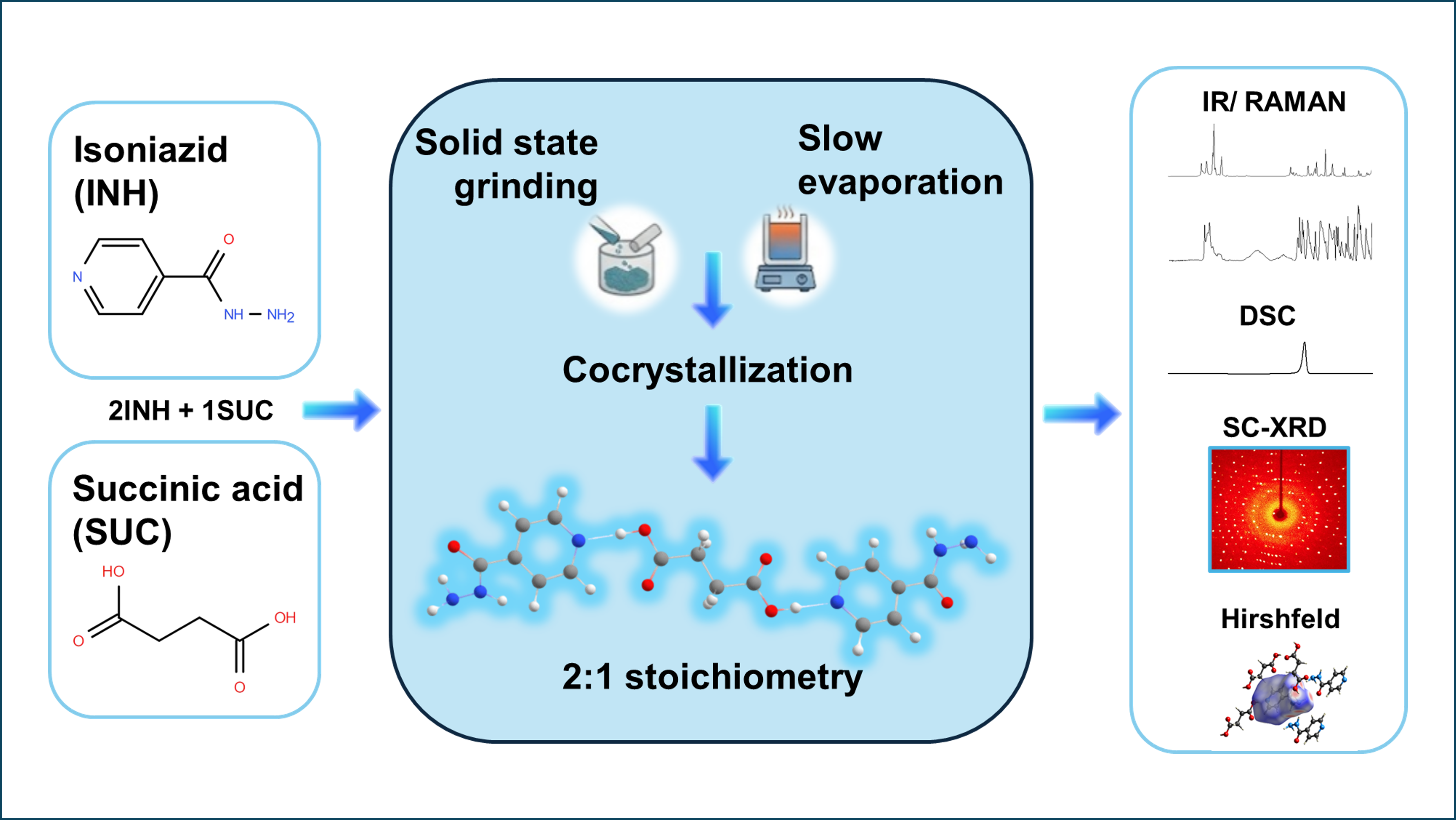
Cocrystals have gained increasing attention as an effective solid-state strategy to modify physicochemical properties through noncovalent intermolecular interactions in keeping or improving pharmacological activity. In this study, a 2:1 isoniazid (INH): succinic acid (SUC) cocrystal, where the coformers are linked through interactions between the carboxylic acid groups of SUC and the pyridine nitrogen and hydrazide moieties of INH, was synthesized and comprehensively analyzed using a combination of crystallographic, thermal, spectroscopic, and computational methods. Single crystal X-ray diffraction (SC-XRD) confirmed the structure of the synthesized material, highlighting the relevance of intermolecular O–H···N, N–H···O and N–H···N interactions to stabilize the cocrystal. Differential scanning calorimetry (DSC) was used to evaluate the thermal behavior of the cocrystal, in a comparative basis with crystals of the pure components. The cocrystal melts at Tm (onset) = 141.3 ± 0.5 °C, with ΔHfus = 27.4 ± 0.5 kJ mol−1 per molecule, i.e., at a significant lower temperature and with a lower enthalpy of fusion compared to the stable polymorphic variations of the pure components (INH (polymorph 1): Tm (onset) = 170.1 ± 0.5 °C, ΔHfus of 29.5 ± 0.5 kJ mol−1; SUC (polymorph β): Tm (onset) = 188.4 ± 0.5 °C, ΔHfus = 31.4 kJ mol−1), but at a considerably higher temperature than that corresponding to the melting of the eutectics (126.0 ± 0.5 °C). Infrared and Raman spectroscopies were employed to characterize vibrationally the cocrystal and to evaluate changes associated with changes in intermolecular interactions in going from the crystals of the pure components to the cocrystal. The experimental results were complemented by density functional theory (DFT) calculations performed on the isolated INH-SUC-INH, SUC-INH-INH-SUC and INH-INH structural units present in the cocrystal as well as on the cocrystal itself using fully-periodic calculations. A detailed investigation of the major intermolecular interactions present in the cocrystal was also undertaken using the Hirshfeld surface analysis approach. The CE-energy decomposition method of Spackman and coworkers was applied to obtain a detailed picture of the cocrystal lattice energetics, also in comparison with those of the crystals of the pure components.
- Open Access
- Article
Synthesis, Structure, Vibrational Spectra and Thermal Analysis of Isoniazid-Succinic Acid Cocrystal
- Neris Inci Inan 1,
- Ainur Syeitkhajy 1,
- Ayberk Yilmaz 1,2,
- Nihal Sarier 1,3,
- Gulce O. Ildiz 1,4,*,
- Rui Fausto 1,4,5
Author Information
Received: 28 Mar 2026 | Revised: 27 Apr 2026 | Accepted: 28 Apr 2026 | Published: 17 Aug 2026
Abstract
Graphical Abstract
Keywords
isoniazid | succinic acid | cocrystal | single crystal X-ray diffraction | FTIR and Raman spectroscopies | DSC | DFT | fully periodic calculations | Hirshfeld analysis | CE-energy decomposition
References
- 1.
Vioglio, P.C.; Chierotti, M.R.; Gobetto, R. Pharmaceutical aspects of salt and cocrystal forms of APIs and characterization challenges. Adv. Drug Deliv. Rev. 2017, 117, 86–110. https://doi.org/10.1016/j.addr.2017.07.001.
- 2.
Bhalani, D.V.; Nutan, B.; Kumar, A.; et al. Bioavailability enhancement techniques for poorly aqueous soluble drugs and therapeutics. Biomedicines 2022, 10, 2055. https://doi.org/10.3390/biomedicines10092055.
- 3.
Guo, M.; Sun, X.; Chen, J.; et al. Pharmaceutical cocrystals: A review of preparations, physicochemical properties and applications. Acta Pharm. Sin. B 2021, 11, 2537–2564. https://doi.org/10.1016/j.apsb.2021.03.030.
- 4.
Vippagunta, S.R.; Brittain, H.G.; Grant, D.J.W. Crystalline solids. Adv. Drug Deliv. Rev. 2001, 48, 3–26. https://doi.org/10.1016/S0169-409X(01)00097-7.
- 5.
Agostini, S.B.N.; Borges, B.A.; de Araújo, M.B.; et al. Growing interest in pharmaceutical cocrystals: A comprehensive review of applications and trends. ChemistrySelect 2025, 10, e202500831. https://doi.org/10.1002/slct.202500831.
- 6.
Tupe, S.A.; Khandagale, S.P.; Jadhav, A.B. Pharmaceutical cocrystals: An emerging approach to modulate physicochemical properties of active pharmaceutical ingredients. J. Drug Deliv. Ther. 2023, 13, 101–112. https://doi.org/10.22270/jddt.v13i4.6016.
- 7.
Huang, S.; Xu, J.; Peng, Y.; et al. Facile tuning of the photoluminescence and dissolution properties of phloretin through cocrystallization. Cryst. Growth Des. 2019, 19, 6837–6844. https://doi.org/10.1021/acs.cgd.9b01111.
- 8.
Buddhadev, S.S.; Garala, K.C. Pharmaceutical cocrystals—A review. Proceedings 2021, 62, 14. https://doi.org/10.3390/proceedings2020062014.
- 9.
Sopyan, I.; Kurniawansyah, I.; Cikra, I.; et al. Systematic review: Cocrystal as efforts to improve physicochemical and bioavailability properties of oral solid dosage form. Int. J. Appl. Pharm. 2021, 13, 43–52. https://doi.org/10.22159/ijap.2021v13i1.39594.
- 10.
Hema, R.; Ganesh, N.; Vineeth, C. Role of coformers in solubility enhancement of poorly soluble drugs through cocrystallization: An overview. Int. J. Pharm. Sci. Rev. Res. 2022, 74, 148–157. https://doi.org/10.47583/ijpsrr.2022.v74i02.024.
- 11.
Stagg, H.R.; Lipman, M.C.; McHugh, T.D.; et al. Isoniazid-resistant tuberculosis: A cause for concern? Int. J. Tuberc. Lung Dis. 2017, 21, 129–139. https://doi.org/10.5588/ijtld.16.0716.
- 12.
Zumla, A.; Chakaya, J.; Centis, R.; et al. Tuberculosis treatment and management-an update on treatment regimens, trials, new drugs, and adjunct therapies. Lancet Respir. Med. 2015, 3, 220–234. https://doi.org/10.1016/S2213-2600(15)00063-6.
- 13.
Mashhadi, S.M.A.; Batsanov, A.S.; Sajjad, S.A.; et al. Isoniazid-gentisic acid cocrystallization: Solubility, stability, dissolution rate, antioxidant and flowability properties studies. J. Mol. Struct. 2021, 1226, 129388. https://doi.org/10.1016/j.molstruc.2020.129388.
- 14.
Salem, A.; Khanfar, E.; Nagy, S.; et al. Cocrystals of tuberculosis antibiotics: Challenges and missed opportunities. Int. J. Pharm. 2022, 623, 121924. https://doi.org/10.1016/j.ijpharm.2022.121924.
- 15.
Fuliaş, A.; Vlase, G.; Vlase, T.; et al. Screening and characterization of cocrystal formation between carbamazepine and succinic acid. J. Therm. Anal. Calorim. 2015, 121, 1081–1086. https://doi.org/10.1007/s10973-015-4473-8.
- 16.
Saxena, R.K.; Saran, S.; Isar, J.; et al. Production and applications of succinic acid. In Current Developments in Biotechnology and Bioengineering: Production, Isolation and Purification of Industrial Products; Pandey, A., Negi, S., Soccol, C.R., Eds.; Elsevier: Amsterdam, The Netherlands, 2016; Chapter 27, pp. 601–630.
- 17.
Lin, H.L.; Wu, T.K.; Lin, S.Y. Screening and characterization of cocrystal formation of metaxalone with short-chain dicarboxylic acids induced by solvent-assisted grinding approach. Thermochim. Acta 2014, 575, 313–321. https://doi.org/10.1016/j.tca.2013.10.029.
- 18.
Diniz, L.F.; Souza, M.S.; Carvalho, P.S.; et al. Novel isoniazid cocrystals with aromatic carboxylic acids: Crystal engineering, spectroscopy and thermochemical investigations. J. Mol. Struct. 2018, 1153, 58–68. https://doi.org/10.1016/j.molstruc.2017.09.115.
- 19.
DeRuiter, J. Carboxylic Acid Structure and Chemistry: Part 1. In Principles of Drug Action, Auburn University: Auburn, AL, USA, 2005; Volume 1, pp. 1–10.
- 20.
Lemmerer, A.; Bernstein, J.; Kahlenberg, V. One-pot covalent and supramolecular synthesis of pharmaceutical co-crystals using the API isoniazid: A potential supramolecular reagent. CrystEngComm 2010, 12, 2856–2864. https://doi.org/10.1039/c000473a.
- 21.
Lemmerer, A. Covalent assistance to supramolecular synthesis: Modifying the drug functionality of the antituberculosis API isoniazid in situ during co-crystallization with GRAS and API compounds. CrystEngComm 2012, 14, 2465–2478. https://doi.org/10.1039/c1ce06310c.
- 22.
Rosa, J.; Machado, T.C.; da Silva, A.C.; et al. Isoniazid-resveratrol cocrystal: A novel alternative for topical treatment of cutaneous tuberculosis. Cryst. Growth Des. 2019, 19, 5029–5036. https://doi.org/10.1021/acs.cgd.9b00313
- 23.
Hirshfeld, F.L. Bonded-atom fragments for describing molecular charge densities. Theor. Chim. Acta 1977, 44, 129–138. https://doi.org/10.1007/BF00549096.
- 24.
Spackman, M.A.; Byrom, P.G. A novel definition of a molecule in a crystal. Chem. Phys. Lett. 1977, 267, 215–220. https://doi.org/10.1016/S0009-2614(97)00100-0.
- 25.
McKinnon, J.J.; Spackman, M.A.; Mitchell, A.S. Novel tools for visualizing and exploring intermolecular interactions in molecular crystals. Acta Crystallogr. B 2004, 60, 627–668. https://doi.org/10.1107/S0108768104020300.
- 26.
Spackman, M.A.; Jayatilaka, D. Hirshfeld surface analysis. CrystEngComm 2009, 11, 19–32. https://doi.org/10.1039/b818330a.
- 27.
McKinnon, J.J.; Jayatilaka, D.; Spackman, M.A. Towards quantitative analysis of intermolecular interactions with Hirshfeld surfaces. Chem. Commun. 2007, 3814–3816. https://doi.org/10.1039/B704980C.
- 28.
Spackman, M.A.; McKinnon, J.J. Fingerprinting intermolecular interactions in molecular crystals. CrystEngComm 2002, 4, 378–392. https://doi.org/10.1039/B203191B.
- 29.
Turner, M.J.; Grabowsky, S.; Jayatilaka, D.; et al. Accurate and efficient model energies for exploring intermolecular interactions in molecular crystals. J. Phys. Chem. Lett. 2014, 5, 4249–4255. https://doi.org/10.1021/jz502271c.
- 30.
Mackenzie, C.F.; Spackman, P.R.; Jayatilaka, D.; et al. CrystalExplorer model energies and energy frameworks: Extension to metal coordination compounds, organic salts, solvates and open-shell systems. IUCrJ 2017, 4, 575–587. https://doi.org/10.1107/S205225251700848X.
- 31.
Thomas, S.P.; Spackman, P.R.; Jayatilaka, D.; et al. Accurate lattice energies for molecular crystals from experimental crystal structures. J. Chem. Theory Comput. 2018, 14, 1614–1623. https://doi.org/10.1021/acs.jctc.7b01200.
- 32.
Spackman, M.A. Towards the use of experimental electron densities to estimate reliable lattice energies. CrystEngComm 2018, 20, 5340–5347. https://doi.org/10.1039/C8CE01108G.
- 33.
Spackman, P.R.; Turner, M.J.; McKinnon, J.J.; et al. CrystalExplorer: A program for Hirshfeld surface analysis, visualization and quantitative analysis of molecular crystals. J. Appl. Crystallogr. 2021, 54, 1006–1011. https://doi.org/10.1107/S1600576721002910.
- 34.
Bruker AXS Inc. APEX6: Software Suite for Crystallographic Data Collection and Reduction; Bruker AXS Inc.: Madison, WI, USA, 2023.
- 35.
Sheldrick, G.M. SHELXT—Integrated space-group and crystal-structure determination. Acta Crystallogr. A 2015, 71, 3–8. https://doi.org/10.1107/S2053273314026370.
- 36.
Sheldrick, G.M. Crystal structure refinement with SHELXL. Acta Crystallogr. C 2015, 71, 3–8. https://doi.org/10.1107/S2053229614024218.
- 37.
Dolomanov, O.V.; Bourhis, L.J.; Gildea, R.J.; et al. OLEX2: A complete structure solution, refinement and analysis program. J. Appl. Crystallogr. 2009, 42, 339–341. https://doi.org/10.1107/S0021889808042726.
- 38.
Dennington, R.; Keith, T.A.; Millam, J.M. GaussView, Version 6.0.16; Semichem Inc.: Shawnee Mission, KS, USA, 2016.
- 39.
Frisch, M.J.; Trucks, G.W.; Schlegel, H.B.; et al. Gaussian 16, Revision A.03; Gaussian, Inc.: Wallingford, CT, USA, 2016.
- 40.
Lee, C.; Yang, W.; Parr, R.G. Development of the Colle–Salvetti correlation-energy formula into a functional of the electron density. Phys. Rev. B 1988, 37, 785–789. https://doi.org/10.1103/PhysRevB.37.785.
- 41.
Becke, A.D. Density-functional exchange-energy approximation with correct asymptotic behavior. Phys. Rev. A 1988, 38, 3098–3100. https://doi.org/10.1103/PhysRevA.38.3098.
- 42.
Becke, A.D. Density-functional thermochemistry. III. The role of exact exchange. J. Chem. Phys. 1993, 98, 5648–5652. https://doi.org/10.1063/1.464913.
- 43.
McLean, A.D.; Chandler, G.S. Contracted Gaussian basis sets for molecular calculations. I. Second-row atoms, Z = 11–18. J. Chem. Phys. 1980, 72, 5639–5648. https://doi.org/10.1063/1.438980.
- 44.
Krishnan, R.; Binkley, J.S.; Seeger, R.; et al. Self-consistent molecular orbital methods. XX. A basis set for correlated wave functions. J. Chem. Phys. 1980, 72, 650–654. https://doi.org/10.1063/1.438955.
- 45.
Binkley, J.S.; Pople, J.A.; Hehre, W.J. Self-consistent molecular orbital methods. XXI. Small split-valence basis sets for first-row and second-row atoms. J. Am. Chem. Soc. 1980, 102, 939–947. https://doi.org/10.1021/ja00523a008.
- 46.
Dovesi, R.; Erba, A.; Orlando, R.; et al. Quantum-mechanical condensed matter simulations with CRYSTAL17. Wiley Interdiscip. Rev. Comput. Mol. Sci. 2018, 8, e1360. https://doi.org/10.1002/wcms.1360.
- 47.
Hehre, W.J.; Ditchfield, R.; Pople, J.A. Self-consistent molecular orbital methods. XII. Further extensions of Gaussian-type basis sets for use in molecular orbital studies of organic molecules. J. Chem. Phys. 1972, 56, 2257–2261. https://doi.org/10.1063/1.1677527.
- 48.
Hariharan, P.C.; Pople, J.A. The influence of polarization functions on molecular orbital hydrogenation energies. Theor. Chim. Acta 1973, 28, 213–222. https://doi.org/10.1007/BF00533485.
- 49.
Grimme, S.; Ehrlich, S.; Goerigk, L. Effect of the damping function in dispersion-corrected density functional theory. J. Comput. Chem. 2011, 32, 1456–1465. https://doi.org/10.1002/jcc.21759.
- 50.
Williams, R.W.; Schlücker, S.; Hudson, B.S. Inelastic neutron scattering, Raman spectroscopy, vibrational analysis with anharmonic corrections, and scaled quantum mechanical force field for polycrystalline L-alanine. Chem. Phys. 2008, 343, 1–18. https://doi.org/10.1016/j.chemphys.2007.09.063.
- 51.
Williams, R.W.; Malhotra, D. Van der Waals corrections to density functional theory calculations: Methane, ethane, ethylene, benzene, formaldehyde, ammonia, and water. Chem. Phys. 2006, 327, 54–62. https://doi.org/10.1016/j.chemphys.2006.03.037.
- 52.
Williams, R.W. A scaled quantum mechanical force field and vibrational analysis for the γ-glycine crystal polymorph: Hydrogen-bond stretching modes observed. J. Mol. Struct. THEOCHEM 2004, 685, 101–107. https://doi.org/10.1016/j.theochem.2004.04.044.
- 53.
Pulay, P.; Fogarasi, G.; Pang, F.; et al. Systematic ab initio gradient calculation of molecular geometries, force constants, and dipole moment derivatives. J. Am. Chem. Soc. 1979, 101, 2550–2560. https://doi.org/10.1021/ja00504a009.
- 54.
Pongor, G.; Fogarasi, G.; Magdo, I.; et al. Theoretical prediction of vibrational spectra. The a priori scaled quantum mechanical (SQM) force field and vibrational spectra of pyrimidine. Spectrochim. Acta A 1992, 48, 111–119. https://doi.org/10.1016/0584-8539(92)80204-A.
- 55.
Pongor, G. SCALE3 Program Description; Department of Theoretical Chemistry, Eötvös Loránd University: Budapest, Hungary, 1993.
- 56.
Thalladi, V.R.; Nüsse, M.; Boese, R. The melting point alternation in α,ω-alkanedicarboxylic acids. J. Am. Chem. Soc. 2000, 122, 9227–9236. https://doi.org/10.1021/ja0011459.
- 57.
Lucaioli, P.; Nauha, E.; Gimondi, I.; et al. Serendipitous isolation of a disappearing conformational polymorph of succinic acid challenges computational polymorph prediction. CrystEngComm 2018, 20, 3971–3977. https://doi.org/10.1039/C8CE00625C.
- 58.
Price, D.J.; Roberts, J.D.; Jorgensen, W.L. Conformational complexity of succinic acid and its monoanion in the gas phase and in solution: Ab initio calculations and Monte Carlo simulations. J. Am. Chem. Soc. 1998, 120, 9672–9679. https://doi.org/10.1021/ja9812397.
- 59.
Akalin, E.; Akyuz, S. Vibrational structure of free and hydrogen bonded complexes of isoniazid: FT-IR, FT-Raman and DFT study. J. Mol. Struct. 2007, 834–836, 492–497. https://doi.org/10.1016/j.molstruc.2006.12.056.
- 60.
Borba, A.; Gómez-Zavaglia, A.; Fausto, R. Molecular structure, infrared spectra, and photochemistry of isoniazid under cryogenic conditions. J. Phys. Chem. A 2009, 113, 9220–9230. https://doi.org/10.1021/jp9037914.
- 61.
Milovanović, M.R.; Stanković, I.M.; Živković, J.M.; et al. Water: New aspect of hydrogen bonding in the solid state. IUCrJ 2022, 9, 639–647. https://doi.org/10.1107/S2052252522006728.
- 62.
Steiner, T. The hydrogen bond in the solid state. Angew. Chem. Int. Ed. 2002, 41, 48–76. https://doi.org/10.1002/1521-3773(20020104)41:1<48::AID-ANIE48>3.0.CO;2-U.
- 63.
Desiraju, G.R. Hydrogen bridges in crystal engineering: Interactions without borders. Acc. Chem. Res. 2002, 35, 565–573. https://doi.org/10.1021/ar010054t.
- 64.
Li, S.; Bu, R.; Gou, R.J.; et al. Hirshfeld surface method and its application in energetic crystals. Cryst. Growth Des. 2021, 21, 6619–6634. https://doi.org/10.1021/acs.cgd.1c00961.
- 65.
Brinck, T.; Borrfors, A.N. The importance of electrostatics and polarization for noncovalent interactions: Ionic hydrogen bonds vs ionic halogen bonds. J. Mol. Model. 2022, 28, 275. https://doi.org/10.1007/s00894-022-05189-6.
- 66.
Otero-de-la-Roza, A.; Johnson, E.R. A benchmark for non-covalent interactions in solids. J. Chem. Phys. 2012, 137, 054103. https://doi.org/10.1063/1.4738961.
- 67.
Reilly, A.M.; Tkatchenko, A. Understanding the role of vibrations, exact exchange, and many-body van der Waals interactions in the cohesive properties of molecular crystals. J. Chem. Phys. 2013, 139, 024705. https://doi.org/10.1063/1.4812819.
- 68.
Cutini, M.; Civalleri, B.; Corno, M.; et al. Assessment of different quantum mechanical methods for the prediction of structure and cohesive energy of molecular crystals. J. Chem. Theory Comput. 2016, 12, 3340–3352. https://doi.org/10.1021/acs.jctc.6b00304.
- 69.
Gavezzotti, A.; Filippini, G. Energetic aspects of crystal packing: Experiment and computer simulations. In Theoretical Aspects and Computer Modeling of the Molecular Solid State; Gavezzotti, A., Ed.; Wiley: Chichester, UK, 1997; pp. 61–97.
- 70.
Buchholz, H.K.; Hylton, R.K.; Brandenburg, J.G.; et al. Thermochemistry of racemic and enantiopure organic crystals for predicting enantiomer separation. Cryst. Growth Des. 2017, 17, 4676–4686. https://doi.org/10.1021/acs.cgd.7b00582.
- 71.
Perlovich, G.L. Two-component molecular crystals: Evaluation of the formation thermodynamics based on melting points and sublimation data. CrystEngComm 2017, 19, 2870–2883. https://doi.org/10.1039/C7CE00554G.
- 72.
Ribeiro da Silva, M.A.V.; Monte, M.J.S.; Ribeiro, J.R. Thermodynamic study on the sublimation of succinic acid and of methyl- and dimethyl-substituted succinic and glutaric acids. J. Chem. Thermodyn. 2001, 33, 23–31. https://doi.org/10.1006/jcht.2000.0715.
- 73.
Ngilirabanga, J.B.; Aucamp, M.; Pires Rosa, P.; et al. Mechanochemical synthesis and physicochemical characterization of isoniazid and pyrazinamide co-crystals with glutaric acid. Front. Chem. 2020, 8, 595908. https://doi.org/10.3389/fchem.2020.595908.
- 74.
Fernandes, G.F.S.; Salgado, H.R.N.; dos Santos, J.L. Isoniazid: A review of characteristics, properties and analytical methods. Crit. Rev. Anal. Chem. 2017, 47, 298–308. https://doi.org/10.1080/10408347.2017.1281098.
- 75.
Freire, F.D.; Aragão, C.F.S.; de Lima e Moura, T.F.A.; et al. Thermal studies of isoniazid and mixtures with rifampicin. J. Therm. Anal. Calorim. 2009, 97, 333–336. https://doi.org/10.1007/s10973-009-0084-6.
- 76.
Gong, T.; Han, D.; Chen, Y.; et al. Solubility and data correlation of isoniazid in different pure and binary mixed solvent systems from 283.15 K to 323.15 K. J. Chem. Eng. Data 2018, 63, 4767–4778. https://doi.org/10.1021/acs.jced.8b00785.
- 77.
Zaini, E.; Afriyani; Fitriani, L.; et al. Improved solubility and dissolution rates in novel multicomponent crystals of piperine with succinic acid. Sci. Pharm. 2020, 88, 21. https://doi.org/10.3390/scipharm88020021.
- 78.
Chikhalia, V.; Forbes, R.T.; Storey, R.A.; et al. The effect of crystal morphology and mill type on milling induced crystal disorder. Eur. J. Pharm. Sci. 2006, 27, 19–26. https://doi.org/10.1016/j.ejps.2005.08.013.
- 79.
Schröder, R. Über die Abhängigkeit der Schmelztemperatur eines festen Körpers von der Zusammensetzung des Schmelzflusses. Z. Phys. Chem. 1893, 11, 449–465. https://doi.org/10.1515/zpch-1893-1134.
- 80.
van Laar, J.J. Le point de congélation dans les mélanges. Arch. Néerl. Sci. Exact. Nat. 1908, 13, 1–35.
- 81.
Atkins, P.; de Paula, J. Atkins’ Physical Chemistry, 11th ed.; Oxford University Press: Oxford, UK, 2018.
- 82.
Fandiño, O.E.; McPeake, L.K.; Li, H.; et al. Strategic modulation of isoniazid solubility through cocrystal formation for long-acting microneedle therapy of tuberculosis. ACS Appl. Mater. Interfaces 2025, 17, 54553–54562. https://doi.org/10.1021/acsami.5c13207.
- 83.
Paris Junior, J.A.; Rocchi, A.J.S.; Biagioni, B.T.; et al. Chemical, spectroscopic characterization, molecular modeling and antibacterial activity assays of a silver(I) complex with succinic acid. Eclet. Quim. 2021, 46, 26–35. https://doi.org/10.26850/1678-4618eqj.v46.2.2021.p26-35.
- 84.
Wolfs, I.; Desseyn, H.O. Characteristic vibrational pattern for the cyclic dimer carboxylic acid function in the solid state. Appl. Spectrosc. 1996, 50, 1000–1006. https://doi.org/10.1366/0003702963905394.
- 85.
Ribeiro-Claro, P.J.A.; Vaz, P.D.; Nolasco, M.M.; et al. Understanding the vibrational spectra of crystalline isoniazid: Raman, IR and INS spectroscopy and solid-state DFT study. Spectrochim. Acta A Mol. Biomol. Spectrosc. 2018, 204, 452–459. https://doi.org/10.1016/j.saa.2018.06.073.
- 86.
Suzuki, M.; Shimanouchi, T. Infrared and Raman spectra of succinic acid crystal. J. Mol. Spectrosc. 1968, 28, 394–410. https://doi.org/10.1016/0022-2852(68)90083-0.
- 87.
Pallipurath, A.R.; Flandrin, P.B.; Wayment, L.E.; et al. In situ non-invasive Raman spectroscopic characterisation of succinic acid polymorphism during segmented flow crystallisation. Mol. Syst. Des. Eng. 2020, 5, 294–303. https://doi.org/10.1039/c9me00103d.


This work is licensed under a Creative Commons Attribution 4.0 International License.

 Suite 4002 Level 4, 447 Collins Street, Melbourne, Victoria 3000, Australia
 General Inquiries: info@sciltp.com