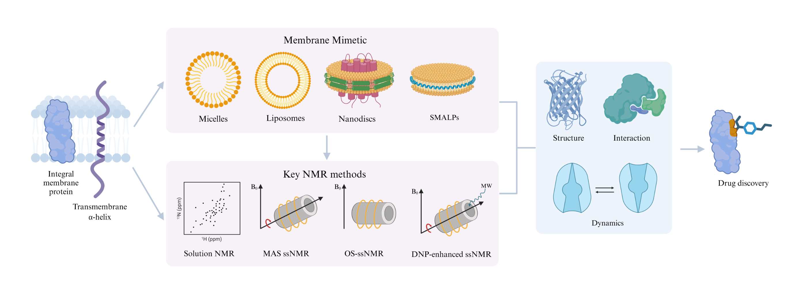
As the functional core of biological membranes, membrane proteins mediate nearly all critical cellular processes, and their dysfunction is closely associated with the onset and progression of various major diseases. However, the inherent hydrophobicity of transmembrane domains, dependence on native lipid microenvironments, and prominent structural dynamics of membrane proteins pose substantial challenges to their structural and functional research. Although X-ray crystallography and cryo-electron microscopy (cryo-EM) are mainstream structural biology techniques, they each have limitations in membrane protein studies. Nuclear magnetic resonance (NMR) spectroscopy, as a powerful complementary approach, enables atomic-resolution structural analysis of membrane proteins in selected native-like membrane environments or in situ membrane systems, and can sensitively characterize conformational dynamics across multiple timescales as well as intermolecular interactions, offering unique advantages in membrane protein research. This review focuses on the core advances of NMR technology in membrane protein studies, systematically summarizes the key membrane-mimetic systems adapted for NMR analysis and the methodological developments of solution NMR, solid-state NMR (including magic-angle spinning, oriented-sample NMR, and dynamic nuclear polarization-enhanced NMR), as well as in situ/intracellular NMR. It further elaborates on the application achievements of NMR in membrane protein structure determination, molecular interaction analysis, and conformational dynamics research, aiming to provide a comprehensive reference for the in-depth investigation of membrane protein structure and function, and the development of membrane protein-targeted drugs.
- Open Access
- Review
Advances in NMR Studies of Membrane Protein Structure and Function
Author Information
Received: 07 June 2026 | Revised: 08 September 2026 | Accepted: 08 September 2026 | Published: 18 November 2026
Abstract
Graphical Abstract
Keywords
NMR | membrane proteins | MAS ssNMR | OS-ssNMR | DNP NMR
References
- 1.
Gromiha, M.M.; Ou, Y.Y. Bioinformatics approaches for functional annotation of membrane proteins. Brief. Bioinform. 2014, 15, 155–168. https://doi.org/10.1093/bib/bbt015.
- 2.
Myers, J.K.; Beihoffer, L.A.; Sanders, C.R. Phenotology of disease-linked proteins. Hum. Mutat. 2005, 25, 90–97. https://doi.org/10.1002/humu.20118.
- 3.
Lundstrom, K. Structural genomics and drug discovery. J. Cell. Mol. Med. 2007, 11, 224–238. https://doi.org/10.1111/j.1582-4934.2007.00028.x.
- 4.
Srivastava, A.; Nagai, T.; Srivastava, A.; Miyashita, O.; Tama, F. Role of Computational Methods in Going beyond X-ray Crystallography to Explore Protein Structure and Dynamics. Int. J. Mol. Sci. 2018, 19, 3401. https://doi.org/10.3390/ijms19113401.
- 5.
Smyth, M.S.; Martin, J.H.J. X-ray crystallography. Mol. Pathol. 2000, 53, 8–14. https://doi.org/10.1136/mp.53.1.8.
- 6.
Boulos, I.; Jabbour, J.; Khoury, S.; Mikhael, N.; Tishkova, V.; Candoni, N.; Ghadieh, H.E.; Veesler, S.; Bassim, Y.; Azar, S.; et al. Exploring the World of Membrane Proteins: Techniques and Methods for Understanding Structure, Function, and Dynamics. Molecules 2023, 28, 7176. https://doi.org/10.3390/molecules28207176.
- 7.
Earl, L.A.; Falconieri, V.; Milne, J.L.S.; Subramaniam, S. Cryo-EM: Beyond the microscope. Curr. Opin. Struct. Biol. 2017, 46, 71–78. https://doi.org/10.1016/j.sbi.2017.06.002.
- 8.
Murata, K.; Wolf, M. Cryo-electron microscopy for structural analysis of dynamic biological macromolecules. Biochim. Biophys. Acta Gen. Subj. 2018, 1862, 324–334. https://doi.org/10.1016/j.bbagen.2017.07.020.
- 9.
Yip, K.M.; Fischer, N.; Paknia, E.; Chari, A.; Stark, H. Atomic-resolution protein structure determination by cryo-EM. Nature 2020, 587, 157–161. https://doi.org/10.1038/s41586-020-2833-4.
- 10.
Ahmed, R.; Forman-Kay, J.D. NMR insights into dynamic, multivalent interactions of intrinsically disordered regions: From discrete complexes to condensates. Essays Biochem. 2022, 66, 863–873. https://doi.org/10.1042/ebc20220056.
- 11.
Wagner, S.; Bader, M.L.; Drew, D.; de Gier, J.W. Rationalizing membrane protein overexpression. Trends Biotechnol. 2006, 24, 364–371. https://doi.org/10.1016/j.tibtech.2006.06.008.
- 12.
Pandey, A.; Shin, K.; Patterson, R.E.; Liu, X.Q.; Rainey, J.K. Current strategies for protein production and purification enabling membrane protein structural biology. Biochem. Cell Biol. 2016, 94, 507–527. https://doi.org/10.1139/bcb-2015-0143.
- 13.
Marley, J.; Lu, M.; Bracken, C. A method for efficient isotopic labeling of recombinant proteins. J. Biomol. NMR 2001, 20, 71–75. https://doi.org/10.1023/a:1011254402785.
- 14.
Danmaliki, G.I.; Hwang, P.M. Solution NMR spectroscopy of membrane proteins. Biochim. Biophys. Acta Biomembr. 2020, 1862, 183356. https://doi.org/10.1016/j.bbamem.2020.183356.
- 15.
Klammt, C.; Schwarz, D.; Löhr, F.; Schneider, B.; Dötsch, V.; Bernhard, F. Cell-free expression as an emerging technique for the large scale production of integral membrane protein. FEBS J. 2006, 273, 4141–4153. https://doi.org/10.1111/j.1742-4658.2006.05432.x.
- 16.
Pacull, E.M.; Sendker, F.; Bernhard, F.; Scheidt, H.A.; Schmidt, P.; Huster, D.; Krug, U. Integration of Cell-Free Expression and Solid-State NMR to Investigate the Dynamic Properties of Different Sites of the Growth Hormone Secretagogue Receptor. Front. Pharmacol. 2020, 11, 562113. https://doi.org/10.3389/fphar.2020.562113.
- 17.
Rose-Sperling, D.; Tran, M.A.; Lauth, L.M.; Goretzki, B.; Hellmich, U.A. 19F NMR as a versatile tool to study membrane protein structure and dynamics. Biol. Chem. 2019, 400, 1277–1288. https://doi.org/10.1515/hsz-2018-0473.
- 18.
Drewniak, P.; Su, C.Y.C.; Francisco, C.B.; Prosser, R.S. Recent (19)F NMR Applications to the Study of Membrane Proteins and Protein Complexes. J. Mol. Biol. 2025, 437, 169434. https://doi.org/10.1016/j.jmb.2025.169434.
- 19.
LaGuerre, A.; Löhr, F.; Bernhard, F.; Dötsch, V. Labeling of membrane proteins by cell-free expression. Methods Enzymol. 2015, 565, 367–388. https://doi.org/10.1016/bs.mie.2015.06.001.
- 20.
Sobhanifar, S.; Reckel, S.; Junge, F.; Schwarz, D.; Kai, L.; Karbyshev, M.; Löhr, F.; Bernhard, F.; Dötsch, V. Cell-free expression and stable isotope labelling strategies for membrane proteins. J. Biomol. NMR 2010, 46, 33–43. https://doi.org/10.1007/s10858-009-9364-5.
- 21.
Yadav, R.; Gupta, D.P.; Singh, C. Lock and key: Quest to find the most compatible membrane mimetic for studying membrane proteins in native environment. Biochim. Biophys. Acta Biomembr. 2025, 1867, 184414. https://doi.org/10.1016/j.bbamem.2025.184414.
- 22.
Majeed, S.; Ahmad, A.B.; Sehar, U.; Georgieva, E.R. Lipid Membrane Mimetics in Functional and Structural Studies of Integral Membrane Proteins. Membranes 2021, 11, 685. https://doi.org/10.3390/membranes11090685.
- 23.
Puthenveetil, R.; Vinogradova, O. Solution NMR: A powerful tool for structural and functional studies of membrane proteins in reconstituted environments. J. Biol. Chem. 2019, 294, 15914–15931. https://doi.org/10.1074/jbc.rev119.009178.
- 24.
Zhang, Q.; Horst, R.; Geralt, M.; Ma, X.; Hong, W.X.; Finn, M.G.; Stevens, R.C.; Wüthrich, K. Microscale NMR screening of new detergents for membrane protein structural biology. J. Am. Chem. Soc. 2008, 130, 7357–7363. https://doi.org/10.1021/ja077863d.
- 25.
Gutmann, D.A.P.; Mizohata, E.; Newstead, S.; Ferrandon, S.; Henderson, P.J.F.; van Veen, H.W.; Byrne, B. A high-throughput method for membrane protein solubility screening: The ultracentrifugation dispersity sedimentation assay. Protein Sci. 2007, 16, 1422–1428. https://doi.org/10.1110/ps.072759907.
- 26.
Yeh, V.; Goode, A.; Bonev, B.B. Membrane Protein Structure Determination and Characterisation by Solution and Solid-State NMR. Biology 2020, 9, 396. https://doi.org/10.3390/biology9110396.
- 27.
Porcelli, F.; Ramamoorthy, A.; Barany, G.; Veglia, G. On the role of NMR spectroscopy for characterization of antimicrobial peptides. Methods Mol. Biol. 2013, 1063, 159–180. https://doi.org/10.1007/978-1-62703-583-5_9.
- 28.
Klöpfer, K.; Hagn, F. Beyond detergent micelles: The advantages and applications of non-micellar and lipid-based membrane mimetics for solution-state NMR. Prog. Nucl. Magn. Reson. Spectrosc. 2019, 114–115, 271–283. https://doi.org/10.1016/j.pnmrs.2019.08.001.
- 29.
Galiakhmetov, A.R.; Shah, A.A.; Lane, A.; Davern, C.M.; Proulx, C.; Nevzorov, A.A. Peptoid-based macrodiscs of variable lipid composition for structural studies of membrane proteins by oriented-sample solid-state NMR. J. Struct. Biol. X 2024, 9, 100095. https://doi.org/10.1016/j.yjsbx.2023.100095.
- 30.
Poget, S.F.; Girvin, M.E. Solution NMR of membrane proteins in bilayer mimics: Small is beautiful, but sometimes bigger is better. Biochim. Biophys. Acta 2007, 1768, 3098–3106. https://doi.org/10.1016/j.bbamem.2007.09.006.
- 31.
Fu, Q.; Shaik, M.M.; Cai, Y.; Ghantous, F.; Piai, A.; Peng, H.; Rits-Volloch, S.; Liu, Z.; Harrison, S.C.; Seaman, M.S.; et al. Structure of the membrane proximal external region of HIV-1 envelope glycoprotein. Proc. Natl. Acad. Sci. USA 2018, 115, E8892–E8899. https://doi.org/10.1073/pnas.1807259115.
- 32.
Fu, Q.; Fu, T.M.; Cruz, A.C.; Sengupta, P.; Thomas, S.K.; Wang, S.; Siegel, R.M.; Wu, H.; Chou, J.J. Structural Basis and Functional Role of Intramembrane Trimerization of the Fas/CD95 Death Receptor. Mol. Cell 2016, 61, 602–613. https://doi.org/10.1016/j.molcel.2016.01.009.
- 33.
Kroncke, B.M.; Van Horn, W.D.; Smith, J.; Kang, C.; Welch, R.C.; Song, Y.; Nannemann, D.P.; Taylor, K.C.; Sisco, N.J.; George, A.L.; et al. Structural basis for KCNE3 modulation of potassium recycling in epithelia. Sci. Adv. 2016, 2, e1501228. https://doi.org/10.1126/sciadv.1501228.
- 34.
Morrison, E.A.; Henzler-Wildman, K.A. Reconstitution of integral membrane proteins into isotropic bicelles with improved sample stability and expanded lipid composition profile. Biochim. Biophys. Acta 2012, 1818, 814–820. https://doi.org/10.1016/j.bbamem.2011.12.020.
- 35.
Kononova, P.A.; Selyutina, O.Y.; Polyakov, N.E. The Interaction of the Transmembrane Domain of SARS-CoV-2 E-Protein with Glycyrrhizic Acid in Lipid Bilayer. Membranes 2023, 13, 505. https://doi.org/10.3390/membranes13050505.
- 36.
Carlier, L.; Samson, D.; Khemtemourian, L.; Joliot, A.; Fuchs, P.F.J.; Lequin, O. Anionic lipids induce a fold-unfold transition in the membrane-translocating Engrailed homeodomain. Biochim. Biophys. Acta Biomembr 2022, 1864, 184030. https://doi.org/10.1016/j.bbamem.2022.184030.
- 37.
Bayburt, T.H.; Carlson, J.W.; Sligar, S.G. Reconstitution and imaging of a membrane protein in a nanometer-size phospholipid bilayer. J. Struct. Biol. 1998, 123, 37–44. https://doi.org/10.1006/jsbi.1998.4007.
- 38.
Liang, B.; Tamm, L.K. NMR as a tool to investigate the structure, dynamics and function of membrane proteins. Nat. Struct. Mol. Biol. 2016, 23, 468–474. https://doi.org/10.1038/nsmb.3226.
- 39.
Denisov, I.G.; Sligar, S.G. Nanodiscs for the study of membrane proteins. Curr. Opin. Struct. Biol. 2024, 87, 102844. https://doi.org/10.1016/j.sbi.2024.102844.
- 40.
Hagn, F.; Etzkorn, M.; Raschle, T.; Wagner, G. Optimized phospholipid bilayer nanodiscs facilitate high-resolution structure determination of membrane proteins. J. Am. Chem. Soc. 2013, 135, 1919–1925. https://doi.org/10.1021/ja310901f.
- 41.
Puthenveetil, R.; Nguyen, K.; Vinogradova, O. Nanodiscs and Solution NMR: Preparation, application and challenges. Nanotechnol. Rev. 2017, 6, 111–126. https://doi.org/10.1515/ntrev-2016-0076.
- 42.
Jamshad, M.; Grimard, V.; Idini, I.; Knowles, T.J.; Dowle, M.R.; Schofield, N.; Sridhar, P.; Lin, Y.; Finka, R.; Wheatley, M.; et al. Structural analysis of a nanoparticle containing a lipid bilayer used for detergent-free extraction of membrane proteins. Nano Res. 2015, 8, 774–789. https://doi.org/10.1007/s12274-014-0560-6.
- 43.
Vinogradova, O. Nanodiscs and solution nuclear magnetic resonance. Curr. Opin. Struct. Biol. 2025, 93, 103067. https://doi.org/10.1016/j.sbi.2025.103067.
- 44.
Yasuhara, K.; Arakida, J.; Ravula, T.; Ramadugu, S.K.; Sahoo, B.; Kikuchi, J.I.; Ramamoorthy, A. Spontaneous Lipid Nanodisc Fomation by Amphiphilic Polymethacrylate Copolymers. J. Am. Chem. Soc. 2017, 139, 18657–18663. https://doi.org/10.1021/jacs.7b10591.
- 45.
Ravula, T.; Hardin, N.Z.; Ramadugu, S.K.; Cox, S.J.; Ramamoorthy, A. Formation of pH-Resistant Monodispersed Polymer-Lipid Nanodiscs. Angew. Chem. Int. Ed. 2018, 57, 1342–1345. https://doi.org/10.1002/anie.201712017.
- 46.
Walters, S.H.; Birchfield, A.S.; Fuglestad, B. Advances in utilizing reverse micelles to investigate membrane proteins. Biochem. Soc. Trans. 2024, 52, 2499–2511. https://doi.org/10.1042/bst20240830.
- 47.
Marques, B.S.; Nucci, N.V.; Dodevski, I.; Wang, K.W.C.; Athanasoula, E.A.; Jorge, C.; Wand, A.J. Measurement and control of pH in the aqueous interior of reverse micelles. J. Phys. Chem. B 2014, 118, 2020–2031. https://doi.org/10.1021/jp4103349.
- 48.
Dodevski, I.; Nucci, N.V.; Valentine, K.G.; Sidhu, G.K.; O’Brien, E.S.; Pardi, A.; Wand, A.J. Optimized reverse micelle surfactant system for high-resolution NMR spectroscopy of encapsulated proteins and nucleic acids dissolved in low viscosity fluids. J. Am. Chem. Soc. 2014, 136, 3465–3474. https://doi.org/10.1021/ja410716w.
- 49.
Labrecque, C.L.; Nolan, A.L.; Develin, A.M.; Castillo, A.J.; Offenbacher, A.R.; Fuglestad, B. Membrane-Mimicking Reverse Micelles for High-Resolution Interfacial Study of Proteins and Membranes. Langmuir 2022, 38, 3676–3686. https://doi.org/10.1021/acs.langmuir.1c03085.
- 50.
Howarth, G.S.; McDermott, A.E. High-Resolution Magic Angle Spinning NMR of KcsA in Liposomes: The Highly Mobile C-Terminus. Biomolecules 2022, 12, 1122. https://doi.org/10.3390/biom12081122.
- 51.
Ong, Y.S.; Lakatos, A.; Becker-Baldus, J.; Pos, K.M.; Glaubitz, C. Detecting substrates bound to the secondary multidrug efflux pump EmrE by DNP-enhanced solid-state NMR. J. Am. Chem. Soc. 2013, 135, 15754–15762. https://doi.org/10.1021/ja402605s.
- 52.
Williamson, M.P. Using chemical shift perturbation to characterise ligand binding. Prog. Nucl. Magn. Reson. Spectrosc. 2013, 73, 1–16. https://doi.org/10.1016/j.pnmrs.2013.02.001.
- 53.
Luchinat, E.; Banci, L. In-cell NMR: From target structure and dynamics to drug screening. Curr. Opin. Struct. Biol. 2022, 74, 102374. https://doi.org/10.1016/j.sbi.2022.102374.
- 54.
Zuiderweg, E.R.P. Mapping protein-protein interactions in solution by NMR spectroscopy. Biochemistry 2002, 41, 1–7. https://doi.org/10.1021/bi011870b.
- 55.
Pervushin, K.; Riek, R.; Wider, G.; Wüthrich, K. Attenuated T2 relaxation by mutual cancellation of dipole-dipole coupling and chemical shift anisotropy indicates an avenue to NMR structures of very large biological macromolecules in solution. Proc. Natl. Acad. Sci. USA 1997, 94, 12366–12371. https://doi.org/10.1073/pnas.94.23.12366.
- 56.
McCoy, M.A.; Wyss, D.F. Spatial localization of ligand binding sites from electron current density surfaces calculated from NMR chemical shift perturbations. J. Am. Chem. Soc. 2002, 124, 11758–11763. https://doi.org/10.1021/ja026166c.
- 57.
Creutznacher, R.; Maass, T.; Veselkova, B.; Ssebyatika, G.; Krey, T.; Empting, M.; Tautz, N.; Frank, M.; Kölbel, K.; Uetrecht, C.; et al. NMR Experiments Provide Insights into Ligand-Binding to the SARS-CoV-2 Spike Protein Receptor-Binding Domain. J. Am. Chem. Soc. 2022, 144, 13060–13065. https://doi.org/10.1021/jacs.2c05603.
- 58.
Maass, T.; Ssebyatika, G.; Brückner, M.; Breckwoldt, L.; Krey, T.; Mallagaray, A.; Peters, T.; Frank, M.; Creutznacher, R. Binding of Glycans to the SARS CoV-2 Spike Protein, an Open Question: NMR Data on Binding Site Localization, Affinity, and Selectivity. Chem. Eur. J. 2022, 28, e202202614. https://doi.org/10.1002/chem.202202614.
- 59.
Gautier, A.; Mott, H.R.; Bostock, M.J.; Kirkpatrick, J.P.; Nietlispach, D. Structure determination of the seven-helix transmembrane receptor sensory rhodopsin II by solution NMR spectroscopy. Nat. Struct. Mol. Biol. 2010, 17, 768–774. https://doi.org/10.1038/nsmb.1807.
- 60.
Reckel, S.; Gottstein, D.; Stehle, J.; Löhr, F.; Verhoefen, M.K.; Takeda, M.; Silvers, R.; Kainosho, M.; Glaubitz, C.; Wachtveitl, J.; et al. Solution NMR Structure of Proteorhodopsin. Angew. Chem. Int. Ed. 2011, 50, 11942–11946. https://doi.org/10.1002/anie.201105648.
- 61.
Gopinath, T.; Weber, D.; Wang, S.; Larsen, E.; Veglia, G. Solid-State NMR of Membrane Proteins in Lipid Bilayers: To Spin or Not to Spin? Acc. Chem. Res. 2021, 54, 1430–1439. https://doi.org/10.1021/acs.accounts.0c00670.
- 62.
Hagn, F.; Wagner, G. Structure refinement and membrane positioning of selectively labeled OmpX in phospholipid nanodiscs. J. Biomol. NMR 2015, 61, 249–260. https://doi.org/10.1007/s10858-014-9883-6.
- 63.
Lesovoy, D.M.; Mineev, K.S.; Bragin, P.E.; Bocharova, O.V.; Bocharov, E.V.; Arseniev, A.S. NMR relaxation parameters of methyl groups as a tool to map the interfaces of helix-helix interactions in membrane proteins. J. Biomol. NMR 2017, 69, 165–179. https://doi.org/10.1007/s10858-017-0146-1.
- 64.
Eddy, M.T.; Su, Y.; Silvers, R.; Andreas, L.; Clark, L.; Wagner, G.; Pintacuda, G.; Emsley, L.; Griffin, R.G. Lipid bilayer-bound conformation of an integral membrane beta barrel protein by multidimensional MAS NMR. J. Biomol. NMR 2015, 61, 299–310. https://doi.org/10.1007/s10858-015-9903-1.
- 65.
Mandala, V.S.; Hong, M. High-sensitivity protein solid-state NMR spectroscopy. Curr. Opin. Struct. Biol. 2019, 58, 183–190. https://doi.org/10.1016/j.sbi.2019.03.027.
- 66.
Zhou, D.H.; Nieuwkoop, A.J.; Berthold, D.A.; Comellas, G.; Sperling, L.J.; Tang, M.; Shah, G.J.; Brea, E.J.; Lemkau, L.R.; Rienstra, C.M. Solid-state NMR analysis of membrane proteins and protein aggregates by proton detected spectroscopy. J. Biomol. NMR 2012, 54, 291–305. https://doi.org/10.1007/s10858-012-9672-z.
- 67.
Nimerovsky, E.; Movellan, K.T.; Zhang, X.C.; Forster, M.C.; Najbauer, E.; Xue, K.; Dervişoǧlu, R.; Giller, K.; Griesinger, C.; Becker, S.; et al. Proton Detected Solid-State NMR of Membrane Proteins at 28 Tesla (1.2 GHz) and 100 kHz Magic-Angle Spinning. Biomolecules 2021, 11, 752. https://doi.org/10.3390/biom11050752.
- 68.
Weingarth, M.; van der Cruijsen, E.A.W.; Ostmeyer, J.; Lievestro, S.; Roux, B.; Baldus, M. Quantitative analysis of the water occupancy around the selectivity filter of a K+ channel in different gating modes. J. Am. Chem. Soc. 2014, 136, 2000–2007. https://doi.org/10.1021/ja411450y.
- 69.
Lalli, D.; Idso, M.N.; Andreas, L.B.; Hussain, S.; Baxter, N.; Han, S.; Chmelka, B.F.; Pintacuda, G. Proton-Based Structural Analysis of a Heptahelical Transmembrane Protein in Lipid Bilayers. J. Am. Chem. Soc. 2017, 139, 13006–13012. https://doi.org/10.1021/jacs.7b05269.
- 70.
Sun, Z.; Ollier, C.; Rancz, A.; Thienpont, B.; Grohe, K.; Becker, L.; Purea, A.; Engelke, F.; Wegner, S.; Sturgis, J.; et al. Pushing the Boundaries of Resolution in Solid-State Nuclear Magnetic Resonance of Biomolecules with 160 kHz Magic-Angle Spinning. J. Am. Chem. Soc. 2025, 147, 19433–19437. https://doi.org/10.1021/jacs.5c02466.
- 71.
Forster, M.C.; Tekwani Movellan, K.; Najbauer, E.E.; Becker, S.; Andreas, L.B. Magic-angle spinning NMR structure of Opa60 in lipid bilayers. J. Struct. Biol. X 2024, 9, 100098. https://doi.org/10.1016/j.yjsbx.2024.100098.
- 72.
Hallock, K.J.; Henzler Wildman, K.; Lee, D.K.; Ramamoorthy, A. An innovative procedure using a sublimable solid to align lipid bilayers for solid-state NMR studies. Biophys. J. 2002, 82, 2499–2503. https://doi.org/10.1016/s0006-3495(02)75592-6.
- 73.
Sanders, C.R.; Landis, G.C. Reconstitution of membrane proteins into lipid-rich bilayered mixed micelles for NMR studies. Biochemistry 1995, 34, 4030–4040. https://doi.org/10.1021/bi00012a022.
- 74.
Kamihira, M.; Vosegaard, T.; Mason, A.J.; Straus, S.K.; Nielsen, N.C.; Watts, A. Structural and orientational constraints of bacteriorhodopsin in purple membranes determined by oriented-sample solid-state NMR spectroscopy. J. Struct. Biol. 2005, 149, 7–16. https://doi.org/10.1016/j.jsb.2004.10.002.
- 75.
Park, S.H.; Mrse, A.A.; Nevzorov, A.A.; Mesleh, M.F.; Oblatt-Montal, M.; Montal, M.; Opella, S.J. Three-dimensional structure of the channel-forming trans-membrane domain of virus protein “u” (Vpu) from HIV-1. J. Mol. Biol. 2003, 333, 409–424. https://doi.org/10.1016/j.jmb.2003.08.048.
- 76.
Gayen, A.; Banigan, J.R.; Traaseth, N.J. Ligand-induced conformational changes of the multidrug resistance transporter EmrE probed by oriented solid-state NMR spectroscopy. Angew. Chem. Int. Ed. 2013, 52, 10321–10324. https://doi.org/10.1002/anie.201303091.
- 77.
Das, N.; Dai, J.; Hung, I.; Rajagopalan, M.; Zhou, H.X.; Cross, T.A. Structure of CrgA, a cell division structural and regulatory protein from Mycobacterium tuberculosis, in lipid bilayers. Proc. Natl. Acad. Sci. USA 2015, 112, E119–26. https://doi.org/10.1073/pnas.1415908112.
- 78.
Biedenbänder, T.; Aladin, V.; Saeidpour, S.; Corzilius, B. Dynamic Nuclear Polarization for Sensitivity Enhancement in Biomolecular Solid-State NMR. Chem. Rev. 2022, 122, 9738–9794. https://doi.org/10.1021/acs.chemrev.1c00776.
- 79.
Kent, J.E.; Ackermann, B.E.; Debelouchina, G.T.; Marassi, F.M. Dynamic Nuclear Polarization Illuminates Key Protein-Lipid Interactions in the Native Bacterial Cell Envelope. Biochemistry 2023, 62, 2252–2256. https://doi.org/10.1021/acs.biochem.3c00262.
- 80.
Spadaccini, R.; Kaur, H.; Becker-Baldus, J.; Glaubitz, C. The effect of drug binding on specific sites in transmembrane helices 4 and 6 of the ABC exporter MsbA studied by DNP-enhanced solid-state NMR. Biochim. Biophys. Acta Biomembr. 2018, 1860, 833–840. https://doi.org/10.1016/j.bbamem.2017.10.017.
- 81.
Jaroniec, C.P.; Kaufman, J.D.; Stahl, S.J.; Viard, M.; Blumenthal, R.; Wingfield, P.T.; Bax, A. Structure and Dynamics of Micelle-Associated Human Immunodeficiency Virus gp41 Fusion Domain. Biochemistry 2005, 44, 16167–16180. https://doi.org/10.1021/bi051672a.
- 82.
Pan, L.; Fu, T.M.; Zhao, W.; Zhao, L.; Chen, W.; Qiu, C.; Liu, W.; Liu, Z.; Piai, A.; Fu, Q.; et al. Higher-Order Clustering of the Transmembrane Anchor of DR5 Drives Signaling. Cell 2019, 176, 1477–1489.e14. https://doi.org/10.1016/j.cell.2019.02.001.
- 83.
Liang, B.; Arora, A.; Tamm, L.K. Fast-time scale dynamics of outer membrane protein A by extended model-free analysis of NMR relaxation data. Biochim. Biophys. Acta 2010, 1798, 68–76. https://doi.org/10.1016/j.bbamem.2009.07.022.
- 84.
Fernández, C.; Adeishvili, K.; Wüthrich, K. Transverse relaxation-optimized NMR spectroscopy with the outer membrane protein OmpX in dihexanoyl phosphatidylcholine micelles. Proc. Natl. Acad. Sci. USA 2001, 98, 2358–2363. https://doi.org/10.1073/pnas.051629298.
- 85.
Hiller, S.; Garces, R.G.; Malia, T.J.; Orekhov, V.Y.; Colombini, M.; Wagner, G. Solution structure of the integral human membrane protein VDAC-1 in detergent micelles. Science 2008, 321, 1206–1210. https://doi.org/10.1126/science.1161302.
- 86.
Park, S.H.; Das, B.B.; Casagrande, F.; Tian, Y.; Nothnagel, H.J.; Chu, M.; Kiefer, H.; Maier, K.; De Angelis, A.A.; Marassi, F.M.; et al. Structure of the chemokine receptor CXCR1 in phospholipid bilayers. Nature 2012, 491, 779–783. https://doi.org/10.1038/nature11580.
- 87.
Zhuang, T.; Chen, Q.; Cho, M.K.; Vishnivetskiy, S.A.; Iverson, T.M.; Gurevich, V.V.; Sanders, C.R. Involvement of distinct arrestin-1 elements in binding to different functional forms of rhodopsin. Proc. Natl. Acad. Sci. USA 2013, 110, 942–947. https://doi.org/10.1073/pnas.1215176110.
- 88.
Berardi, M.J.; Shih, W.M.; Harrison, S.C.; Chou, J.J. Mitochondrial uncoupling protein 2 structure determined by NMR molecular fragment searching. Nature 2011, 476, 109–113. https://doi.org/10.1038/nature10257.
- 89.
Mandala, V.S.; Williams, J.K.; Hong, M. Structure and Dynamics of Membrane Proteins from Solid-State NMR. Annu. Rev. Biophys. 2018, 47, 201–222. https://doi.org/10.1146/annurev-biophys-070816-033712.
- 90.
Roos, M.; Wang, T.; Shcherbakov, A.A.; Hong, M. Fast Magic-Angle-Spinning (19)F Spin Exchange NMR for Determining Nanometer (19)F-(19)F Distances in Proteins and Pharmaceutical Compounds. J. Phys. Chem. B 2018, 122, 2900–2911. https://doi.org/10.1021/acs.jpcb.8b00310.
- 91.
Shcherbakov, A.A.; Hong, M. Rapid measurement of long-range distances in proteins by multidimensional (13)C-(19)F REDOR NMR under fast magic-angle spinning. J. Biomol. NMR 2018, 71, 31–43. https://doi.org/10.1007/s10858-018-0187-0.
- 92.
Elkins, M.R.; Sergeyev, I.V.; Hong, M. Determining Cholesterol Binding to Membrane Proteins by Cholesterol (13)C Labeling in Yeast and Dynamic Nuclear Polarization NMR. J. Am. Chem. Soc. 2018, 140, 15437–15449. https://doi.org/10.1021/jacs.8b09658.
- 93.
Barrett, P.J.; Song, Y.; Van Horn, W.D.; Hustedt, E.J.; Schafer, J.M.; Hadziselimovic, A.; Beel, A.J.; Sanders, C.R. The amyloid precursor protein has a flexible transmembrane domain and binds cholesterol. Science 2012, 336, 1168–1171. https://doi.org/10.1126/science.1219988.
- 94.
Eddy, M.T.; Ong, T.C.; Clark, L.; Teijido, O.; van der Wel, P.C.A.; Garces, R.; Wagner, G.; Rostovtseva, T.K.; Griffin, R.G. Lipid dynamics and protein-lipid interactions in 2D crystals formed with the β-barrel integral membrane protein VDAC1. J. Am. Chem. Soc. 2012, 134, 6375–6387. https://doi.org/10.1021/ja300347v.
- 95.
Ueda, T.; Takeuchi, K.; Nishida, N.; Stampoulis, P.; Kofuku, Y.; Osawa, M.; Shimada, I. Cross-saturation and transferred cross-saturation experiments. Q. Rev. Biophys. 2014, 47, 143–187. https://doi.org/10.1017/s0033583514000043.
- 96.
Kofuku, Y.; Yoshiura, C.; Ueda, T.; Terasawa, H.; Hirai, T.; Tominaga, S.; Hirose, M.; Maeda, Y.; Takahashi, H.; Terashima, Y.; et al. Structural basis of the interaction between chemokine stromal cell-derived factor-1/CXCL12 and its G-protein-coupled receptor CXCR4. J. Biol. Chem. 2009, 284, 35240–35250. https://doi.org/10.1074/jbc.m109.024851.
- 97.
Xiang, S.; Pinto, C.; Baldus, M. Divide and Conquer: A Tailored Solid-state NMR Approach to Study Large Membrane Protein Complexes. Angew. Chem. Int. Ed. 2022, 61, e202203319. https://doi.org/10.1002/anie.202203319.
- 98.
Pinto, C.; Mance, D.; Julien, M.; Daniels, M.; Weingarth, M.; Baldus, M. Studying assembly of the BAM complex in native membranes by cellular solid-state NMR spectroscopy. J. Struct. Biol. 2019, 206, 1–11. https://doi.org/10.1016/j.jsb.2017.11.015.
- 99.
Takeuchi, K.; Kofuku, Y.; Imai, S.; Ueda, T.; Tokunaga, Y.; Toyama, Y.; Shiraishi, Y.; Shimada, I. Function-Related Dynamics in Multi-Spanning Helical Membrane Proteins Revealed by Solution NMR. Membranes 2021, 11, 604. https://doi.org/10.3390/membranes11080604.
- 100.
Palmer, A.G. NMR characterization of the dynamics of biomacromolecules. Chem. Rev. 2004, 104, 3623–3640. https://doi.org/10.1021/cr030413t.
- 101.
O’Brien, E.S.; Fuglestad, B.; Lessen, H.J.; Stetz, M.A.; Lin, D.W.; Marques, B.S.; Gupta, K.; Fleming, K.G.; Wand, A.J. Membrane Proteins Have Distinct Fast Internal Motion and Residual Conformational Entropy. Angew. Chem. Int. Ed. 2020, 59, 11108–11114. https://doi.org/10.1002/anie.202003527.
- 102.
Hwang, P.M.; Bishop, R.E.; Kay, L.E. The integral membrane enzyme PagP alternates between two dynamically distinct states. Proc. Natl. Acad. Sci. USA 2004, 101, 9618–9623. https://doi.org/10.1073/pnas.0402324101.
- 103.
Jaremko, M.; Jaremko, Ł.; Villinger, S.; Schmidt, C.D.; Griesinger, C.; Becker, S.; Zweckstetter, M. High-Resolution NMR Determination of the Dynamic Structure of Membrane Proteins. Angew. Chem. Int. Ed. 2016, 55, 10518–10521. https://doi.org/10.1002/anie.201602639.
- 104.
Brüschweiler, S.; Yang, Q.; Run, C.; Chou, J.J. Substrate-modulated ADP/ATP-transporter dynamics revealed by NMR relaxation dispersion. Nat. Struct. Mol. Biol. 2015, 22, 636–641. https://doi.org/10.1038/nsmb.3059.
- 105.
Zhuang, T.; Chisholm, C.; Chen, M.; Tamm, L.K. NMR-Based Conformational Ensembles Explain pH-Gated Opening and Closing of OmpG Channel. J. Am. Chem. Soc. 2013, 135, 15101–15113. https://doi.org/10.1021/ja408206e.
- 106.
Cho, M.K.; Gayen, A.; Banigan, J.R.; Leninger, M.; Traaseth, N.J. Intrinsic conformational plasticity of native EmrE provides a pathway for multidrug resistance. J. Am. Chem. Soc. 2014, 136, 8072–8080. https://doi.org/10.1021/ja503145x.
- 107.
Iwahashi, Y.; Toyama, Y.; Imai, S.; Itoh, H.; Osawa, M.; Inoue, M.; Shimada, I. Conformational equilibrium shift underlies altered K(+) channel gating as revealed by NMR. Nat. Commun. 2020, 11, 5168. https://doi.org/10.1038/s41467-020-19005-3.
- 108.
Enkavi, G.; Javanainen, M.; Kulig, W.; Róg, T.; Vattulainen, I. Multiscale Simulations of Biological Membranes: The Challenge To Understand Biological Phenomena in a Living Substance. Chem. Rev. 2019, 119, 5607–5774. https://doi.org/10.1021/acs.chemrev.8b00538.
- 109.
Marassi, F.M.; Pintacuda, G. Solid-state NMR of membrane proteins in situ. Curr. Opin. Struct. Biol. 2025, 94, 103129. https://doi.org/10.1016/j.sbi.2025.103129.
- 110.
Ward, M.E.; Wang, S.; Munro, R.; Ritz, E.; Hung, I.; Gor’kov, P.L.; Jiang, Y.; Liang, H.; Brown, L.S.; Ladizhansky, V. In situ structural studies of Anabaena sensory rhodopsin in the E. coli membrane. Biophys. J. 2015, 108, 1683–1696. https://doi.org/10.1016/j.bpj.2015.02.018.
- 111.
Kumar, K.; Sebastiao, M.; Arnold, A.A.; Bourgault, S.; Warschawski, D.E.; Marcotte, I. In situ solid-state NMR study of antimicrobial peptide interactions with erythrocyte membranes. Biophys. J. 2022, 121, 1512–1524. https://doi.org/10.1016/j.bpj.2022.03.009.
- 112.
Ding, X.; Sun, C.; Cui, H.; Chen, S.; Gao, Y.; Yang, Y.; Wang, J.; He, X.; Iuga, D.; Tian, F.; et al. Functional roles of tyrosine 185 during the bacteriorhodopsin photocycle as revealed by in situ spectroscopic studies. Biochim. Biophys. Acta Bioenerg. 2018, 1859, 1006–1014. https://doi.org/10.1016/j.bbabio.2018.05.011.
- 113.
Höfurthner, T.; Mateos, B.; Konrat, R. On-Cell NMR Contributions to Membrane Receptor Binding Characterization. Chempluschem 2021, 86, 938–945. https://doi.org/10.1002/cplu.202100134.
- 114.
Narasimhan, S.; Scherpe, S.; Lucini Paioni, A.; van der Zwan, J.; Folkers, G.E.; Ovaa, H.; Baldus, M. DNP-Supported Solid-State NMR Spectroscopy of Proteins Inside Mammalian Cells. Angew. Chem. Int. Ed. 2019, 58, 12969–12973. https://doi.org/10.1002/anie.201903246.
- 115.
Ikari, M.; Yagi, H.; Kasai, T.; Inomata, K.; Ito, M.; Higuchi, K.; Matsuda, N.; Ito, Y.; Kigawa, T. Direct Observation of Membrane-Associated H-Ras in the Native Cellular Environment by In-Cell (19)F-NMR Spectroscopy. JACS Au 2023, 3, 1658–1669. https://doi.org/10.1021/jacsau.3c00108.
- 116.
Separovic, F.; Hofferek, V.; Duff, A.P.; McConville, M.J.; Sani, M.A. In-cell DNP NMR reveals multiple targeting effect of antimicrobial peptide. J. Struct. Biol. X 2022, 6, 100074. https://doi.org/10.1016/j.yjsbx.2022.100074.
- 117.
Warnet, X.L.; Arnold, A.A.; Marcotte, I.; Warschawski, D.E. In-Cell Solid-State NMR: An Emerging Technique for the Study of Biological Membranes. Biophys. J. 2015, 109, 2461–2466. https://doi.org/10.1016/j.bpj.2015.10.041.
- 118.
Palmioli, A.; Ceresa, C.; Tripodi, F.; La Ferla, B.; Nicolini, G.; Airoldi, C. On-cell saturation transfer difference NMR study of Bombesin binding to GRP receptor. Bioorg. Chem. 2020, 99, 103861. https://doi.org/10.1016/j.bioorg.2020.103861.
- 119.
Krishnarjuna, B.; Anantharamaiah, G.M.; Ramamoorthy, A. Peptide nanodiscs: Versatile platforms for membrane protein functional reconstitution and structural studies: A review. Int. J. Biol. Macromol. 2025, 333, 148668. https://doi.org/10.1016/j.ijbiomac.2025.148668.
- 120.
Motov, V.V.; Kot, E.F.; Kislova, S.O.; Bocharov, E.V.; Arseniev, A.S.; Boldyrev, I.A.; Goncharuk, S.A.; Mineev, K.S. On the Properties of Styrene-Maleic Acid Copolymer-Lipid Nanoparticles: A Solution NMR Perspective. Polymers 2024, 16, 3009. https://doi.org/10.3390/polym16213009.
- 121.
Lasorsa, A.; van der Wel, P.C.A. Solid-state NMR protocols for unveiling dynamics and (drug) interactions of membrane-bound proteins. Protein Sci. 2025, 34, e70102. https://doi.org/10.1002/pro.70102.
- 122.
Toke, O. Three Decades of REDOR in Protein Science: A Solid-State NMR Technique for Distance Measurement and Spectral Editing. Int. J. Mol. Sci. 2023, 24, 13637. https://doi.org/10.3390/ijms241713637.
- 123.
Bonaccorsi, M.; Le Marchand, T.; Pintacuda, G. Protein structural dynamics by Magic-Angle Spinning NMR. Curr. Opin. Struct. Biol. 2021, 70, 34–43. https://doi.org/10.1016/j.sbi.2021.02.008.
- 124.
Hauser, A.S.; Attwood, M.M.; Rask-Andersen, M.; Schiöth, H.B.; Gloriam, D.E. Trends in GPCR drug discovery: New agents, targets and indications. Nat. Rev. Drug Discov. 2017, 16, 829–842. https://doi.org/10.1038/nrd.2017.178.
- 125.
Schlessinger, A.; Zatorski, N.; Hutchinson, K.; Colas, C. Targeting SLC transporters: Small molecules as modulators and therapeutic opportunities. Trends Biochem. Sci. 2023, 48, 801–814. https://doi.org/10.1016/j.tibs.2023.05.011.
- 126.
Zhang, Y.; Ma, K.; Fang, X.; Zhang, Y.; Miao, R.; Guan, H.; Tian, J. Targeting ion homeostasis in metabolic diseases: Molecular mechanisms and targeted therapies. Pharmacol. Res. 2025, 212, 107579. https://doi.org/10.1016/j.phrs.2025.107579.
- 127.
Li, G.C.; Castro, M.A.; Ukwaththage, T.; Sanders, C.R. Optimizing NMR fragment-based drug screening for membrane protein targets. J. Struct. Biol. X 2024, 9, 100100. https://doi.org/10.1016/j.yjsbx.2024.100100.


This work is licensed under a Creative Commons Attribution 4.0 International License.